Dermatoscope and Magnifiers
 Diagnostic Kits
 Vital Signs Monitors
 Stethoscopes and Accessories
 Otoscopes, Ophthalmoscopes, and Retinoscopes
 Reflex Hammers and Neurological Tools
 Scales and Measuring Devices
 Spirometers and Pulmonary Function Tests
 Electrosurgical Units and Accessories
 Cutting Instruments
 Grasping and Holding Instruments
 Hemostatic Instruments
 Specialized Surgical Sets
 Single-Use Procedure Trays and Packs
 Surgical Drapes, Gowns, and Covers
 Tissue Unifying Instruments
 Radiation Protection
 X-Ray Machines and Accessories
 Ultrasound Systems and Probes
 MRI and CT Scanners
 Radiology Consumables
 Bone Densitometers
 Fluoroscopy Equipment
 Imaging Tables and Positioning Aids
 Microscopes and Accessories
 Centrifuges and Separators
 Analyzers
 Incubators and Ovens
 Pipettes, Dispensers, and Lab Glassware
 Refrigerators, Freezers, and Storage Units
 Lab Consumables
 Sterilizers and Autoclaves for Lab Use
 Multi-Parameter Monitors
 Ventilators and Respiratory Support Devices
 Defibrillators and AEDs
 Infusion Pumps and IV Systems
 Patient Warmers and Cooling Devices
 Central Monitoring Stations
 Accessories
 Anesthesia Machines and Workstations
 Oxygen Concentrators and Delivery Systems
 Nebulizers and Inhalers
 CPAP/BiPAP Machines
 Airway Management
 Anesthesia Masks, Circuits, and Bags
 Humidifiers and Heaters
 Respiratory Therapy Accessories
 First Aid Kits and Cabinets
 Emergency Resuscitation Equipment
 Trauma Supplies
 Emergency Carts and Crash Carts
 Burn Care Products
 Bleeding Control
 Automated External Defibrillators (AEDs)
 Transport and Evacuation
 Wheelchairs and Accessories
 Walkers, Crutches, and Canes
 Prosthetics and Orthotics
 Physical Therapy Equipment
 Transfer Devices
 Bathroom Safety
 Orthopedic Traction and Tables
 Hot/Cold Therapy Packs and Units
 Beds and Mattresses
 Chairs and Stools
 Tables
 Cabinets and Storage
 Privacy Screens & Curtains
 Stands and Racks
 Linens and Textiles
 Lighting
 Autoclaves and Sterilizers
 Ultrasonic Cleaners
 Disinfectant Solutions and Wipes
 Sterilization Pouches, Wraps, and Indicators
 Instrument Trays and Containers
 UV and Ozone Disinfection Devices
 Washer Disinfectors
 Wound Care
 Gloves
 Masks and Respirators
 Catheters and Tubing
 Swabs, Applicators, and Sponges
 Incontinence Products
 Personal Protective Equipment (PPE)
 Dental Chairs and Units
 Handpieces and Burs
 Instruments
 Consumables
 Sterilization for Dental Use
 Orthodontic Supplies
 Endodontic Tools
 Slit Lamps and Tonometers
 Lensometers and Phoropters
 Ophthalmic Surgical Instruments
 Eyewear Frames and Lenses
 Contact Lens Supplies
 Vision Testing Charts and Devices
 Eye Care Consumables
 Laser Systems for Eye Care
 ENT Exam Chairs and Tables
 Endoscopes
 Audiometers and Hearing Tests
 ENT Instruments
 Nasal and Throat Packs
 Hearing Aids and Accessories
 Otology Supplies
 Fetal Dopplers and Monitors
 Delivery Beds and Tables
 Gynecological Instruments
 Neonatal Incubators and Warmers
 Breast Pumps and Accessories
 Contraceptive Devices
 Maternity Supports and Pads
 Neonatal Consumables
 Cystoscopes and Urethroscopes
 Dialysis Machines and Supplies
 Urological Catheters and Bags
 Lithotripters
 Prostate Treatment Devices
 Urinary Incontinence Products
 Kidney Stone Management Tools
 Consumables & Disposables
 EEG and EMG Machines
 Neurosurgical Instruments
 Nerve Stimulators
 Headrests and Positioning Aids
 Lumbar Puncture Kits
 Seizure Monitoring Devices
 Consumables
 Rehabilitation for Neurological Conditions
 ECG Machines and Accessories
 Holter Monitors
 Stress Test Systems
 Pacemakers and Defibrillator Accessories
 Vascular Access Devices
 Cardiac Catheters and Guidewires
 Blood Flow Meters
 Consumables
 Orthopedic Instruments
 Casts, Splints, and Padding
 Joint Replacement Supplies
 Prosthetic Limbs and Components
 Bone Grafts and Substitutes
 Traction Devices
 Orthopedic Braces and Supports
 Rehabilitation Aids for Orthopedics
 Home Oxygen Therapy
 Hospital Beds for Home Use
 Mobility Aids
 Bathroom and Daily Living Aids
 Wound Care for Home
 Monitoring Devices
 Enteral Feeding Pumps and Tubes
 Hand Sanitizers and Dispensers
 Face Shields and Goggles
 Isolation Gowns and Suits
 Biohazard Waste Containers
 Air Purifiers and HEPA Filters
 Surface Disinfectants
 Sharps Containers
 Protective Barriers
 Cardiovascular & Endurance Training
 Strength Training & Weightlifting
 Functional Training & Core Conditioning
 Physical Therapy & Rehabilitation
 Sports & Outdoor Recreation
 Gym Flooring & Facility Equipment
 Fitness Monitoring & Accessories
 Kids & Novelties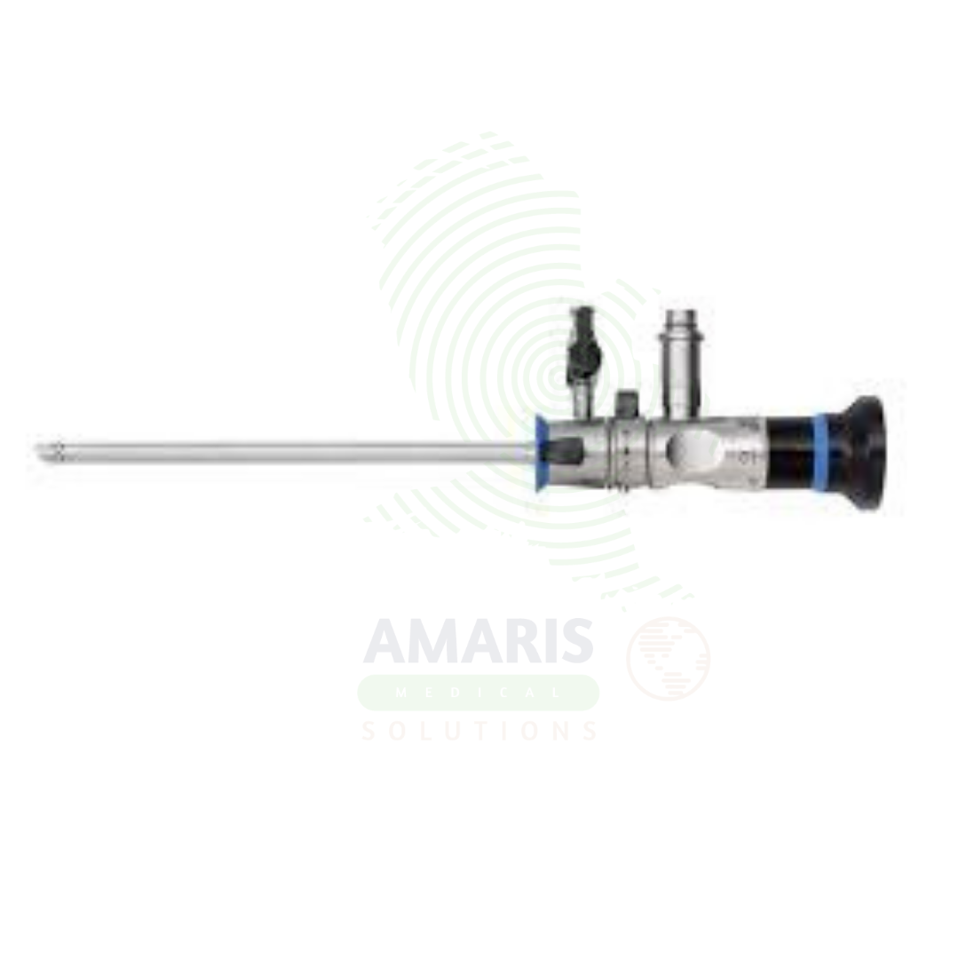
Arthroscope
$1.00
WhatsApp Order
An Arthroscope is a specialized endoscope used for minimally invasive visualization and treatment of joint pathology. Inserted through small incisions, it enables diagnostic examination of cartilage, ligaments, menisci, and synovium, as well as therapeutic procedures including meniscal repair, ligament reconstruction, and cartilage restoration. Used in knee, shoulder, hip, ankle, wrist, and elbow surgery, arthroscopy provides the benefits of minimally invasive surgery: smaller incisions, less pain, faster recovery, and earlier return to function.
Description
Arthroscope
PRIMARY CLINICAL & DIAGNOSTIC USES
1. Diagnostic Visualization of Joint Interiors
-
Primary Use: Provides direct, magnified visualization of the interior of a joint (synovium, articular cartilage, menisci, ligaments, tendons) to diagnose the source of pain, swelling, locking, or instability when non-invasive imaging is inconclusive. The arthroscope is inserted through small incisions (portals) to examine the joint in real time.
-
How it helps: For the orthopedic surgeon, the arthroscope transforms joint diagnosis from indirect imaging to direct visualization—revealing the exact condition of cartilage, the precise location of a meniscal tear, the true state of a ligament, or the source of unexplained synovitis. For the patient suffering from chronic knee pain, shoulder instability, or hip impingement, diagnostic arthroscopy often provides the definitive answer that MRI and X-ray could not, guiding treatment with certainty.
2. Therapeutic Joint Surgery
-
Primary Use: Enables minimally invasive surgical interventions within the joint, performed through small “keyhole” portals. The arthroscope serves as the “eyes” of the surgeon while specialized instruments are passed through additional portals to perform repairs, debridement, and reconstructions.
-
How it helps: For the orthopedic surgeon, the arthroscope is both eyes and instrument—allowing them to repair torn menisci, reconstruct cruciate ligaments, debride damaged cartilage, and stabilize shoulders through incisions measured in millimeters rather than inches. For the athlete sidelined by an ACL tear, the worker unable to lift due to rotator cuff injury, or the aging adult limited by meniscal pathology, arthroscopic surgery means their joint can be repaired through minimally invasive techniques, with less pain, faster rehabilitation, and earlier return to the activities they love.
3. Knee Arthroscopy
-
Primary Use: Performs meniscectomy, meniscal repair, ACL/PCL reconstruction, cartilage debridement, microfracture, loose body removal, and synovectomy through small portals. The arthroscope provides visualization of all compartments of the knee joint.
-
How it helps: For the knee surgeon, the arthroscope provides access to all compartments of this complex joint—visualizing the torn meniscus catching and locking, the absent ACL causing instability, or the cartilage defect causing pain. For the patient with a locked knee from a bucket-handle meniscal tear, or the athlete whose knee gives way from ACL insufficiency, arthroscopic repair offers the chance to restore function and return to activity without the morbidity of open arthrotomy.
4. Shoulder Arthroscopy
-
Primary Use: Performs rotator cuff repair, subacromial decompression, labral repair, biceps tenodesis, and stabilization for recurrent dislocation. The arthroscope navigates the complex anatomy of the glenohumeral joint and subacromial space.
-
How it helps: For the shoulder surgeon, the arthroscope navigates the complex anatomy of the glenohumeral joint and subacromial space—repairing torn rotator cuff tendons, debriding impinging bone spurs, and stabilizing recurrently dislocating shoulders. For the patient unable to lift their arm due to rotator cuff tear, or the young athlete with recurrent shoulder instability, arthroscopic repair offers the hope of restored function and stability without the large incisions and extensive soft tissue dissection of open surgery.
5. Hip Arthroscopy
-
Primary Use: Treats femoroacetabular impingement, labral repair and debridement, loose body removal, and synovial biopsy. The arthroscope provides access to the hip joint through specialized portals.
-
How it helps: For the hip preservation surgeon, arthroscopy enables treatment of femoroacetabular impingement and labral tears that cause groin pain and early arthritis—reshaping bone, repairing damaged labrum, and preserving joint function. For the young, active patient with hip pain, arthroscopic surgery offers the chance to return to sports and activities without the need for joint replacement.
SECONDARY & SUPPORTIVE USES
1. Ankle Arthroscopy: Treatment of osteochondral defects, anterior and posterior ankle impingement, arthrolysis, and synovitis.
2. Wrist and Elbow Arthroscopy: Diagnostic evaluation, loose body removal, synovectomy, and treatment of osteochondritis dissecans.
3. Biopsy Guidance: Allows precise, targeted biopsy of synovial tissue for histopathological diagnosis of conditions like pigmented villonodular synovitis or inflammatory arthritis.
4. Irrigation and Debridement: Used for lavage of septic joints to remove purulent material and debridement in early septic arthritis.
5. Fracture Assessment: Assists in the assessment and reduction of intra-articular fractures such as tibial plateau and radial head fractures.
6. Post-Operative Assessment: Used for second-look procedures to assess healing after meniscal repair or cartilage procedures.
KEY PRODUCT FEATURES
1. BASIC IDENTIFICATION ATTRIBUTES
-
Device Type: A specialized endoscope designed for visualization of joint interiors during diagnostic and therapeutic procedures.
-
Designation: Arthroscope, Joint Scope, Arthroscopic Camera, Arthroscopic System.
-
Key Components:
-
Rod Lens System: High-resolution optics for clear visualization.
-
Light Source: Fiberoptic or LED illumination.
-
Angled Optics: 0°, 30°, 70°, or 90° viewing angles for different joint visualization.
-
Camera: High-definition camera for image capture.
-
Sheath: Protective outer sleeve with irrigation and outflow channels.
-
2. TECHNICAL & PERFORMANCE PROPERTIES
-
Diameter: 1.9 mm, 2.7 mm, 4.0 mm, 5.5 mm depending on joint.
-
Length: 10-20 cm depending on application.
-
Viewing Angle: 0°, 30°, 70°, 90° for different anatomical visualization.
-
Resolution: High-definition (HD) and 4K options.
-
Sterilization: Steam autoclave or low-temperature sterilization.
-
Material: Surgical-grade stainless steel.
3. PHYSICAL & OPERATIONAL PROPERTIES
-
Construction: Precision-machined stainless steel with rod lens optics.
-
Ergonomics: Designed for one-handed manipulation.
-
Sterilization: Autoclavable.
-
Portability: Handheld with light cable and camera connection.
4. SAFETY & COMPLIANCE ATTRIBUTES
-
Regulatory Status: Class II medical device regulated by FDA.
-
Biocompatibility: Materials safe for intra-articular use.
-
Electrical Safety: Compliant for use with light sources and camera systems.
5. STORAGE & HANDLING ATTRIBUTES
-
Storage: Store in protective cases; protect lenses from damage.
-
Cleaning: Thorough cleaning after each use; ultrasonic cleaning recommended.
-
Sterilization: Steam autoclave per manufacturer instructions.
-
Inspection: Inspect for lens damage, scratches, or misalignment before use.
6. LABORATORY & CLINICAL APPLICATIONS
-
Primary Application: Diagnostic and therapeutic procedures in orthopedic surgery.
-
Clinical Role: Essential for minimally invasive joint surgery across multiple joints.
SAFETY HANDLING PRECAUTIONS
1. SAFETY PRECAUTIONS
-
Lens Protection: Handle carefully to avoid scratching the lens.
-
Sterility: Ensure sterility before intra-articular use.
-
Irrigation: Maintain irrigation flow to clear the field.
-
Portal Placement: Proper portal placement to avoid neurovascular structures.
2. FIRST AID MEASURES
-
Instrument Damage: If the arthroscope is dropped or damaged, remove from service; inspect before reuse.
3. FIRE FIGHTING MEASURES
-
Flammability: Metal components are non-flammable; light cables may burn.
-
Extinguishing Media: Use water, foam, or CO₂ as appropriate for surrounding materials.
Related products
AFO & Drop Foot Splint
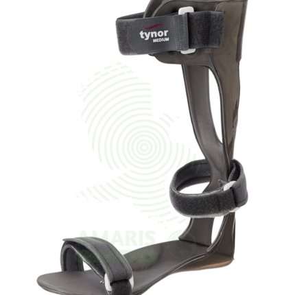
An AFO & Drop Foot Splint is a custom-fabricated brace that stabilizes the ankle and foot to correct foot drop and improve walking safety. By maintaining the foot at a 90-degree angle, it prevents toes from dragging during the swing phase of gait, thereby preventing trips and falls. Essential for patients with neurological conditions (stroke, MS, cerebral palsy), peripheral nerve injuries, or post-surgical instability, it is a prescription device meticulously crafted by an orthotist to provide specific levels of support, flexibility, and control. Daily skin checks and proper footwear are imperative for safe and effective long-term use.
Airprene Shoulder Support
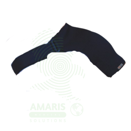
An Airprene Shoulder Support is a neoprene-based shoulder orthosis providing adjustable compression, warmth, and proprioceptive feedback for acute shoulder injuries, rotator cuff tendinopathy, shoulder instability, and post-operative recovery. The perforated neoprene material retains body heat to promote circulation and muscle relaxation while allowing air circulation to reduce moisture buildup. Used in sports medicine, orthopedics, and physical therapy settings, it provides symptomatic relief and functional support during healing and rehabilitation. Proper fit and skin integrity monitoring are essential for safe use.
Ankle Walkers (Fixed/Air)
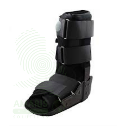
Ankle Walkers (Fixed/Air) are rigid, boot-like orthotic devices designed to immobilize the foot and ankle. Serving as a modern alternative to short leg casts, they are used for treating stable fractures, severe sprains, post-operative conditions, and for offloading diabetic foot ulcers. The "Fixed" type uses foam padding, while the "Air" type incorporates adjustable air bladders for a custom fit and superior swelling management. Both types feature a rocker-bottom sole to facilitate walking and multiple straps for secure immobilization. Daily skin inspection and proper fit are critical for safe and effective use, especially for patients with compromised sensation.
Cotton Arm Sling
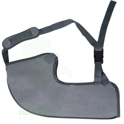
A Cotton Arm Sling is a soft, fabric support used to immobilize and support the arm following upper extremity injuries, fractures, and surgery. The sling supports the weight of the arm, reduces pain by limiting movement, and elevates the extremity to control swelling. Made from breathable cotton fabric with an adjustable neck strap, it provides comfortable, lightweight support for a wide range of conditions including wrist fractures, elbow sprains, hand injuries, and post-operative recovery. Proper positioning and circulation monitoring are essential for safe use.
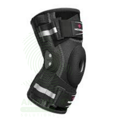
Hinged & Stabilizer Knee Braces
Hinged & Stabilizer Knee Braces are rigid or semi-rigid orthotic devices featuring side hinges, designed to provide mechanical stability, control motion, and protect the knee joint. They are essential for post-operative rehabilitation (e.g., after ACL reconstruction), managing chronic ligament instability, treating acute sprains, and, in the form of unloader braces, alleviating pain from osteoarthritis. Their effectiveness relies on a precise fit with proper hinge alignment, an adjustable strapping system, and selection of the correct brace type for the specific clinical indication. They are a cornerstone of non-surgical and post-surgical knee management.
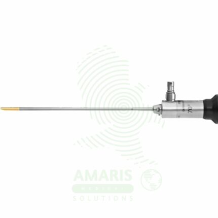
Orthopedic Arthroscope
An Orthopedic Arthroscope is a rigid, rod-lens telescope used for the visualization and surgical treatment of joint interiors. As the cornerstone of minimally invasive joint surgery, it provides a bright, magnified view of structures like cartilage, menisci, and ligaments through tiny incisions. Available in key diameters (2.7mm, 4.0mm) and viewing angles (0°, 30°, 70°), its optical quality is paramount for surgical precision. Mandatory pre-sterilization leak testing and careful autoclaving are required to maintain sterility and integrity. Proper handling is essential to prevent costly damage to the optics and to ensure patient safety during diagnostic and therapeutic arthroscopic procedures.
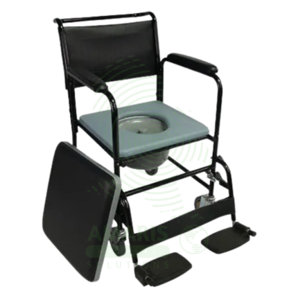
Orthopedic Commode Chair
An Orthopedic Commode Chair is a specialized hybrid device designed for patients with lower limb injuries or surgeries requiring non-weight-bearing status. It combines an extended-frame orthopedic wheelchair with an elevating leg rest and a commode seat, allowing for simultaneous fracture management (leg elevation/immobilization), pressure sore prevention (heel off-loading), and safe, hygienic toileting without dangerous transfers. Its use mandates the constant engagement of anti-tip levers due to a high risk of backwards tipping and requires careful navigation of its substantial length.