Dermatoscope and Magnifiers
 Diagnostic Kits
 Vital Signs Monitors
 Stethoscopes and Accessories
 Otoscopes, Ophthalmoscopes, and Retinoscopes
 Reflex Hammers and Neurological Tools
 Scales and Measuring Devices
 Spirometers and Pulmonary Function Tests
 Electrosurgical Units and Accessories
 Cutting Instruments
 Grasping and Holding Instruments
 Hemostatic Instruments
 Specialized Surgical Sets
 Single-Use Procedure Trays and Packs
 Surgical Drapes, Gowns, and Covers
 Tissue Unifying Instruments
 Radiation Protection
 X-Ray Machines and Accessories
 Ultrasound Systems and Probes
 MRI and CT Scanners
 Radiology Consumables
 Bone Densitometers
 Fluoroscopy Equipment
 Imaging Tables and Positioning Aids
 Microscopes and Accessories
 Centrifuges and Separators
 Analyzers
 Incubators and Ovens
 Pipettes, Dispensers, and Lab Glassware
 Refrigerators, Freezers, and Storage Units
 Lab Consumables
 Sterilizers and Autoclaves for Lab Use
 Multi-Parameter Monitors
 Ventilators and Respiratory Support Devices
 Defibrillators and AEDs
 Infusion Pumps and IV Systems
 Patient Warmers and Cooling Devices
 Central Monitoring Stations
 Accessories
 Anesthesia Machines and Workstations
 Oxygen Concentrators and Delivery Systems
 Nebulizers and Inhalers
 CPAP/BiPAP Machines
 Airway Management
 Anesthesia Masks, Circuits, and Bags
 Humidifiers and Heaters
 Respiratory Therapy Accessories
 First Aid Kits and Cabinets
 Emergency Resuscitation Equipment
 Trauma Supplies
 Emergency Carts and Crash Carts
 Burn Care Products
 Bleeding Control
 Automated External Defibrillators (AEDs)
 Transport and Evacuation
 Wheelchairs and Accessories
 Walkers, Crutches, and Canes
 Prosthetics and Orthotics
 Physical Therapy Equipment
 Transfer Devices
 Bathroom Safety
 Orthopedic Traction and Tables
 Hot/Cold Therapy Packs and Units
 Beds and Mattresses
 Chairs and Stools
 Tables
 Cabinets and Storage
 Privacy Screens & Curtains
 Stands and Racks
 Linens and Textiles
 Lighting
 Autoclaves and Sterilizers
 Ultrasonic Cleaners
 Disinfectant Solutions and Wipes
 Sterilization Pouches, Wraps, and Indicators
 Instrument Trays and Containers
 UV and Ozone Disinfection Devices
 Washer Disinfectors
 Wound Care
 Gloves
 Masks and Respirators
 Catheters and Tubing
 Swabs, Applicators, and Sponges
 Incontinence Products
 Personal Protective Equipment (PPE)
 Dental Chairs and Units
 Handpieces and Burs
 Instruments
 Consumables
 Sterilization for Dental Use
 Orthodontic Supplies
 Endodontic Tools
 Slit Lamps and Tonometers
 Lensometers and Phoropters
 Ophthalmic Surgical Instruments
 Eyewear Frames and Lenses
 Contact Lens Supplies
 Vision Testing Charts and Devices
 Eye Care Consumables
 Laser Systems for Eye Care
 ENT Exam Chairs and Tables
 Endoscopes
 Audiometers and Hearing Tests
 ENT Instruments
 Nasal and Throat Packs
 Hearing Aids and Accessories
 Otology Supplies
 Fetal Dopplers and Monitors
 Delivery Beds and Tables
 Gynecological Instruments
 Neonatal Incubators and Warmers
 Breast Pumps and Accessories
 Contraceptive Devices
 Maternity Supports and Pads
 Neonatal Consumables
 Cystoscopes and Urethroscopes
 Dialysis Machines and Supplies
 Urological Catheters and Bags
 Lithotripters
 Prostate Treatment Devices
 Urinary Incontinence Products
 Kidney Stone Management Tools
 Consumables & Disposables
 EEG and EMG Machines
 Neurosurgical Instruments
 Nerve Stimulators
 Headrests and Positioning Aids
 Lumbar Puncture Kits
 Seizure Monitoring Devices
 Consumables
 Rehabilitation for Neurological Conditions
 ECG Machines and Accessories
 Holter Monitors
 Stress Test Systems
 Pacemakers and Defibrillator Accessories
 Vascular Access Devices
 Cardiac Catheters and Guidewires
 Blood Flow Meters
 Consumables
 Orthopedic Instruments
 Casts, Splints, and Padding
 Joint Replacement Supplies
 Prosthetic Limbs and Components
 Bone Grafts and Substitutes
 Traction Devices
 Orthopedic Braces and Supports
 Rehabilitation Aids for Orthopedics
 Home Oxygen Therapy
 Hospital Beds for Home Use
 Mobility Aids
 Bathroom and Daily Living Aids
 Wound Care for Home
 Monitoring Devices
 Enteral Feeding Pumps and Tubes
 Hand Sanitizers and Dispensers
 Face Shields and Goggles
 Isolation Gowns and Suits
 Biohazard Waste Containers
 Air Purifiers and HEPA Filters
 Surface Disinfectants
 Sharps Containers
 Protective Barriers
 Cardiovascular & Endurance Training
 Strength Training & Weightlifting
 Functional Training & Core Conditioning
 Physical Therapy & Rehabilitation
 Sports & Outdoor Recreation
 Gym Flooring & Facility Equipment
 Fitness Monitoring & Accessories
 Kids & Novelties
Bloodlines for Dialysis
$1.00
WhatsApp Order
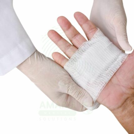
Bloodlines for Dialysis are sterile, single-use tubing sets that form the extracorporeal circuit for hemodialysis and other blood purification therapies. Transporting blood from the patient to the dialyzer and back, they include pressure monitoring ports, air trap chambers, and medication administration sites. Essential for dialysis centers, hospitals, and home hemodialysis.
Description
Bloodlines for Dialysis
PRIMARY CLINICAL & DIAGNOSTIC USES
1. Blood Conduit Between Patient and Dialyzer
-
Primary Use: Provides sterile, single-use tubing sets that transport the patient’s blood from the vascular access to the dialyzer for purification and then return the cleaned blood back to the patient. The bloodline forms the closed circuit essential for hemodialysis treatment.
-
How it helps: For the dialysis nurse and nephrologist, the bloodline set provides the sterile, disposable conduit that enables the extracorporeal circuit—allowing the dialyzer to remove toxins and excess fluid from the patient’s blood. For the patient undergoing dialysis, this means safe, effective treatment with a new, sterile bloodline for each session.
2. Arterial and Venous Pressure Monitoring
-
Primary Use: Integrated pressure monitoring ports allow continuous measurement of arterial and venous pressures during dialysis, providing critical information about vascular access function, circuit integrity, and the effectiveness of the treatment.
-
How it helps: For the dialysis clinician, pressure monitoring ports provide real-time data on circuit pressures—alerting to potential issues such as access obstruction, needle malposition, or clotting. For the patient, this monitoring ensures that treatment proceeds safely and that problems are detected early.
3. Blood Flow Control and Regulation
-
Primary Use: Designed with specific lumen diameters and flow characteristics to accommodate the high blood flow rates required for effective dialysis (typically 200-500 mL/min), ensuring consistent, turbulent-free flow through the circuit.
-
How it helps: For the dialysis team, bloodlines are engineered to maintain consistent flow rates—ensuring that the prescribed blood flow is delivered to the dialyzer for optimal clearance. For the patient, this means efficient treatment that removes adequate toxins and fluid within the prescribed treatment time.
4. Air Detection and Safety Mechanisms
-
Primary Use: Bloodlines are designed with integrated air traps and chambers that prevent air emboli from reaching the patient, working in conjunction with the dialysis machine’s air detection sensors to ensure patient safety.
-
How it helps: For the dialysis nurse, the air trap chambers provide a visible means of monitoring for air in the circuit and protecting against air embolus. For the patient, this safety feature is critical—preventing a potentially fatal complication of extracorporeal circulation.
5. Saline and Medication Administration Ports
-
Primary Use: Includes injection ports and saline administration sites that allow for priming of the circuit, delivery of medications (e.g., heparin, erythropoietin, iron), and saline flushes during the dialysis procedure.
-
How it helps: For the dialysis clinician, integrated ports simplify medication administration and circuit management—allowing for heparinization, saline boluses, and medication delivery without disrupting the sterile circuit. For the patient, this means medications can be administered safely during treatment without additional needle sticks.
SECONDARY & SUPPORTIVE USES
1. Hemoperfusion: Used with hemoperfusion cartridges for toxin removal in poisonings.
2. Plasmapheresis: Provides blood circuit for plasma exchange procedures.
3. Hemofiltration: Used in continuous renal replacement therapy and hemofiltration.
4. Pediatric Dialysis: Smaller bloodlines available for pediatric patients.
5. Home Hemodialysis: Used by patients performing dialysis at home.
6. Apheresis Procedures: Bloodlines for therapeutic apheresis and cell collection.
KEY PRODUCT FEATURES
1. BASIC IDENTIFICATION ATTRIBUTES
-
Product Type: Sterile, single-use tubing sets for extracorporeal blood circulation during dialysis.
-
Designation: Bloodlines for Dialysis, Dialysis Bloodlines, Hemodialysis Tubing Set, Extracorporeal Circuit.
-
Key Components:
-
Arterial Line: Tubing that carries blood from patient to dialyzer.
-
Venous Line: Tubing that returns cleaned blood to patients.
-
Pressure Monitoring Ports: For arterial and venous pressure measurement.
-
Air Trap Chamber: Prevents air emboli from reaching patients.
-
Injection Ports: For heparin, saline, and medication administration.
-
Blood Pump Segment: Reinforced tubing designed for peristaltic pump operation.
-
Connectors: Standard dialysis connectors for patient access and dialyzer.
-
2. TECHNICAL & PERFORMANCE PROPERTIES
-
Material: Medical-grade PVC; non-toxic, biocompatible.
-
Tubing Diameter: Designed for blood flow rates of 200-500 mL/min.
-
Length: 100-200 cm depending on configuration.
-
Priming Volume: 50-150 mL depending on circuit design.
-
Sterility: Ethylene oxide or gamma irradiation sterilized.
-
Latex-Free: Manufactured without natural rubber latex.
-
DEHP-Free: Available in DEHP-free options.
3. PHYSICAL & OPERATIONAL PROPERTIES
-
Construction: Flexible, kink-resistant tubing.
-
Transparency: Clear for visualization of blood flow and air bubbles.
-
Connectors: Standard dialysis connectors (Luer, ISO, or proprietary).
-
Packaging: Sterile, single-use; individually packaged.
-
Disposal: Dispose as medical waste after use.
4. SAFETY & COMPLIANCE ATTRIBUTES
-
Regulatory Status: Class II medical device regulated by FDA.
-
Sterility Assurance: Validated sterilization process.
-
Biocompatibility: Materials safe for blood contact.
-
Air Detection: Designed to work with machine air detection systems.
-
Pressure Ratings: Tested for pressure ranges encountered during dialysis.
5. STORAGE & HANDLING ATTRIBUTES
-
Storage: Store in cool, dry location; protect from heat and direct sunlight.
-
Sterility Maintenance: Do not use it if the package is opened, damaged, or wet.
-
Expiration: Check expiration date before use; do not use after expiration.
-
Single-Use Only: Intended for single patient use; do not resterilize or reuse.
6. LABORATORY & CLINICAL APPLICATIONS
-
Primary Application: Hemodialysis and other extracorporeal blood purification therapies.
-
Clinical Role: Essential equipment for dialysis centers, hospitals, and home hemodialysis.
SAFETY HANDLING PRECAUTIONS
1. SAFETY PRECAUTIONS
-
Priming: Prime circuit thoroughly with saline before connecting to the patient.
-
Air Detection: Monitor air trap chambers; ensure machine air detection is functional.
-
Connections: Ensure all connections are secure to prevent blood loss or air entry.
-
Pressure Monitoring: Monitor arterial and venous pressures throughout treatment.
-
Single-Use: Do not reuse; discard after each treatment.
-
Disposal: Dispose as biohazardous waste after use.
2. FIRST AID MEASURES
-
Air Embolism: If air embolism is suspected, clamp lines; place patients in left lateral decubitus; administer oxygen; call emergency.
-
Blood Loss: If line disconnect occurs, clamp immediately; apply pressure to access site; replace circuit.
-
Circuit Clotting: If clotting occurs, discontinue treatment; do not flush clots back to the patient.
3. FIRE FIGHTING MEASURES
-
Flammability: PVC tubing is combustible; an oxygen-enriched environment increases fire risk.
-
Extinguishing Media: For fire, disconnect from patient; use appropriate extinguisher.
Related products
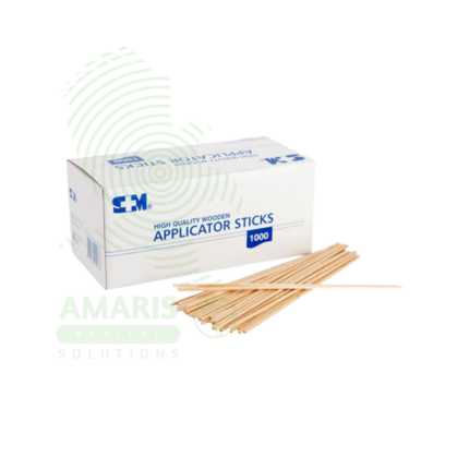
Applicator Sticks
Applicator Sticks are single-use medical devices with absorbent tips (cotton, rayon, flocked polyester) on wood, plastic, or paper shafts for collecting clinical specimens (throat, nasal, wound, cervical, urethral, rectal) for microbiological culture, rapid testing, and molecular diagnostics. Sterile individually wrapped or non-sterile bulk formats. Flocked swabs provide >90% specimen release for PCR testing. Primary applications include specimen collection for infection diagnosis, wound care and antiseptic application, gynecological sampling (Pap smears, STI testing), eye/ear specimen collection, laboratory slide preparation, and forensic evidence collection. Critical precautions include single-use only, sterile technique, proper collection method, and biohazard disposal. Essential consumable in all clinical settings.

Hepatitis C Test Kit
The Hepatitis C Test Kit is an in vitro diagnostic device for the detection of Hepatitis C virus antibodies or RNA in human blood, serum, or plasma samples. Available in rapid, laboratory immunoassay, and molecular formats, it screens for HCV infection, confirms active infection with RNA testing, and monitors treatment response. With high sensitivity and specificity exceeding 99%, it is essential for identifying infected individuals, linking them to curative direct-acting antiviral therapy, and confirming cure. Essential applications include risk-based screening of high-risk populations, birth cohort screening of baby boomers, evaluation of acute hepatitis, management of occupational exposures, and monitoring of antiviral therapy that can cure over 95% of infections. All patient samples must be handled with universal precautions, and positive antibody results require confirmatory testing.
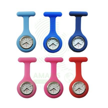
Nurse Watch
A Nurse Watch is a professional-grade analog wristwatch specifically designed for clinical use. Its hallmark is a built-in "pulsometer" scale that allows for rapid, on-the-spot calculation of a patient's heart rate by timing pulses over a short interval. Featuring a high-contrast dial with a clear sweep second hand, durable water-resistant construction, and a cleanable band, it is engineered for accuracy, legibility, and infection control in healthcare settings. It is an indispensable personal tool for nurses, enabling precise measurement of vital signs, timing of medications and procedures, and accurate clinical documentation throughout the shift.
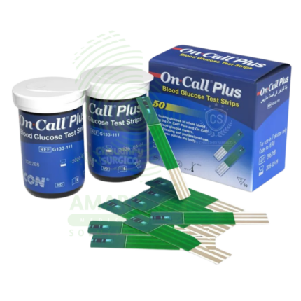
On Call Blood Sugar Strips
On Call Blood Sugar Strips are single-use, disposable electrochemical test strips (ISO 15197:2013 compliant) designed exclusively for use with On Call glucose meters to measure capillary blood glucose from 0.5-1.0 µL samples in 5-7 seconds. Each strip contains glucose oxidase or glucose dehydrogenase reagents that generate electrical current proportional to glucose concentration (20-600 mg/dL range). Packaging options include multi-strip vials (25-100 strips) with desiccant for daily use or individually foil-wrapped strips for travel. Strips feature no-coding automatic calibration, hematocrit compensation (20-60%), and interference resistance. Primary clinical applications include daily blood glucose testing for Type 1, Type 2, and gestational diabetes management, hypoglycemia and hyperglycemia detection, pre- and post-meal glucose assessment, and hospital point-of-care testing. Critical safety precautions include single-use only, immediate vial closure after strip removal to prevent humidity damage, storage at 2-30°C away from bathrooms and temperature extremes, never using expired strips, proper hand hygiene before testing, and disposal in sharps containers. Essential consumable for diabetes self-management when used with On Call meters.
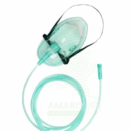
Oxygen Mask (Adult/Infant/Paediatric)
An Oxygen Mask is a disposable, single-use interface used to deliver supplemental oxygen from a medical gas supply to a patient's airways. Available in types for specific clinical needs—Simple Mask for low-flow therapy, Venturi Mask for precise FiO2 control (especially in COPD), and Non-Rebreather Mask for emergency high-flow delivery—and in sizes for adults, children, and infants. Correct selection and application, including ensuring the proper oxygen flow rate for the mask type, are essential for effective therapy and patient safety. Adherence to strict fire safety protocols is non-negotiable due to the combustion risk posed by enriched oxygen environments.
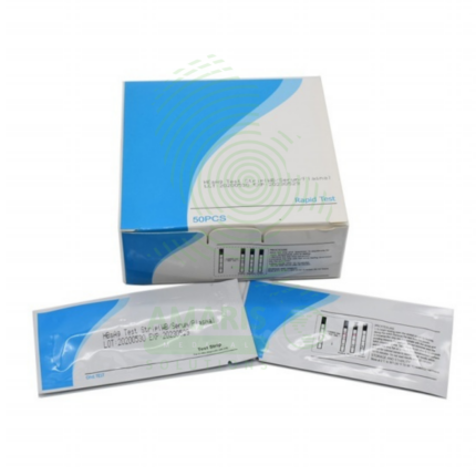
Rapid Test Kits (HIV, HBV, HCV, Malaria)
Rapid Test Kits for HIV, HBV, HCV, and Malaria are single-use, immunochromatographic devices designed for the quick, preliminary screening of these critical infectious diseases at the point of care. By detecting specific antibodies or antigens in a small blood sample (or oral fluid for some HIV tests), they provide a visual result within 15-30 minutes without the need for laboratory equipment. Their primary role is to expand access to testing in community and resource-limited settings, enabling immediate counseling, triage, and referral for confirmatory testing and treatment. As essential tools in public health, they are characterized by their simplicity, speed, and critical importance in early diagnosis and outbreak response.
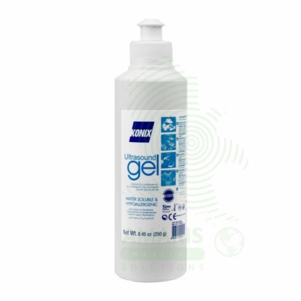
Ultrasound Gel
Ultrasound Gel is a water-based, hypoallergenic, acoustically conductive coupling medium essential for diagnostic and therapeutic ultrasound procedures. It eliminates air pockets between the transducer and skin, creating an efficient acoustic pathway for sound wave transmission and reception. Formulated to match the acoustic impedance of human tissue, it ensures high-quality imaging while providing lubrication for smooth probe movement and patient comfort. Available in standard, sterile, and hypoallergenic formulations, it is a single-use consumable critical for all ultrasound examinations. Proper storage, handling, and selection of appropriate sterile versus non-sterile formulations are essential for patient safety and image quality.