Dermatoscope and Magnifiers
 Diagnostic Kits
 Vital Signs Monitors
 Stethoscopes and Accessories
 Otoscopes, Ophthalmoscopes, and Retinoscopes
 Reflex Hammers and Neurological Tools
 Scales and Measuring Devices
 Spirometers and Pulmonary Function Tests
 Electrosurgical Units and Accessories
 Cutting Instruments
 Grasping and Holding Instruments
 Hemostatic Instruments
 Specialized Surgical Sets
 Single-Use Procedure Trays and Packs
 Surgical Drapes, Gowns, and Covers
 Tissue Unifying Instruments
 Radiation Protection
 X-Ray Machines and Accessories
 Ultrasound Systems and Probes
 MRI and CT Scanners
 Radiology Consumables
 Bone Densitometers
 Fluoroscopy Equipment
 Imaging Tables and Positioning Aids
 Microscopes and Accessories
 Centrifuges and Separators
 Analyzers
 Incubators and Ovens
 Pipettes, Dispensers, and Lab Glassware
 Refrigerators, Freezers, and Storage Units
 Lab Consumables
 Sterilizers and Autoclaves for Lab Use
 Multi-Parameter Monitors
 Ventilators and Respiratory Support Devices
 Defibrillators and AEDs
 Infusion Pumps and IV Systems
 Patient Warmers and Cooling Devices
 Central Monitoring Stations
 Accessories
 Anesthesia Machines and Workstations
 Oxygen Concentrators and Delivery Systems
 Nebulizers and Inhalers
 CPAP/BiPAP Machines
 Airway Management
 Anesthesia Masks, Circuits, and Bags
 Humidifiers and Heaters
 Respiratory Therapy Accessories
 First Aid Kits and Cabinets
 Emergency Resuscitation Equipment
 Trauma Supplies
 Emergency Carts and Crash Carts
 Burn Care Products
 Bleeding Control
 Automated External Defibrillators (AEDs)
 Transport and Evacuation
 Wheelchairs and Accessories
 Walkers, Crutches, and Canes
 Prosthetics and Orthotics
 Physical Therapy Equipment
 Transfer Devices
 Bathroom Safety
 Orthopedic Traction and Tables
 Hot/Cold Therapy Packs and Units
 Beds and Mattresses
 Chairs and Stools
 Tables
 Cabinets and Storage
 Privacy Screens & Curtains
 Stands and Racks
 Linens and Textiles
 Lighting
 Autoclaves and Sterilizers
 Ultrasonic Cleaners
 Disinfectant Solutions and Wipes
 Sterilization Pouches, Wraps, and Indicators
 Instrument Trays and Containers
 UV and Ozone Disinfection Devices
 Washer Disinfectors
 Wound Care
 Gloves
 Masks and Respirators
 Catheters and Tubing
 Swabs, Applicators, and Sponges
 Incontinence Products
 Personal Protective Equipment (PPE)
 Dental Chairs and Units
 Handpieces and Burs
 Instruments
 Consumables
 Sterilization for Dental Use
 Orthodontic Supplies
 Endodontic Tools
 Slit Lamps and Tonometers
 Lensometers and Phoropters
 Ophthalmic Surgical Instruments
 Eyewear Frames and Lenses
 Contact Lens Supplies
 Vision Testing Charts and Devices
 Eye Care Consumables
 Laser Systems for Eye Care
 ENT Exam Chairs and Tables
 Endoscopes
 Audiometers and Hearing Tests
 ENT Instruments
 Nasal and Throat Packs
 Hearing Aids and Accessories
 Otology Supplies
 Fetal Dopplers and Monitors
 Delivery Beds and Tables
 Gynecological Instruments
 Neonatal Incubators and Warmers
 Breast Pumps and Accessories
 Contraceptive Devices
 Maternity Supports and Pads
 Neonatal Consumables
 Cystoscopes and Urethroscopes
 Dialysis Machines and Supplies
 Urological Catheters and Bags
 Lithotripters
 Prostate Treatment Devices
 Urinary Incontinence Products
 Kidney Stone Management Tools
 Consumables & Disposables
 EEG and EMG Machines
 Neurosurgical Instruments
 Nerve Stimulators
 Headrests and Positioning Aids
 Lumbar Puncture Kits
 Seizure Monitoring Devices
 Consumables
 Rehabilitation for Neurological Conditions
 ECG Machines and Accessories
 Holter Monitors
 Stress Test Systems
 Pacemakers and Defibrillator Accessories
 Vascular Access Devices
 Cardiac Catheters and Guidewires
 Blood Flow Meters
 Consumables
 Orthopedic Instruments
 Casts, Splints, and Padding
 Joint Replacement Supplies
 Prosthetic Limbs and Components
 Bone Grafts and Substitutes
 Traction Devices
 Orthopedic Braces and Supports
 Rehabilitation Aids for Orthopedics
 Home Oxygen Therapy
 Hospital Beds for Home Use
 Mobility Aids
 Bathroom and Daily Living Aids
 Wound Care for Home
 Monitoring Devices
 Enteral Feeding Pumps and Tubes
 Hand Sanitizers and Dispensers
 Face Shields and Goggles
 Isolation Gowns and Suits
 Biohazard Waste Containers
 Air Purifiers and HEPA Filters
 Surface Disinfectants
 Sharps Containers
 Protective Barriers
 Cardiovascular & Endurance Training
 Strength Training & Weightlifting
 Functional Training & Core Conditioning
 Physical Therapy & Rehabilitation
 Sports & Outdoor Recreation
 Gym Flooring & Facility Equipment
 Fitness Monitoring & Accessories
 Kids & Novelties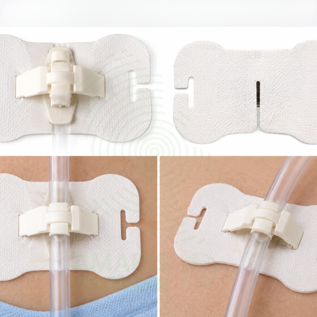

Body surface conduit fixing device – Type B
WhatsApp Order
The Body Surface Conduit Fixing Device – Type B is a sterile, adhesive catheter stabilization device specifically engineered for the securement of epidural catheters, central venous catheters (CVCs), and large-bore drainage tubes . Unlike standard tape or suture methods, the Type B device features a rigid locking cradle or mechanical clamp integrated into a breathable, transparent adhesive dressing .
For the clinician (anesthesiologist, intensivist, or nurse), Type B devices offer a “hands-free” solution to catheter fixation. The specialized clamp holds the catheter wings or hub firmly in place, preventing pistoning (back-and-forth movement) that introduces bacteria into the bloodstream, thereby reducing Catheter-Related Bloodstream Infections (CRBSI) . The transparent film backing allows for continuous visual inspection of the insertion site without disturbing the dressing, facilitating early detection of phlebitis or drainage .
For the patient, the primary benefit is safety and comfort. Elimination of silk sutures removes the “pinching” and “pulling” sensation at the insertion site, which is often a major source of pain for patients with long-term central lines (e.g., oncology or dialysis patients). The hypoallergenic, waterproof adhesive allows patients to shower without fear of the dressing falling off, significantly improving quality of life. Type B devices are anatomically versatile, used on the chest for PICCs, the back for epidurals, and the abdomen for drainage tubes, offering a universal, standardized solution for device stabilization across the hospital .
Description
Body Surface Conduit Fixing Device – Type B
PRIMARY CLINICAL & DIAGNOSTIC USES
1. Securement of Epidural and Central Venous Catheters
-
Primary Use: Provides a sterile, single-use adhesive device with a specialized mechanical fixation clamp (or locking cradle) designed to securely anchor epidural catheters, central venous catheters (CVCs), and peripherally inserted central catheters (PICCs) to the patient’s skin without sutures .
-
How it helps: For the anesthesiologist, interventional radiologist, and critical care nurse, the Type B device features a rigid or semi-rigid locking mechanism that securely grips the catheter hub or wings, preventing pistoning (back-and-forth movement) and accidental dislodgement . Unlike standard tape, the fixation clamp holds the catheter without compressing the lumen, maintaining flow integrity. For the patient receiving epidural analgesia, chemotherapy, or long-term antibiotics, this secure fixation eliminates the pain of suture removal and reduces the risk of catheter-related bloodstream infections (CRBSI) by preventing skin flora migration along the catheter tract.
2. Universal Compatibility with Multiple Conduit Types
-
Primary Use: Engineered with an adjustable fixation cradle that accommodates a wide range of catheter diameters and connector sizes, including central lines, drainage catheters, arterial lines, and feeding tubes .
-
How it helps: For the hospital supply chain manager and intensive care unit (ICU) clinician, the Type B device reduces inventory complexity by using a single fixation device across multiple catheter types. The adjustable cradle (often utilizing a “snap-on” or “click-lock” design) fits various manufacturers’ catheter wings without modification. For the patient, this means the nursing staff always has the correct securement device readily available, reducing delays in catheter dressing changes and minimizing the risk of accidental line pulls during patient turning or transport.
3. Skin-Friendly Adhesive Base with Hydrocolloid Protection
-
Primary Use: Incorporates a soft, hypoallergenic adhesive pad (typically polyurethane film or non-woven fabric) that adheres to the percutaneously, providing a stable base for the fixation clamp while protecting the fragile skin of critically ill or irradiated patients .
-
How it helps: For the wound care nurse and dermatology liaison, the Type B device features a gentle acrylic or silicone adhesive that minimizes the risk of Medical Adhesive-Related Skin Injury (MARSI). The integrated hydrocolloid or foam layer wicks moisture away from the insertion site, reducing maceration associated with sweat or serous drainage. For the patient with sensitive skin, history of radiation therapy, or long-term central access (e.g., oncology patients), this gentle but secure adhesion prevents painful skin tears and contact dermatitis, allowing for extended wear times (up to 7 days) without the need for painful re-taping.
4. Integrated Strain Relief for High-Flow and Large-Bore Catheters
-
Primary Use: Features a reinforced strain relief channel that redirects tubing stress vectors away from the catheter insertion site, preventing kinking and pullout of large-bore hemodialysis or apheresis catheters .
-
How it helps: For the dialysis nurse and apheresis specialist, the Type B device includes a built-in “J-loop” or “stress-relief” channel that absorbs the torque generated by heavy IV tubing. This prevents the catheter from being pulled out of the vessel during patient position changes. For the patient undergoing renal replacement therapy or stem cell collection, this strain relief prevents the sudden, painful sensation of the catheter tugging at the insertion site, allowing them to sleep or sit comfortably during long procedures (often 4-6 hours).
5. Visual Site Inspection Window for Infection Surveillance
-
Primary Use: Manufactured with a transparent film or fenestrated window that allows clinicians to directly visualize the catheter insertion site for early signs of infection (erythema, drainage, swelling) without removing the securement device .
-
How it helps: For the infection control practitioner and bedside nurse, the transparent backing provides continuous visibility of the “VAD” site. This facilitates compliance with the “Daily Review of Necessity” protocols and allows for instant assessment of site integrity during hourly rounding. For the patient, this transparency means the device does not need to be changed unnecessarily just to check the skin, reducing the frequency of dressing changes, which can be both painful and costly.
SECONDARY & SUPPORTIVE USES
1. Peripheral Nerve Catheter Stabilization: Used in regional anesthesia to secure catheters for continuous peripheral nerve blocks (femoral, sciatic, brachial plexus), preventing dislodgement that would interrupt post-operative pain control .
2. Chest Tube and Surgical Drain Management: Secures thoracostomy tubes and Jackson-Pratt (JP) drains to the chest or abdomen, preventing pullout that could lead to pneumothorax or surgical site infection .
3. Endotracheal Tube Fixation (Oral/Nasal): Modified Type B devices are used in anesthesia and critical care to anchor endotracheal tubes to the face, replacing traditional tape and reducing facial skin breakdown .
4. Nasogastric and Orogastric Tube Securement: Used to affix feeding tubes to the nose or cheek, preventing accidental removal which requires radiographic confirmation for re-placement .
5. Tracheostomy Tube Holder: Functions as a securement device for tracheostomy flanges, preventing tube dislodgement and reducing the risk of tracheal injury .
6. Hemodialysis Catheter Anchor: Specifically designed for large-bore dialysis catheters to withstand the high flow rates and patient movement during treatment, preventing venous stenosis caused by mechanical irritation .
7. Cardiovascular Monitoring Line Fixation: Used to hold arterial lines (A-lines) and pulmonary artery catheters in place in the ICU to ensure continuous hemodynamic monitoring accuracy .
8. Urinary Catheter Stabilization: Secures Foley catheters to the patient’s leg, preventing urethral traction and reducing the risk of catheter-associated urinary tract infections (CAUTI) .
9. Wound Drainage Tube Management: Attaches to surgical drains (e.g., Hemovac, Blakes) exiting the wound to prevent tugging and tension on the fresh incision line .
10. Interventional Radiology Access Site Care: Used to fix sheaths and dilators during interventional procedures to maintain vascular access without manual holding .
11. Pediatric Catheter Securement: Provides a secure, low-profile anchor for pediatric patients with small vasculature and highly mobile skin, reducing the risk of extravasation from dislodged IVs .
12. Patient-Controlled Analgesia (PCA) Line Protection: Holds PCA tubing securely in place so that the patient does not dislodge the line during sleep or ambulation .
13. Cerebral Spinal Fluid (CSF) Drainage System Securement: For patients with external ventricular drains (EVDs) or lumbar drains, the Type B device ensures the critical zero-level reference point remains stable to prevent over-drainage .
14. No-Sting Removal Protocols: Designed to be used in conjunction with silicone-based adhesive removers to facilitate pain-free device changes for burn or graft patients .
15. Home Healthcare Long-Term Line Management: Used by visiting nurses for patients with Port-a-Caths or PICC lines receiving home infusions, ensuring secure access without requiring professional suture removal .
16. Pre-hospital and Emergency Room Securement: Quickly applied in trauma bays to stabilize emergency lines (chest tubes, central lines) before the patient is transported to radiology or the ICU .
17. Magnetic Resonance Imaging (MRI) Compatibility: Type B devices are typically manufactured without metal components, making them safe to wear during MRI scans .
KEY PRODUCT FEATURES
1. BASIC IDENTIFICATION ATTRIBUTES
-
Device Type: A sterile, single-use adhesive securing device consisting of a skin-friendly base patch integrated with a mechanical locking clamp or gel pad designed to fixate medical conduit to the body .
-
Designation: Body Surface Conduit Fixing Device - Type B, Catheter Fixation Device, Central Line Holder, Epidural Catheter Anchor, Tube Securement Device, Catheter Stabilization Device, Dressing with Fixation .
-
Key Components:
o Adhesive Base (Skin Patch): Polyurethane film or breathable non-woven fabric; may include a hydrocolloid moisture-wicking layer .
o Fixation Mechanism: Locking clamp, "Christmas tree" adapter, or hook-and-eye closure; often designed with a low profile to prevent snagging .
o Catheter Channel: Pre-formed groove or cradle (sizes range from 5 Fr to 18 Fr) to seat the conduit.
o Strain Relief Tab: Extended portion of the device to tape down the tubing loop, preventing direct pull on the clamp.
o Absorbent Pad: Some versions include an iodine or silver-infused pad directly at the exit site to provide light antimicrobial protection.
o Reinforced Hub Gripper: Specific molded features designed to grip the wings of central venous catheters (CVCs).
2. TECHNICAL & PERFORMANCE PROPERTIES
-
Material Composition: Backing: Polyurethane (PU) or Polyethylene (PE); Adhesive: Acrylic or Silicone; Clamp: Medical-grade ABS plastic or Nylon; Latex-Free.
-
Adhesion Strength: Stronger than standard medical tape, often designed to withstand high pull forces (e.g., > 2 lbs or antimicrobial dressings) .
-
Clamp Versatility: Type B models typically cover a range of 5 Fr to 12 Fr catheters; some are universal, while others are size-specific.
-
Fluid Resistance: Waterproof backing protects the insertion site from external moisture during showering.
-
Wear Time: Up to 7 days per application, provided the site remains clean, dry, and intact.
-
Moisture Vapor Transmission Rate (MVTR): High (breathable) to prevent maceration of the skin under the patch.
-
Sterility: Sterile; Ethylene Oxide (EtO) or Gamma sterilized.
-
HCPCS Code: A4450 or A4452 (Tape, non-waterproof / waterproof, per 18 inches), depending on local coverage specifics.
3. PHYSICAL & OPERATIONAL PROPERTIES
-
Construction: Multi-layer design: PET release liner / Acrylic adhesive / PU backing / Foam block / Plastic clamp .
-
Application: "Peel and stick" base plate application followed by "snap-down" clamp closure.
-
Visibility: Fully transparent base to allow for continuous site assessment (ISO 10993 tested) .
-
Portability: Individually packed in a peel-pouch maintaining sterility; available in bulk boxes of 50 or 100.
-
Texture: The backing is often slightly textured to prevent slipping and to provide a surface for writing the date of insertion.
-
Disposal: Discard as medical waste; do not recycle due to potential biohazard contamination.
4. SAFETY & COMPLIANCE ATTRIBUTES
-
Regulatory Status: Class I Medical Device (21 CFR 880.5090 - Catheter Securement Device) or Class II with specific infection prevention claims .
-
Latex-Free: Standard for modern securement devices .
-
Biocompatibility: ISO 10993 tested for cytotoxicity, sensitization, and irritation.
-
MRI Compatibility: Non-ferromagnetic components; labeled as "MR Safe" or "MR Conditional."
-
Phthalate-Free: Free of DEHP (Di(2-ethylhexyl)phthalate).
-
Adhesion Type: Pressure-sensitive adhesive (PSA) that requires activation by firm finger pressure.
5. STORAGE & HANDLING ATTRIBUTES
-
Storage: Store in a cool, dry place (15-30°C / 59-86°F). Keep away from direct sunlight and extreme heat, which may melt the adhesive.
-
Shelf Life: Typically 2 to 3 years from date of manufacture.
-
Skin Prep: Requires clean, dry, alcohol-free skin (if using silicone adhesive) or thoroughly dried Chlorhexidine prep (if using acrylic adhesive).
-
Inspection: Do not use if the package is torn, opened, or if the adhesive appears yellowed or dry.
-
Removal: Requires an adhesive remover wipe to prevent skin stripping; remove by gently pulling the film parallel to the skin surface (low-and-slow technique).
6. LABORATORY & CLINICAL APPLICATIONS
-
Primary Application: To non-invasively secure and immobilize invasive medical lines (central lines, epidurals, chest tubes) to the skin surface to prevent dislodgement and infection .
-
Clinical Role: An essential infection control and patient safety supply used in Intensive Care Units (ICUs), Operating Rooms (ORs), Emergency Departments (EDs), Oncology Clinics, and Home Healthcare settings.
SAFETY HANDLING PRECAUTIONS
1. SAFETY PRECAUTIONS
-
Avoid Tension: Ensure the tubing has a "service loop" (slack) between the fixation clamp and the patient's clothing to prevent accidental pull-outs .
-
Do Not Cover the Clamp: Ensure the mechanical clamp itself is not covered by secondary tape that could put tension on the release mechanism.
-
Site Assessment: Remove and replace immediately if signs of phlebitis (redness, heat, swelling) or purulent drainage are visible through the viewing window .
-
Do Not Reuse: The mechanical clamp is designed for single patient use only; reuse risks cross-contamination and clamp fatigue.
-
Pressure Injury Prevention: For patients with edema or thin skin, do not overtighten the clamp; ensure the foam block provides cushioning without blanching the skin.
2. FIRST AID MEASURES
-
Contact Dermatitis: If the skin under the patch becomes red, weepy, or itchy, remove the device, cleanse the skin, and consult the wound care team about switching to silicone-based adhesive devices.
-
Adhesive Residue: Use a silicone-based medical adhesive remover (wipes or spray) to remove sticky residue from the skin; do not use alcohol on fragile skin as it causes drying and cracking.
-
Bleeding at Insertion Site: If the catheter site bleeds when the device is removed, apply sterile gauze with direct pressure for 5 minutes and assess for coagulopathy.
3. FIRE FIGHTING MEASURES
-
Flammability: The plastic components (backing and clamp) are slow to ignite but will melt if exposed to open flame.
-
Extinguishing Media: Use Water, Foam, Dry Chemical, or CO₂ for fires involving the packaging.
Related products
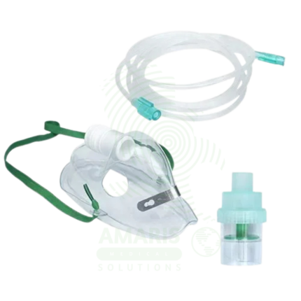
Adult & Pediatric Nebulizer Kit
An Adult & Pediatric Nebulizer Kit is a complete set of patient-contact components required for aerosol medication delivery, including a nebulizer cup, connecting tubing, and both an adult mouthpiece and a pediatric mask (or universal fit). Designed for use with a compressor, it provides the delivery circuit for treating asthma, COPD, and other respiratory conditions. Available in disposable (single-patient-use) formats for infection control in clinics or reusable formats for home care, its proper use, coupled with rigorous cleaning for reusable kits, is essential for effective therapy and preventing respiratory infections. The kit's universal connectors ensure compatibility with standard compressors.
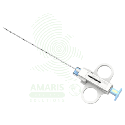
Biopsy Needles
Biopsy Needles are sterile, single-use precision medical devices designed to obtain tissue or cellular specimens from internal organs, masses, and lesions for histopathological or cytological diagnosis. Available in core biopsy (automatic spring-loaded, semi-automatic, vacuum-assisted, coaxial) and fine needle aspiration configurations, with gauges ranging from 14G-27G and lengths from 6-25 cm. Core biopsy needles obtain intact tissue cores preserving histological architecture; FNA needles aspirate cellular material for cytological evaluation. Specialized variants include bone marrow biopsy needles (Jamshidi, Illinois), echogenic-tip needles for ultrasound guidance, and MRI-compatible needles (nitinol, titanium). Critical safety requirements include strict image guidance for non-palpable lesions, verification of coagulation status, adherence to single-use protocols, and immediate post-procedure monitoring for complications (hemorrhage, pneumothorax, organ injury). An indispensable tool in interventional radiology, oncology, and minimally invasive diagnosis.
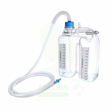
Chest Bottle
A Chest Bottle (Underwater Seal Drainage System) is a three-chamber disposable device used to drain air and fluid from the pleural space via a chest tube. Its critical component is a water seal chamber that acts as a one-way valve, preventing air from re-entering the chest while allowing drainage. It is used to treat pneumothorax, hemothorax, and pleural effusions, and is mandatory post-thoracic surgery. The system must always be kept upright and below the patient's chest level. It provides essential clinical information through tidaling in the water seal (indicating tube patency and lung expansion) and bubbling (indicating an air leak), making it both a therapeutic and diagnostic tool.
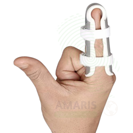
Finger Cot
A Finger Cot is a small, finger-shaped protective covering used to protect minor finger wounds, diabetic ulcers, surgical incisions, and amputation stumps, as well as to provide barrier protection for healthcare workers and laboratory personnel. Made from latex, nitrile, vinyl, or silicone, it provides a comfortable, secure fit while protecting the finger from contamination, moisture, friction, and irritants. Available in various sizes, materials, and sterile or non-sterile versions. Latex-free options are available for individuals with latex allergy.
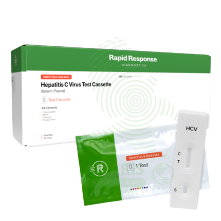
Hepatitis C Test Kit
The Hepatitis C Test Kit is an in vitro diagnostic device for the detection of Hepatitis C virus antibodies or RNA in human blood, serum, or plasma samples. Available in rapid, laboratory immunoassay, and molecular formats, it screens for HCV infection, confirms active infection with RNA testing, and monitors treatment response. With high sensitivity and specificity exceeding 99%, it is essential for identifying infected individuals, linking them to curative direct-acting antiviral therapy, and confirming cure. Essential applications include risk-based screening of high-risk populations, birth cohort screening of baby boomers, evaluation of acute hepatitis, management of occupational exposures, and monitoring of antiviral therapy that can cure over 95% of infections. All patient samples must be handled with universal precautions, and positive antibody results require confirmatory testing.
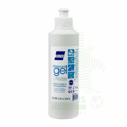
Ultrasound Gel
Ultrasound Gel is a water-based, hypoallergenic, acoustically conductive coupling medium essential for diagnostic and therapeutic ultrasound procedures. It eliminates air pockets between the transducer and skin, creating an efficient acoustic pathway for sound wave transmission and reception. Formulated to match the acoustic impedance of human tissue, it ensures high-quality imaging while providing lubrication for smooth probe movement and patient comfort. Available in standard, sterile, and hypoallergenic formulations, it is a single-use consumable critical for all ultrasound examinations. Proper storage, handling, and selection of appropriate sterile versus non-sterile formulations are essential for patient safety and image quality.
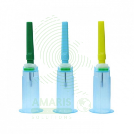
Vacutainer Needle Holder
A Vacutainer Needle Holder is a disposable plastic device (Class I/II medical device) used to securely hold double-ended blood collection needles and evacuated tubes during venipuncture, enabling safe and efficient collection of multiple blood specimens from a single venipuncture. Made from medical-grade ABS plastic or polypropylene, latex-free, approximately 12 cm length, with threaded end for needle attachment and flanged opening with flexible fingers to securely hold standard evacuated tubes (13 × 75 mm, 16 × 100 mm). Compatible with all standard double-ended needles and safety-engineered needles. Primary clinical applications include blood collection for laboratory testing (hematology, chemistry, coagulation, blood banking), multiple tube collection from single venipuncture, standardized venipuncture procedures, infection control (works with safety needles), and phlebotomy training. Critical safety precautions include single-use only (never reuse), secure needle attachment, immediate disposal of an entire unit with a needle in a sharps container, never recapping needles, and inspection for damage before use. Essential components of blood collection systems in hospitals, clinics, and laboratories.