Dermatoscope and Magnifiers
 Diagnostic Kits
 Vital Signs Monitors
 Stethoscopes and Accessories
 Otoscopes, Ophthalmoscopes, and Retinoscopes
 Reflex Hammers and Neurological Tools
 Scales and Measuring Devices
 Spirometers and Pulmonary Function Tests
 Electrosurgical Units and Accessories
 Cutting Instruments
 Grasping and Holding Instruments
 Hemostatic Instruments
 Specialized Surgical Sets
 Single-Use Procedure Trays and Packs
 Surgical Drapes, Gowns, and Covers
 Tissue Unifying Instruments
 Radiation Protection
 X-Ray Machines and Accessories
 Ultrasound Systems and Probes
 MRI and CT Scanners
 Radiology Consumables
 Bone Densitometers
 Fluoroscopy Equipment
 Imaging Tables and Positioning Aids
 Microscopes and Accessories
 Centrifuges and Separators
 Analyzers
 Incubators and Ovens
 Pipettes, Dispensers, and Lab Glassware
 Refrigerators, Freezers, and Storage Units
 Lab Consumables
 Sterilizers and Autoclaves for Lab Use
 Multi-Parameter Monitors
 Ventilators and Respiratory Support Devices
 Defibrillators and AEDs
 Infusion Pumps and IV Systems
 Patient Warmers and Cooling Devices
 Central Monitoring Stations
 Accessories
 Anesthesia Machines and Workstations
 Oxygen Concentrators and Delivery Systems
 Nebulizers and Inhalers
 CPAP/BiPAP Machines
 Airway Management
 Anesthesia Masks, Circuits, and Bags
 Humidifiers and Heaters
 Respiratory Therapy Accessories
 First Aid Kits and Cabinets
 Emergency Resuscitation Equipment
 Trauma Supplies
 Emergency Carts and Crash Carts
 Burn Care Products
 Bleeding Control
 Automated External Defibrillators (AEDs)
 Transport and Evacuation
 Wheelchairs and Accessories
 Walkers, Crutches, and Canes
 Prosthetics and Orthotics
 Physical Therapy Equipment
 Transfer Devices
 Bathroom Safety
 Orthopedic Traction and Tables
 Hot/Cold Therapy Packs and Units
 Beds and Mattresses
 Chairs and Stools
 Tables
 Cabinets and Storage
 Privacy Screens & Curtains
 Stands and Racks
 Linens and Textiles
 Lighting
 Autoclaves and Sterilizers
 Ultrasonic Cleaners
 Disinfectant Solutions and Wipes
 Sterilization Pouches, Wraps, and Indicators
 Instrument Trays and Containers
 UV and Ozone Disinfection Devices
 Washer Disinfectors
 Wound Care
 Gloves
 Masks and Respirators
 Catheters and Tubing
 Swabs, Applicators, and Sponges
 Incontinence Products
 Personal Protective Equipment (PPE)
 Dental Chairs and Units
 Handpieces and Burs
 Instruments
 Consumables
 Sterilization for Dental Use
 Orthodontic Supplies
 Endodontic Tools
 Slit Lamps and Tonometers
 Lensometers and Phoropters
 Ophthalmic Surgical Instruments
 Eyewear Frames and Lenses
 Contact Lens Supplies
 Vision Testing Charts and Devices
 Eye Care Consumables
 Laser Systems for Eye Care
 ENT Exam Chairs and Tables
 Endoscopes
 Audiometers and Hearing Tests
 ENT Instruments
 Nasal and Throat Packs
 Hearing Aids and Accessories
 Otology Supplies
 Fetal Dopplers and Monitors
 Delivery Beds and Tables
 Gynecological Instruments
 Neonatal Incubators and Warmers
 Breast Pumps and Accessories
 Contraceptive Devices
 Maternity Supports and Pads
 Neonatal Consumables
 Cystoscopes and Urethroscopes
 Dialysis Machines and Supplies
 Urological Catheters and Bags
 Lithotripters
 Prostate Treatment Devices
 Urinary Incontinence Products
 Kidney Stone Management Tools
 Consumables & Disposables
 EEG and EMG Machines
 Neurosurgical Instruments
 Nerve Stimulators
 Headrests and Positioning Aids
 Lumbar Puncture Kits
 Seizure Monitoring Devices
 Consumables
 Rehabilitation for Neurological Conditions
 ECG Machines and Accessories
 Holter Monitors
 Stress Test Systems
 Pacemakers and Defibrillator Accessories
 Vascular Access Devices
 Cardiac Catheters and Guidewires
 Blood Flow Meters
 Consumables
 Orthopedic Instruments
 Casts, Splints, and Padding
 Joint Replacement Supplies
 Prosthetic Limbs and Components
 Bone Grafts and Substitutes
 Traction Devices
 Orthopedic Braces and Supports
 Rehabilitation Aids for Orthopedics
 Home Oxygen Therapy
 Hospital Beds for Home Use
 Mobility Aids
 Bathroom and Daily Living Aids
 Wound Care for Home
 Monitoring Devices
 Enteral Feeding Pumps and Tubes
 Hand Sanitizers and Dispensers
 Face Shields and Goggles
 Isolation Gowns and Suits
 Biohazard Waste Containers
 Air Purifiers and HEPA Filters
 Surface Disinfectants
 Sharps Containers
 Protective Barriers
 Cardiovascular & Endurance Training
 Strength Training & Weightlifting
 Functional Training & Core Conditioning
 Physical Therapy & Rehabilitation
 Sports & Outdoor Recreation
 Gym Flooring & Facility Equipment
 Fitness Monitoring & Accessories
 Kids & Novelties
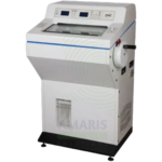
Fully Automated Microtome
WhatsApp Order
A Fully Automated Microtome is a precision instrument for sectioning paraffin-embedded tissue blocks for histopathological examination. Motorized specimen advance and programmable settings ensure consistent section thickness, reduce operator fatigue, and improve throughput in high-volume histology laboratories. Integrated safety features and ergonomic design protect operators from injury while producing high-quality sections essential for accurate pathological diagnosis.
Description
Fully Automated Microtome
PRIMARY CLINICAL & DIAGNOSTIC USES
1. Precision Tissue Sectioning for Histopathology
-
Primary Use: Provides fully automated, precision sectioning of paraffin-embedded tissue blocks for histopathological examination. The automated microtome ensures consistent section thickness, reduces operator fatigue, and improves throughput in high-volume histology laboratories.
-
How it helps: For the histotechnologist and pathologist, the fully automated microtome delivers consistent, high-quality sections essential for accurate diagnosis—reducing variability between operators and minimizing the need for repeat sections. For the patient, consistent, high-quality sections ensure that the pathologist can make an accurate diagnosis from the tissue sample, guiding appropriate treatment.
2. High-Throughput Sectioning for Busy Laboratories
-
Primary Use: Designed for high-volume histology laboratories, the automated microtome processes multiple tissue blocks efficiently, with automated sectioning, collection, and sometimes integrated slide mounting. This increases laboratory productivity and reduces turnaround time for pathology results.
-
How it helps: For the laboratory manager, automated microtomes increase efficiency and throughput—reducing staffing requirements for repetitive sectioning tasks and enabling faster reporting of pathology results. For the patient, this means shorter waiting times for biopsy results and quicker initiation of treatment.
3. Motorized Sectioning with Programmable Settings
-
Primary Use: Features motorized specimen advance and cutting mechanisms with programmable settings for section thickness, cutting speed, and sectioning modes (continuous, single, or step). This ensures consistent, reproducible sectioning across different operators and specimens.
-
How it helps: For the histotechnologist, programmable settings reduce the physical demands of manual sectioning and ensure consistent results regardless of operator experience. For the patient, reproducibility means that diagnostic quality does not depend on the skill level of the individual technician.
4. Section Collection and Integration with Slide Preparation
-
Primary Use: Some fully automated microtomes integrate with section collection systems and automated slide preparation equipment, allowing seamless transfer of sections to slides for staining and coverslipping, reducing manual handling and potential errors.
-
How it helps: For the laboratory, integrated systems reduce manual handling of delicate tissue sections—minimizing the risk of tearing, folding, or loss of tissue. For the patient, this integration improves the quality and reliability of the final slide preparation.
5. User Safety and Ergonomic Design
-
Primary Use: Automated microtomes incorporate safety features such as automatic blade retraction, handwheel locks, and protective covers to reduce the risk of injury. Ergonomic design reduces repetitive strain injuries associated with manual sectioning.
-
How it helps: For the histotechnologist, automated safety features reduce the risk of blade injuries and repetitive strain injuries—creating a safer, more ergonomic work environment. For the laboratory, this reduces worker compensation claims and staff turnover.
SECONDARY & SUPPORTIVE USES
1. Research Applications: Sectioning of research tissue blocks for histology, immunohistochemistry, and in situ hybridization.
2. Quality Control: Consistent sectioning for quality control programs in histology laboratories.
3. Training: Automated microtomes used in training programs to teach sectioning principles without the learning curve of manual techniques.
4. Digital Pathology Integration: Sectioning for digital slide scanning and whole slide imaging.
5. Special Stains: Preparation of sections for special staining procedures.
6. Immunohistochemistry: Sectioning for IHC testing.
KEY PRODUCT FEATURES
1. BASIC IDENTIFICATION ATTRIBUTES
-
Device Type: A fully automated microtome for precision sectioning of paraffin-embedded tissue blocks.
-
Designation: Fully Automated Microtome, Automatic Microtome, Motorized Microtome, Rotary Microtome.
-
Key Components:
-
Motorized Specimen Advance: Automated feed mechanism.
-
Cutting Mechanism: Precision blade holder with adjustable angle.
-
Control Panel: Digital interface for section thickness and speed.
-
Specimen Holder: Adjustable chuck for tissue blocks.
-
Section Collection: Integrated collection system (optional).
-
Safety Features: Blade retraction, handwheel lock, protective covers.
-
2. TECHNICAL & PERFORMANCE PROPERTIES
-
Section Thickness: 0.5-100 microns adjustable.
-
Sectioning Speed: 0.5-10 mm/s variable.
-
Specimen Size: Up to 60 x 50 mm typical.
-
Motorized Advance: Automatic specimen feed.
-
Sectioning Modes: Continuous, single, step, and programmable.
-
Blade Angle: Adjustable 0-45 degrees.
-
Memory: Programmable settings for routine protocols.
3. PHYSICAL & OPERATIONAL PROPERTIES
-
Construction: Durable metal construction; corrosion-resistant.
-
Dimensions: Benchtop unit.
-
Controls: Touchscreen or digital interface.
-
Portability: Stationary benchtop installation.
-
Cleaning: Easy-clean surfaces; removable components.
4. SAFETY & COMPLIANCE ATTRIBUTES
-
Regulatory Status: Class I or Class II medical device.
-
Electrical Safety: Compliant with electrical safety standards.
-
Blade Safety: Automatic blade retraction; blade disposal system.
-
Ergonomics: Designed to reduce repetitive strain injuries.
5. STORAGE & HANDLING ATTRIBUTES
-
Storage: Stored on laboratory benchtop.
-
Cleaning: Regular cleaning of blade holder and specimen clamp.
-
Maintenance: Regular calibration; blade replacement; lubrication.
-
Blade Disposal: Dispose of used blades in sharps containers.
6. LABORATORY & CLINICAL APPLICATIONS
-
Primary Application: Precision tissue sectioning for histopathology, immunohistochemistry, and research.
-
Clinical Role: Essential equipment in histology laboratories, pathology departments, and research facilities.
SAFETY HANDLING PRECAUTIONS
1. SAFETY PRECAUTIONS
-
Blade Handling: Use extreme caution when handling microtome blades; use blade remover; dispose in sharps container.
-
Locking Mechanisms: Engage locks when changing blades or performing maintenance.
-
Cleaning: Clean after each use; decontaminate after infectious specimens.
-
Training: Operators must be trained on safe operation.
2. FIRST AID MEASURES
-
Blade Cut: If cut occurs, clean wound; apply pressure; seek medical attention if needed.
-
Electrical Shock: If shock occurs, disconnect power; seek medical attention.
3. FIRE FIGHTING MEASURES
-
Flammability: Electrical components may pose fire risk.
-
Extinguishing Media: For electrical fire, use CO₂ or dry chemical extinguisher.
Related products
Binocular Microscope
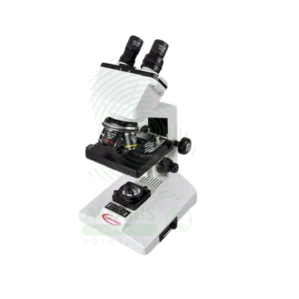
A Binocular Microscope is a high-precision, compound optical instrument fundamental to clinical diagnostics. Featuring dual eyepieces for comfortable viewing and a suite of parfocal objective lenses (4x, 10x, 40x, 100x oil), it provides magnifications from 40x to 1000x. Its integrated LED illumination, mechanical stage, and Abbe condenser enable the detailed examination of stained blood films, tissue sections, microbiological specimens, and cytological preparations. As the primary tool for pathologists and laboratory scientists, it is indispensable for definitive diagnoses in hematology, histopathology, microbiology, and urinalysis, demanding skilled operation and meticulous maintenance for optimal performance.
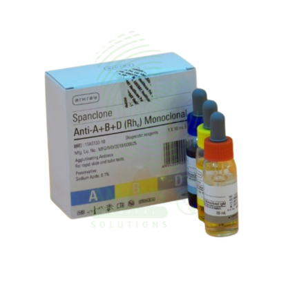
Blood Grouping kit
A Blood Grouping Kit is a Class II medical device (FDA-cleared/CE-marked) containing monoclonal antibodies (Anti-A, Anti-B, Anti-D) and control reagents for determining ABO blood group and Rh factor by agglutination method. The kit includes color-coded reagent vials, reaction cards or slides, mixing sticks, lancets, and capillary pipettes. Test time 2-5 minutes (slide method) or 15-30 minutes (tube method). Interpretation based on agglutination pattern: Anti-A positive = group A; Anti-B positive = group B; both positive = group AB; both negative = group O; Anti-D positive = Rh positive; Anti-D negative = Rh negative. Primary clinical applications include pre-transfusion compatibility testing, prenatal and neonatal testing (Rh incompatibility prevention), preoperative blood typing, blood donor screening, and emergency blood typing. Critical safety precautions include refrigerated storage (2-8°C), proper disposal of lancets in sharps containers, universal precautions for blood samples, and confirmation with reverse typing when possible. Essential kit for transfusion medicine, obstetrics, surgery, and emergency care.
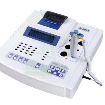
Coagulation Analyzer
A Coagulation Analyzer is a compact, automated instrument designed for performing essential tests that evaluate the blood's clotting ability. It accurately measures key parameters such as Prothrombin Time (PT/INR), Activated Partial Thromboplastin Time (aPTT), Thrombin Time, and Fibrinogen, which are critical for monitoring anticoagulant therapy (warfarin, heparin), diagnosing bleeding disorders, and assessing surgical risk. With its photometric detection and user-friendly operation, it delivers reliable results for small to medium-volume laboratories, clinics, and hospital wards. Its role in ensuring safe and effective patient management in thrombosis and hemostasis makes it a vital tool in clinical diagnostics.
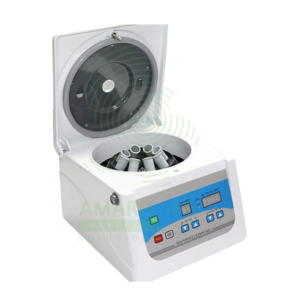
Electric Centrifuge
An Electric Centrifuge is a Class I medical device that uses rapid rotation to separate fluids of different densities, essential in clinical laboratories for processing blood, urine, and other biological specimens. Available in benchtop, refrigerated, microcentrifuge, hematocrit, and high-speed models with fixed-angle, swinging-bucket, or vertical rotors. Speed range 1,000-30,000+ RPM with RCF up to 65,000+ x g. Features include brushless induction motor, digital controls, imbalance detection, lid lock safety, and programmable protocols. Primary clinical applications include separation of serum/plasma for chemistry and hematology testing, urine sediment analysis for microscopy, platelet-rich plasma (PRP) preparation for regenerative medicine, concentration of bacteria for microbiology, cytology fluid analysis, blood banking, and molecular biology sample processing. Critical safety precautions include proper balancing of loads (weight matching), use of sealed rotors for biohazardous samples, regular rotor inspection for corrosion or cracks, never exceeding maximum rated speed, and never opening lid during operation. Essential equipment in clinical, research, and teaching laboratories.
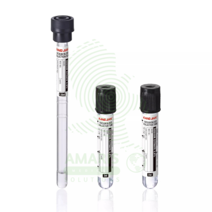
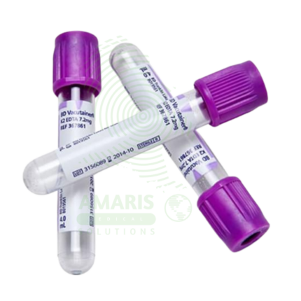
ESR Tubes Glass
ESR Tubes Glass are specialized glass tubes (300 mm length, 2.55 mm bore diameter, graduated 0-200 mm in 1 mm intervals) meeting ICSH and CLSI Westergren specifications for measuring erythrocyte sedimentation rate, a non-specific marker of inflammation. Used with citrated or diluted EDTA blood, filled to exactly 200 mm, placed vertically in Westergren stand, and read at 60 minutes. Primary clinical applications include determination of ESR for diagnosing and monitoring inflammatory conditions (infections, autoimmune disorders, malignancies), monitoring disease activity in rheumatoid arthritis, assessment of temporal arteritis and polymyalgia rheumatica, evaluation of infection and inflammatory response, screening for occult inflammatory conditions, and monitoring chronic inflammatory diseases (SLE, IBD, vasculitis). Critical safety precautions include handling glass tubes carefully to avoid breakage (sharps hazard), proper disposal in sharps containers as biohazardous waste, ensuring vertical alignment and constant temperature (18-25°C), reading at exactly 60 minutes, and avoiding air bubbles and vibration. Essential consumable for reference Westergren ESR testing in clinical laboratories.
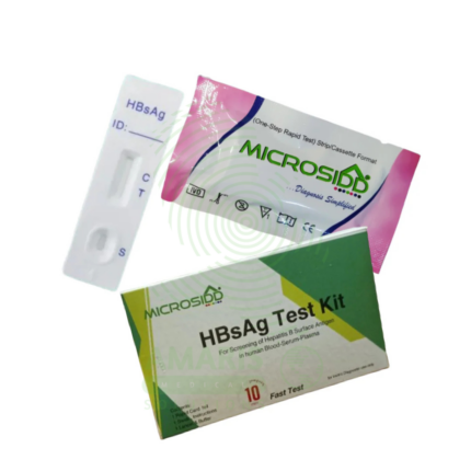
Hepatitis B Test Kit
The Hepatitis B Test Kit is an in vitro diagnostic device for the detection of Hepatitis B virus markers in human blood samples, available in rapid, ELISA, and molecular formats. It detects specific antigens (HBsAg) and antibodies (anti-HBc, anti-HBs) to diagnose acute and chronic infection, monitor treatment response, screen blood donations, and determine immunity status. With high sensitivity (>98%) and specificity (>99%), it provides results in 15 minutes to 4 hours depending on format. Essential applications include prenatal screening to prevent mother-to-child transmission, evaluation of acute hepatitis, pre-immunosuppression screening, and occupational health testing. All patient samples must be handled with universal precautions, and testing must include appropriate quality controls.

Hepatitis C Test Kit
The Hepatitis C Test Kit is an in vitro diagnostic device for the detection of Hepatitis C virus antibodies or RNA in human blood, serum, or plasma samples. Available in rapid, laboratory immunoassay, and molecular formats, it screens for HCV infection, confirms active infection with RNA testing, and monitors treatment response. With high sensitivity and specificity exceeding 99%, it is essential for identifying infected individuals, linking them to curative direct-acting antiviral therapy, and confirming cure. Essential applications include risk-based screening of high-risk populations, birth cohort screening of baby boomers, evaluation of acute hepatitis, management of occupational exposures, and monitoring of antiviral therapy that can cure over 95% of infections. All patient samples must be handled with universal precautions, and positive antibody results require confirmatory testing.