Dermatoscope and Magnifiers
 Diagnostic Kits
 Vital Signs Monitors
 Stethoscopes and Accessories
 Otoscopes, Ophthalmoscopes, and Retinoscopes
 Reflex Hammers and Neurological Tools
 Scales and Measuring Devices
 Spirometers and Pulmonary Function Tests
 Electrosurgical Units and Accessories
 Cutting Instruments
 Grasping and Holding Instruments
 Hemostatic Instruments
 Specialized Surgical Sets
 Single-Use Procedure Trays and Packs
 Surgical Drapes, Gowns, and Covers
 Tissue Unifying Instruments
 Radiation Protection
 X-Ray Machines and Accessories
 Ultrasound Systems and Probes
 MRI and CT Scanners
 Radiology Consumables
 Bone Densitometers
 Fluoroscopy Equipment
 Imaging Tables and Positioning Aids
 Microscopes and Accessories
 Centrifuges and Separators
 Analyzers
 Incubators and Ovens
 Pipettes, Dispensers, and Lab Glassware
 Refrigerators, Freezers, and Storage Units
 Lab Consumables
 Sterilizers and Autoclaves for Lab Use
 Multi-Parameter Monitors
 Ventilators and Respiratory Support Devices
 Defibrillators and AEDs
 Infusion Pumps and IV Systems
 Patient Warmers and Cooling Devices
 Central Monitoring Stations
 Accessories
 Anesthesia Machines and Workstations
 Oxygen Concentrators and Delivery Systems
 Nebulizers and Inhalers
 CPAP/BiPAP Machines
 Airway Management
 Anesthesia Masks, Circuits, and Bags
 Humidifiers and Heaters
 Respiratory Therapy Accessories
 First Aid Kits and Cabinets
 Emergency Resuscitation Equipment
 Trauma Supplies
 Emergency Carts and Crash Carts
 Burn Care Products
 Bleeding Control
 Automated External Defibrillators (AEDs)
 Transport and Evacuation
 Wheelchairs and Accessories
 Walkers, Crutches, and Canes
 Prosthetics and Orthotics
 Physical Therapy Equipment
 Transfer Devices
 Bathroom Safety
 Orthopedic Traction and Tables
 Hot/Cold Therapy Packs and Units
 Beds and Mattresses
 Chairs and Stools
 Tables
 Cabinets and Storage
 Privacy Screens & Curtains
 Stands and Racks
 Linens and Textiles
 Lighting
 Autoclaves and Sterilizers
 Ultrasonic Cleaners
 Disinfectant Solutions and Wipes
 Sterilization Pouches, Wraps, and Indicators
 Instrument Trays and Containers
 UV and Ozone Disinfection Devices
 Washer Disinfectors
 Wound Care
 Gloves
 Masks and Respirators
 Catheters and Tubing
 Swabs, Applicators, and Sponges
 Incontinence Products
 Personal Protective Equipment (PPE)
 Dental Chairs and Units
 Handpieces and Burs
 Instruments
 Consumables
 Sterilization for Dental Use
 Orthodontic Supplies
 Endodontic Tools
 Slit Lamps and Tonometers
 Lensometers and Phoropters
 Ophthalmic Surgical Instruments
 Eyewear Frames and Lenses
 Contact Lens Supplies
 Vision Testing Charts and Devices
 Eye Care Consumables
 Laser Systems for Eye Care
 ENT Exam Chairs and Tables
 Endoscopes
 Audiometers and Hearing Tests
 ENT Instruments
 Nasal and Throat Packs
 Hearing Aids and Accessories
 Otology Supplies
 Fetal Dopplers and Monitors
 Delivery Beds and Tables
 Gynecological Instruments
 Neonatal Incubators and Warmers
 Breast Pumps and Accessories
 Contraceptive Devices
 Maternity Supports and Pads
 Neonatal Consumables
 Cystoscopes and Urethroscopes
 Dialysis Machines and Supplies
 Urological Catheters and Bags
 Lithotripters
 Prostate Treatment Devices
 Urinary Incontinence Products
 Kidney Stone Management Tools
 Consumables & Disposables
 EEG and EMG Machines
 Neurosurgical Instruments
 Nerve Stimulators
 Headrests and Positioning Aids
 Lumbar Puncture Kits
 Seizure Monitoring Devices
 Consumables
 Rehabilitation for Neurological Conditions
 ECG Machines and Accessories
 Holter Monitors
 Stress Test Systems
 Pacemakers and Defibrillator Accessories
 Vascular Access Devices
 Cardiac Catheters and Guidewires
 Blood Flow Meters
 Consumables
 Orthopedic Instruments
 Casts, Splints, and Padding
 Joint Replacement Supplies
 Prosthetic Limbs and Components
 Bone Grafts and Substitutes
 Traction Devices
 Orthopedic Braces and Supports
 Rehabilitation Aids for Orthopedics
 Home Oxygen Therapy
 Hospital Beds for Home Use
 Mobility Aids
 Bathroom and Daily Living Aids
 Wound Care for Home
 Monitoring Devices
 Enteral Feeding Pumps and Tubes
 Hand Sanitizers and Dispensers
 Face Shields and Goggles
 Isolation Gowns and Suits
 Biohazard Waste Containers
 Air Purifiers and HEPA Filters
 Surface Disinfectants
 Sharps Containers
 Protective Barriers
 Cardiovascular & Endurance Training
 Strength Training & Weightlifting
 Functional Training & Core Conditioning
 Physical Therapy & Rehabilitation
 Sports & Outdoor Recreation
 Gym Flooring & Facility Equipment
 Fitness Monitoring & Accessories
 Kids & Novelties
Vein Finder
WhatsApp Order
A Vein Finder is a non-invasive medical imaging device that uses near-infrared light to visualize subcutaneous veins in real time and project a map of the vascular network directly onto the patient’s skin. Primarily used to facilitate difficult venipuncture and IV access in challenging patient populations (pediatric, obese, elderly, dark-skinned), it enhances first-stick success rates, improves patient comfort, and reduces procedure time. As a handheld, portable aid, it complements the clinician’s skill by providing clear visual guidance, making it a valuable tool in emergency rooms, operating theaters, infusion centers, and phlebotomy services.
Description
Vein Finder
PRIMARY CLINICAL & DIAGNOSTIC USES
1. Enhanced Venous Access for Difficult Sticks:
-
Primary Use: The primary and most critical use is to locate and map subcutaneous veins in patients who present challenges for routine venipuncture or intravenous (IV) cannulation. This includes pediatric and neonatal patients, elderly patients, obese patients, patients with dark skin tone, patients with a history of IV drug use, and those who are dehydrated or in hypovolemic shock.
-
How it helps: Takes the guesswork out of finding veins in patients where veins are hard to see or feel, transforming a frustrating, painful experience into a quick, successful procedure for both patient and healthcare provider.
2. Improving First-Attempt Success Rates:
-
Primary Use: Used by phlebotomists, nurses, and anesthetists to significantly increase the likelihood of successful venipuncture or IV cannulation on the first attempt, reducing patient discomfort, anxiety, and the need for multiple needle sticks.
-
How it helps: Spares patients the pain and anxiety of multiple needle sticks, especially for those who require frequent blood draws or IV placements, making each encounter less traumatic and more efficient.
3. Reducing Procedure Time and Resource Use:
-
Primary Use: By providing clear visual guidance, it can speed up the process of establishing venous access, which is crucial in emergency departments, operating rooms, and critical care settings where time is of the essence.
-
How it helps: Saves precious minutes in emergencies when every second counts, allowing healthcare providers to establish IV access quickly and move on to other life-saving interventions.
4. Planning for Vascular Procedures:
-
Primary Use: Assists in pre-procedural planning for the placement of peripherally inserted central catheters (PICCs), midline catheters, or for identifying suitable veins for surgical arteriovenous (AV) fistula creation in dialysis patients.
-
How it helps: Helps vascular access specialists identify the best veins for long-term access, ensuring that patients who need extended IV therapy or dialysis have reliable, durable access that will serve them well.
SECONDARY & SUPPORTIVE USES
1. Patient Education and Anxiety Reduction: Showing the patient a clear map of their veins can build trust, reduce anxiety, and increase cooperation during the procedure, transforming a stressful experience into a collaborative one.
2. Training Healthcare Professionals: Serves as an excellent educational tool for teaching students and trainees the principles of venous anatomy and venipuncture technique, helping the next generation of healthcare providers develop this essential skill.
3. Dermatological and Cosmetic Procedures: Some models are used to visualize superficial vascular lesions (like telangiectasias or spider veins) prior to sclerotherapy or laser treatment, helping dermatologists and cosmetic surgeons plan treatments with precision.
KEY PRODUCT FEATURES
1. BASIC IDENTIFICATION ATTRIBUTES
-
Device Type: Non-invasive, transillumination or projection-based vascular imaging device.
-
Technology Variants:
-
Near-Infrared (NIR) Imaging: The most common. Emits harmless near-infrared light (700-900 nm) which is absorbed by hemoglobin in the blood but scattered by surrounding tissue. A camera detects the contrast and projects a real-time enhanced vein map onto the skin surface.
-
Multispectral Imaging: Combines NIR with other wavelengths for potentially better contrast, especially in patients with higher melanin content.
-
LED-based Transillumination: Uses bright, cool LED light pressed against the skin to transilluminate (light up) veins from beneath, often in a handheld format.
-
-
Form Factors: Handheld devices (most common), overhead mounted systems for procedure rooms, and wearable clip-on devices.
2. TECHNICAL & PERFORMANCE PROPERTIES
-
Imaging Depth: Typically visualizes superficial veins up to 10-15 mm beneath the skin's surface—the depth relevant for peripheral venous access.
-
Contrast and Resolution: The quality of the projected image, including the clarity of vein edges and the ability to distinguish veins from arteries or other structures. Advanced image processing algorithms (e.g., edge enhancement, noise reduction) improve usability.
-
Field of View: The area of skin that can be imaged at once. Handheld devices often have an adjustable field.
-
Projection Method: Projects a real-time, high-contrast map of the vasculature directly onto the patient's skin. Veins are typically displayed as dark lines on a light background, or in color (e.g., blue or green lines).
-
Adjustability: Features to adjust image brightness, contrast, and inversion (making veins appear light on dark) to optimize visualization for different skin tones and ambient light conditions.
3. PHYSICAL & OPERATIONAL PROPERTIES
-
Portability: Handheld models are lightweight, battery-operated (rechargeable Li-ion), and designed for easy cleaning and transport between patients and departments.
-
Ergonomics: Designed for one-handed operation, allowing the clinician to hold the device while preparing for the procedure with the other hand.
-
Durability: Housed in medical-grade plastics designed to withstand frequent cleaning with hospital-grade disinfectants.
4. SAFETY & COMPLIANCE ATTRIBUTES
-
Regulatory Approvals: Classified as a Class I or Class IIa medical device (low to moderate risk). Must carry CE Marking and/or FDA 510(k) clearance.
-
Laser/LED Safety: The light sources must be eye-safe (typically Class 1 or Class 2M laser products or LED-based) and pose no thermal risk to the skin.
-
Electrical Safety: Complies with IEC 60601-1 standards for medical electrical equipment.
5. STORAGE & HANDLING ATTRIBUTES
-
Storage: Store in a clean, dry charging dock or protective case when not in use. Protect from drops and impacts.
-
Cleaning and Disinfection: The part of the device that contacts or is held near the patient must be cleaned between uses. Wipe with a soft cloth dampened with a hospital-grade, non-abrasive disinfectant (e.g., 70% isopropyl alcohol). Do not immerse in liquid.
-
Battery Care: Follow manufacturer instructions for charging to maintain battery health. Have a charged backup available in high-use areas.
6. LABORATORY & CLINICAL APPLICATIONS
-
Primary Application: An assistive technology to improve the efficacy, efficiency, and patient experience of peripheral venous access procedures across all healthcare settings.
-
Adjunctive Tool: It is a visual aid, not an autonomous device. Successful cannulation still depends on the clinician's skill in needle insertion and technique.
SAFETY HANDLING PRECAUTIONS
1. SAFETY PRECAUTIONS
-
Not a Substitute for Skill: The device shows where veins are, but the clinician must still assess vein suitability (size, fragility, proximity to valves/arteries) and use proper aseptic technique.
-
Skin Preparation: The device should be used after skin has been cleaned with an antiseptic (e.g., chlorhexidine/alcohol) and allowed to dry. Do not use the device over uncleaned skin.
-
Ambient Light: Performance is best in subdued ambient light. Direct, bright overhead lights can wash out the projected image.
-
Contraindications: Not for use over open wounds, rashes, or areas with suspected deep vein thrombosis (DVT).
2. FIRST AID MEASURES
-
General: The device is non-invasive and low-risk. In the event of an unrelated needle-stick injury during the procedure, follow standard exposure control protocols.
-
Eye Exposure: Although light is eye-safe, avoid shining directly into eyes for extended periods.
-
Device Malfunction: If the device is damaged, stops working, or emits unusual heat/smell, discontinue use and remove from service.
3. FIRE FIGHTING MEASURES
-
Flammability: Plastic housing, electronic components, and lithium-ion battery are combustible.
-
Extinguishing Media: For electrical fires, use a CO₂ or dry chemical (Class C) fire extinguisher. A lithium battery fire may require a Class D extinguisher or large amounts of water to cool.
-
Firefighter Instructions: Be aware of potential battery thermal runaway.
Related products
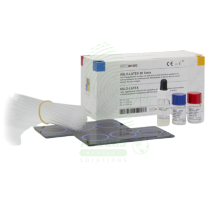
Anti-Streptol Olysin O Titer (ASOT)
Anti-Streptol Olysin O Titer (ASOT) is a quantitative or semi-quantitative serological test (latex agglutination, turbidimetry, nephelometry, or ELISA) for detecting antibodies against streptolysin O, an exotoxin produced by Group A Streptococcus. Elevated or rising titers indicate recent streptococcal infection and are essential for diagnosing post-streptococcal sequelae including acute rheumatic fever (Jones criteria) and post-streptococcal glomerulonephritis. The test requires serum samples; acute and convalescent (2-4 weeks apart) with fourfold rise confirms recent infection. Reference range typically <200-250 Todd units/mL (adults), varies by age and population. Primary clinical applications include diagnosis of Group A streptococcal infections, acute rheumatic fever evaluation, post-streptococcal glomerulonephritis diagnosis, differentiation of acute vs. past infection, evaluation of unexplained arthritis or carditis, pediatric inflammatory conditions (PANDAS), and monitoring disease activity in rheumatic fever. Critical safety precautions include proper timing of acute and convalescent samples, awareness of false negatives/positives, clinical correlation for diagnosis, and standard biohazard precautions. Essential test for rheumatology, nephrology, cardiology, and infectious disease practice.
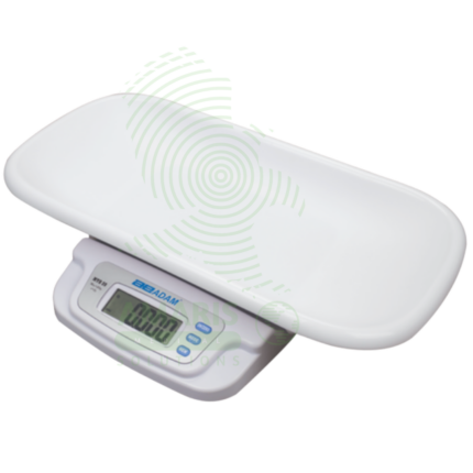
Digital Baby Weighing Scale
A Digital Baby Weighing Scale is a high-precision medical device designed exclusively for the safe and accurate measurement of infant and toddler weight. Featuring a stable, hygienic platform with gram-level precision, it is indispensable in hospitals (especially NICUs), pediatric clinics, and community health settings. Its core functions—such as hold, tare, and zeroing—ensure reliable readings even with a moving child. Accurate weight data is foundational for assessing growth, calculating medication doses, managing nutrition, and detecting early signs of health issues, making this scale a critical tool for safeguarding infant health and development.
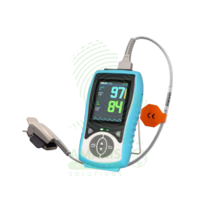
Handheld Pulse Oximeter
A Handheld Pulse Oximeter is a professional-grade, portable medical device used by healthcare providers for accurate and reliable monitoring of blood oxygen saturation (SpO2) and pulse rate. It consists of a durable, feature-rich monitor unit and separate, interchangeable sensors for use on fingers, toes, or ears. Superior to basic fingertip models, it offers advanced functions like configurable alarms, detailed plethysmograph display, trend memory, and data connectivity. Its robust design, clinical accuracy under challenging conditions (motion, low perfusion), and versatility make it an essential tool in hospitals, clinics, ambulances, and home health for patient assessment and monitoring.
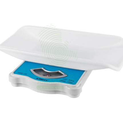
Manual Baby Weighing Scale
A Manual Baby Weighing Scale is a simple, mechanical device used to measure infant weight without the need for electricity or batteries. Operating on a spring or balance principle, it features a suspended sling or seat and a dial display. Its primary advantage is durability and portability for use in community health, outreach programs, and areas with unreliable power. While less precise than digital scales, it provides a reliable and accessible means to monitor growth trends and screen for undernutrition in resource-limited settings, making it a vital tool for basic pediatric care in the field.
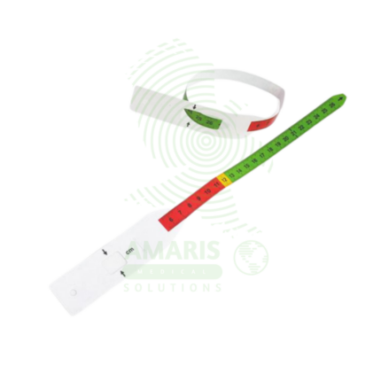
MUAC Tape
A MUAC Tape (Mid-Upper Arm Circumference) is a specialized, color-coded measuring tape used globally as the primary field tool for screening children aged 6-59 months for acute malnutrition. Its simple design—featuring red, yellow, and green zones corresponding to Severe Acute Malnutrition, Moderate Acute Malnutrition, and normal nutritional status—allows for instant, non-invasive assessment without the need for scales, calculation, or literacy. Durable, portable, and inexpensive, it is the cornerstone of community-based nutrition programs, emergency relief efforts, and public health surveillance, enabling early detection and life-saving intervention for malnourished children in even the most resource-constrained settings.
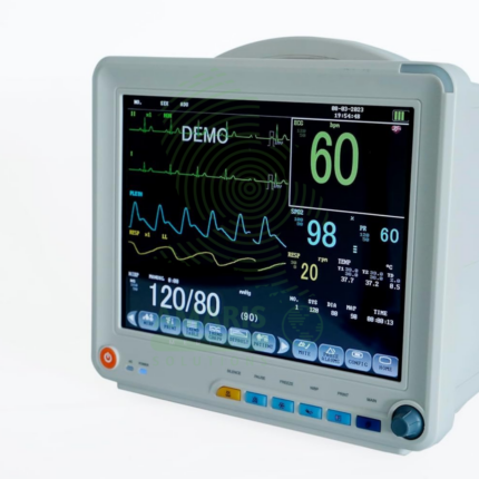
Patient Monitor
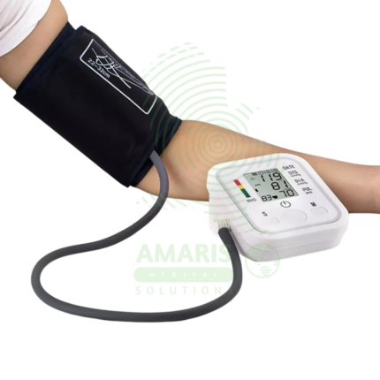
The Patient Monitor (6 Parameters BCCMS8000) is a versatile multi-parameter monitor designed for continuous surveillance of core vital signs in various clinical settings. It tracks six essential parameters—ECG, SpO2, Non-Invasive Blood Pressure (NIBP), Respiration, Temperature, and Pulse Rate—providing clinicians with real-time waveforms and numerical data on a clear color display. With its robust alarm system, battery backup for transport, and reliable performance, it is a fundamental tool for ensuring patient safety on general hospital wards, during procedures, and in emergency departments. Its design balances comprehensive monitoring capability with user-friendly operation.
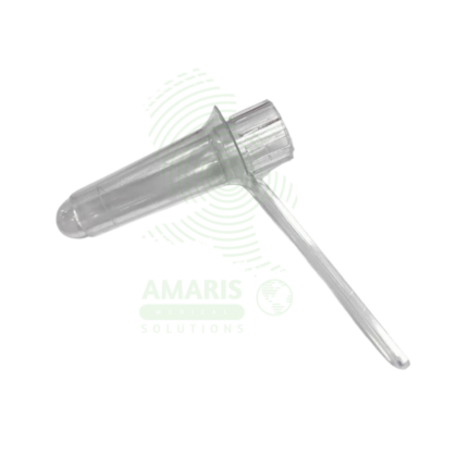
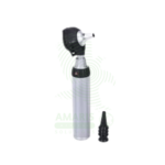
Proctoscope
A Proctoscope is a rigid, straight-tube endoscope specifically designed for the examination and treatment of the anal canal and distal rectum. It enables direct visualization to diagnose conditions like hemorrhoids, fissures, and proctitis, and serves as a conduit for therapeutic procedures such as band ligation. Available in reusable (autoclavable metal) or disposable (single-use plastic) formats, it is a fundamental tool in colorectal practice. Its effective use requires proper patient preparation, gentle insertion technique, and stringent adherence to sterilization protocols to ensure patient safety and diagnostic accuracy.