Dermatoscope and Magnifiers
 Diagnostic Kits
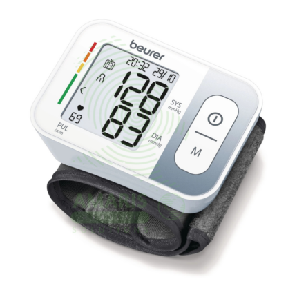
 Vital Signs Monitors
 Stethoscopes and Accessories
 Otoscopes, Ophthalmoscopes, and Retinoscopes
 Reflex Hammers and Neurological Tools
 Scales and Measuring Devices
 Spirometers and Pulmonary Function Tests
 Electrosurgical Units and Accessories
 Cutting Instruments
 Grasping and Holding Instruments
 Hemostatic Instruments
 Specialized Surgical Sets
 Single-Use Procedure Trays and Packs
 Surgical Drapes, Gowns, and Covers
 Tissue Unifying Instruments
 Radiation Protection
 X-Ray Machines and Accessories
 Ultrasound Systems and Probes
 MRI and CT Scanners
 Radiology Consumables
 Bone Densitometers
 Fluoroscopy Equipment
 Imaging Tables and Positioning Aids
 Microscopes and Accessories
 Centrifuges and Separators
 Analyzers
 Incubators and Ovens
 Pipettes, Dispensers, and Lab Glassware
 Refrigerators, Freezers, and Storage Units
 Lab Consumables
 Sterilizers and Autoclaves for Lab Use
 Multi-Parameter Monitors
 Ventilators and Respiratory Support Devices
 Defibrillators and AEDs
 Infusion Pumps and IV Systems
 Patient Warmers and Cooling Devices
 Central Monitoring Stations
 Accessories
 Anesthesia Machines and Workstations
 Oxygen Concentrators and Delivery Systems
 Nebulizers and Inhalers
 CPAP/BiPAP Machines
 Airway Management
 Anesthesia Masks, Circuits, and Bags
 Humidifiers and Heaters
 Respiratory Therapy Accessories
 First Aid Kits and Cabinets
 Emergency Resuscitation Equipment
 Trauma Supplies
 Emergency Carts and Crash Carts
 Burn Care Products
 Bleeding Control
 Automated External Defibrillators (AEDs)
 Transport and Evacuation
 Wheelchairs and Accessories
 Walkers, Crutches, and Canes
 Prosthetics and Orthotics
 Physical Therapy Equipment
 Transfer Devices
 Bathroom Safety
 Orthopedic Traction and Tables
 Hot/Cold Therapy Packs and Units
 Beds and Mattresses
 Chairs and Stools
 Tables
 Cabinets and Storage
 Privacy Screens & Curtains
 Stands and Racks
 Linens and Textiles
 Lighting
 Autoclaves and Sterilizers
 Ultrasonic Cleaners
 Disinfectant Solutions and Wipes
 Sterilization Pouches, Wraps, and Indicators
 Instrument Trays and Containers
 UV and Ozone Disinfection Devices
 Washer Disinfectors
 Wound Care
 Gloves
 Masks and Respirators
 Catheters and Tubing
 Swabs, Applicators, and Sponges
 Incontinence Products
 Personal Protective Equipment (PPE)
 Dental Chairs and Units
 Handpieces and Burs
 Instruments
 Consumables
 Sterilization for Dental Use
 Orthodontic Supplies
 Endodontic Tools
 Slit Lamps and Tonometers
 Lensometers and Phoropters
 Ophthalmic Surgical Instruments
 Eyewear Frames and Lenses
 Contact Lens Supplies
 Vision Testing Charts and Devices
 Eye Care Consumables
 Laser Systems for Eye Care
 ENT Exam Chairs and Tables
 Endoscopes
 Audiometers and Hearing Tests
 ENT Instruments
 Nasal and Throat Packs
 Hearing Aids and Accessories
 Otology Supplies
 Fetal Dopplers and Monitors
 Delivery Beds and Tables
 Gynecological Instruments
 Neonatal Incubators and Warmers
 Breast Pumps and Accessories
 Contraceptive Devices
 Maternity Supports and Pads
 Neonatal Consumables
 Cystoscopes and Urethroscopes
 Dialysis Machines and Supplies
 Urological Catheters and Bags
 Lithotripters
 Prostate Treatment Devices
 Urinary Incontinence Products
 Kidney Stone Management Tools
 Consumables & Disposables
 EEG and EMG Machines
 Neurosurgical Instruments
 Nerve Stimulators
 Headrests and Positioning Aids
 Lumbar Puncture Kits
 Seizure Monitoring Devices
 Consumables
 Rehabilitation for Neurological Conditions
 ECG Machines and Accessories
 Holter Monitors
 Stress Test Systems
 Pacemakers and Defibrillator Accessories
 Vascular Access Devices
 Cardiac Catheters and Guidewires
 Blood Flow Meters
 Consumables
 Orthopedic Instruments
 Casts, Splints, and Padding
 Joint Replacement Supplies
 Prosthetic Limbs and Components
 Bone Grafts and Substitutes
 Traction Devices
 Orthopedic Braces and Supports
 Rehabilitation Aids for Orthopedics
 Home Oxygen Therapy
 Hospital Beds for Home Use
 Mobility Aids
 Bathroom and Daily Living Aids
 Wound Care for Home
 Monitoring Devices
 Enteral Feeding Pumps and Tubes
 Hand Sanitizers and Dispensers
 Face Shields and Goggles
 Isolation Gowns and Suits
 Biohazard Waste Containers
 Air Purifiers and HEPA Filters
 Surface Disinfectants
 Sharps Containers
 Protective Barriers
 Cardiovascular & Endurance Training
 Strength Training & Weightlifting
 Functional Training & Core Conditioning
 Physical Therapy & Rehabilitation
 Sports & Outdoor Recreation
 Gym Flooring & Facility Equipment
 Fitness Monitoring & Accessories
 Kids & Novelties
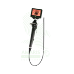
Video Laryngoscope
WhatsApp Order
A Video Laryngoscope is an intubation device combining a laryngoscope blade with an integrated video camera and display, providing indirect visualization of the glottis for difficult airway management, emergency intubation, cervical spine precautions, and teaching applications. Available with reusable or disposable blades (Macintosh, Miller, hyperangulated) in sizes 0 (neonatal) to 4 (large adult). Features high-resolution camera (640×480 to 1920×1080), LED illumination, anti-fog system, and 2–7-inch display with recording capability. Primary clinical applications include difficult airway management (anatomical variations, obesity, cervical spine injury), emergency intubation in trauma and critical care, intubation with cervical spine precautions (minimal neck movement), teaching and training (real-time instructor view), documentation of airway anatomy, bariatric patient intubation, and neonatal/pediatric intubation. Class II medical device requiring FDA clearance. Critical safety considerations include anti-fog preparation, clearance of oral secretions, appropriate blade selection, maintaining direct laryngoscopy skills as backup, gentle tissue handling, infection control (disposable blades or sterilization), and battery verification before use.
Description
Video Laryngoscope
PRIMARY CLINICAL & DIAGNOSTIC USES
1. Difficult Airway Management:
-
Primary Use: Provides enhanced visualization of the glottis and vocal cords in patients with difficult airways due to anatomical variations, obesity, cervical spine immobilization, or head and neck pathology. The video camera on the blade transmits images to a screen, allowing intubation without requiring direct line-of-sight alignment.
-
How it helps: Transforms impossible airways into manageable ones by letting doctors see around anatomical obstacles, ensuring that even patients with challenging anatomy can have their airway secured safely.
2. Emergency Intubation in Trauma and Critical Care:
-
Primary Use: Used in emergency departments, intensive care units, and prehospital settings for rapid sequence intubation in critically ill or injured patients where traditional direct laryngoscopy may be challenging or contraindicated.
-
How it helps: Gives emergency physicians and critical care teams a reliable tool when seconds matter most, allowing them to secure an airway quickly in patients who are bleeding, in shock, or in respiratory failure.
3. Cervical Spine Precautions:
-
Primary Use: Enables intubation with minimal cervical spine movement in patients with suspected or confirmed cervical spine injuries, as the video technology allows glottic visualization without the need for head extension or manipulation.
-
How it helps: Protects patients with neck injuries from further spinal cord damage during intubation, allowing doctors to secure the airway without moving the cervical spine.
4. Teaching and Training:
-
Primary Use: Allows instructors to view the intubation procedure in real-time on the screen, providing immediate feedback to trainees while maintaining patient safety. The ability to record procedures facilitates debriefing and education.
-
How it helps: Revolutionizes airway education by letting instructors see exactly what trainees are seeing, providing targeted feedback that accelerates learning and builds competence.
5. Documentation of Airway Anatomy:
-
Primary Use: Records images of airway anatomy and intubation attempts for medical records, quality improvement, and medicolegal purposes.
-
How it helps: Creates a permanent record of airway findings and intubation attempts, supporting quality improvement efforts and providing documentation for medical-legal purposes.
6. Bariatric Patient Intubation:
-
Primary Use: Facilitates intubation in morbidly obese patients where traditional laryngoscopy may be hindered by large tongue, redundant pharyngeal tissues, and restricted mouth opening.
-
How it helps: Provides a clear view past the anatomical challenges of obesity, ensuring that patients of all sizes receive safe, effective airway management.
7. Neonatal and Pediatric Intubation:
-
Primary Use: Specialized small-diameter video laryngoscopes allow visualization of the pediatric airway, improving success rates in neonatal and pediatric intubations where anatomical differences make direct laryngoscopy challenging.
-
How it helps: Protects the smallest patients by providing specially sized equipment that gives pediatric specialists a clear view of tiny airways, ensuring safe intubation of children from premature infants to adolescents.
SECONDARY & SUPPORTIVE USES
1. Airway Assessment: Provides detailed visualization of airway anatomy for preoperative assessment and planning, helping anesthesiologists anticipate challenges before they occur.
2. Foreign Body Removal: Assists in visualization and removal of foreign bodies from the airway, helping retrieve objects that are blocking breathing.
3. Bronchoscopy Assistance: Can be used to guide bronchoscope placement in combined procedures, expanding diagnostic and therapeutic options.
4. Extubation Assessment: Evaluates airway patency and vocal cord function prior to extubation, ensuring patients are ready to breathe on their own before removing the breathing tube.
5. Research and Quality Improvement: Records airway management procedures for research studies and quality improvement initiatives, advancing the science of airway management and improving patient care.
KEY PRODUCT FEATURES
1. BASIC IDENTIFICATION ATTRIBUTES
-
Device Type: Laryngoscope with integrated video camera and display for indirect visualization of the glottis during intubation.
-
Common Names: Video Laryngoscope, Video Laryngoscopy System, VL Scope.
-
Components:
-
Handle: Reusable or disposable handle with controls and battery.
-
Blade: Reusable or disposable blade with integrated camera at tip.
-
Display: Handheld screen or separate monitor showing camera view.
-
Camera: High-resolution CMOS or CCD camera at blade tip with LED illumination.
-
Recording Capability: Some models allow image and video capture.
-
-
Blade Types: Macintosh (curved), Miller (straight), hyperangulated, and pediatric sizes.
2. TECHNICAL & PERFORMANCE PROPERTIES
-
Camera Resolution: 640×480 to 1920×1080 pixels; higher resolution provides clearer anatomical detail.
-
Field of View: 60-90 degrees; wider field improves visualization of surrounding structures.
-
Illumination: LED lights at blade tip; white balance adjustment for true tissue color.
-
Display Size: 2-7 inches; handheld (integrated) or separate monitor.
-
Anti-Fog System: Built-in heating element or chemical coating to prevent lens fogging.
-
Video Output: HDMI, USB, or wireless transmission for external monitors and recording.
-
Recording: Still image and video capture; internal memory or SD card storage.
-
Battery Life: 60-180 minutes continuous use; rechargeable lithium-ion batteries.
-
Blade Size Range: 0 (neonatal) to 4 (large adult); length 70-160 mm.
3. PHYSICAL & OPERATIONAL PROPERTIES
-
Handle Material: Medical-grade ABS plastic, aluminum, or stainless steel.
-
Blade Material: Medical-grade plastic (disposable) or stainless steel (reusable).
-
Weight: 8-24 ounces depending on configuration.
-
Water Resistance: IPX4 or higher for cleaning and disinfection.
-
Display: Adjustable brightness; anti-glare screen; some models touchscreen.
-
Controls: Buttons for power, image capture, recording, and brightness adjustment.
-
Storage: Carrying case for system components; blade storage rack.
4. SAFETY & COMPLIANCE ATTRIBUTES
-
Regulatory Status: Class II medical device requiring FDA 510(k) clearance.
-
Intended Use: Indicated for oral or nasal intubation providing visualization of the airway.
-
Electrical Safety: Compliant with IEC 60601-1 for medical electrical equipment; Type BF applied part.
-
Biocompatibility: Blade materials must be biocompatible for airway contact (ISO 10993).
-
Sterility: Reusable blades require high-level disinfection or sterilization; disposable blades sterile single-use.
-
Cleaning Validation: Reusable components must have validated cleaning and disinfection protocols.
-
Battery Safety: Lithium-ion batteries must meet transportation and safety standards (UN38.3).
5. STORAGE & HANDLING ATTRIBUTES
-
Storage: Store system in clean, dry environment; protect from impact; maintain battery charge.
-
Cleaning (Reusable Blades): High-level disinfection or sterilization per manufacturer instructions. Clean camera and handle with approved disinfectant wipes.
-
Disposable Blades: Single-use only; discard after procedure; do not resterilize.
-
Battery Maintenance: Recharge after each use; remove battery if storing long-term.
-
Calibration: Annual preventive maintenance and calibration verification recommended.
-
Inspection: Check camera clarity, illumination, and controls before each use; verify anti-fog function.
6. LABORATORY & CLINICAL APPLICATIONS
-
Primary Application: Facilitates tracheal intubation in difficult airway situations, emergency settings, cervical spine precautions, and teaching environments by providing indirect video visualization of the glottis.
-
Limitation: Requires adequate mouth opening for blade insertion; may be difficult in patients with blood, secretions, or fogging obscuring the lens.
SAFETY HANDLING PRECAUTIONS
1. SAFETY PRECAUTIONS
-
Anti-Fog Preparation: Activate anti-fog system or apply anti-fog solution before use to prevent lens fogging.
-
Oral Secretions: Suction pharynx before intubation to clear secretions that may obscure camera view.
-
Blade Selection: Choose appropriate blade type and size for patient anatomy; hyperangulated blades require stylets for tube delivery.
-
Direct vs. Video: Maintain ability to perform direct laryngoscopy; video system may fail.
-
Tube Delivery: Visualize tube passage through cords; do not rely solely on screen if view obscured.
-
Tissue Trauma: Insert blade gently; avoid pressure on soft tissues; video view does not eliminate trauma risk.
-
Infection Control: Use disposable blades or sterilize reusable blades per protocol; clean handle and screen after each use.
-
Battery Check: Verify adequate battery charge before procedure; have backup available.
-
Emergency Backup: Have conventional laryngoscope available in case of video system failure.
2. FIRST AID MEASURES
-
Lens Fogging: Remove blade; reapply anti-fog; have backup blade available.
-
Battery Failure: Switch to backup device or conventional laryngoscope.
-
Equipment Malfunction: Discontinue use; switch to alternative airway device; document malfunction.
3. FIRE FIGHTING MEASURES
-
Flammability: Plastic components and batteries are combustible.
-
Extinguishing Media: For electrical fire, use CO₂ or dry chemical (Class C) extinguisher.
-
Battery Fire: Damaged lithium battery may ignite; use Class D extinguisher or sand.
Related products
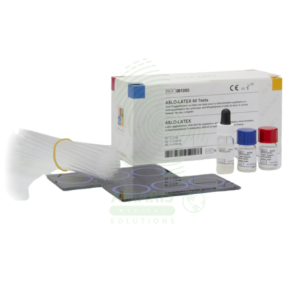
Anti-Streptol Olysin O Titer (ASOT)
Anti-Streptol Olysin O Titer (ASOT) is a quantitative or semi-quantitative serological test (latex agglutination, turbidimetry, nephelometry, or ELISA) for detecting antibodies against streptolysin O, an exotoxin produced by Group A Streptococcus. Elevated or rising titers indicate recent streptococcal infection and are essential for diagnosing post-streptococcal sequelae including acute rheumatic fever (Jones criteria) and post-streptococcal glomerulonephritis. The test requires serum samples; acute and convalescent (2-4 weeks apart) with fourfold rise confirms recent infection. Reference range typically <200-250 Todd units/mL (adults), varies by age and population. Primary clinical applications include diagnosis of Group A streptococcal infections, acute rheumatic fever evaluation, post-streptococcal glomerulonephritis diagnosis, differentiation of acute vs. past infection, evaluation of unexplained arthritis or carditis, pediatric inflammatory conditions (PANDAS), and monitoring disease activity in rheumatic fever. Critical safety precautions include proper timing of acute and convalescent samples, awareness of false negatives/positives, clinical correlation for diagnosis, and standard biohazard precautions. Essential test for rheumatology, nephrology, cardiology, and infectious disease practice.
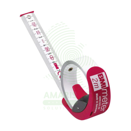
BMI Tape
A BMI Tape is a rapid screening tool that estimates an individual's Body Mass Index (BMI) category by measuring body circumference, most commonly the waist. It features a color-coded scale that directly displays BMI ranges (Underweight, Normal, Overweight, Obese), eliminating the need for separate height and weight measurements and manual calculation. While highly useful for community health screenings, wellness programs, and patient education due to its portability and simplicity, it is an approximate method. Its accuracy is limited compared to BMI calculated from precise clinical measurements, and it should be used as an initial screening indicator rather than a definitive diagnostic tool.
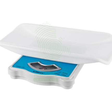
Manual Baby Weighing Scale
A Manual Baby Weighing Scale is a simple, mechanical device used to measure infant weight without the need for electricity or batteries. Operating on a spring or balance principle, it features a suspended sling or seat and a dial display. Its primary advantage is durability and portability for use in community health, outreach programs, and areas with unreliable power. While less precise than digital scales, it provides a reliable and accessible means to monitor growth trends and screen for undernutrition in resource-limited settings, making it a vital tool for basic pediatric care in the field.
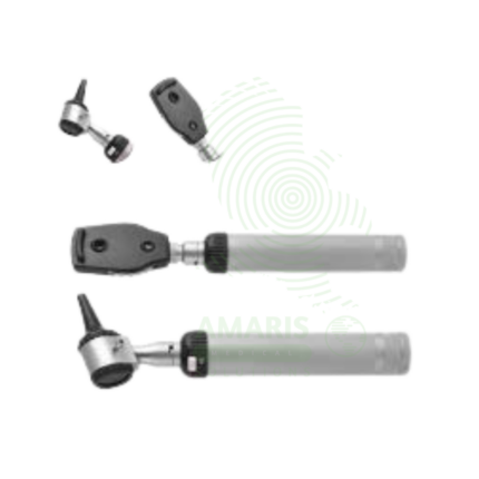
Ophthalmoscope
An Ophthalmoscope is a handheld diagnostic instrument used to examine the interior of the eye, specifically the fundus (retina, optic disc, blood vessels, and macula). By providing illuminated magnification, it serves as a vital screening tool for detecting ocular diseases like glaucoma and retinal disorders, and more importantly, for identifying signs of systemic conditions such as diabetes, hypertension, and increased intracranial pressure. Its operation involves selecting appropriate apertures and compensating lenses to obtain a clear view. While offering a detailed but narrow field of vision, mastery of the direct ophthalmoscope remains a cornerstone skill in general medicine, neurology, and ophthalmology for assessing vascular and neurological health.
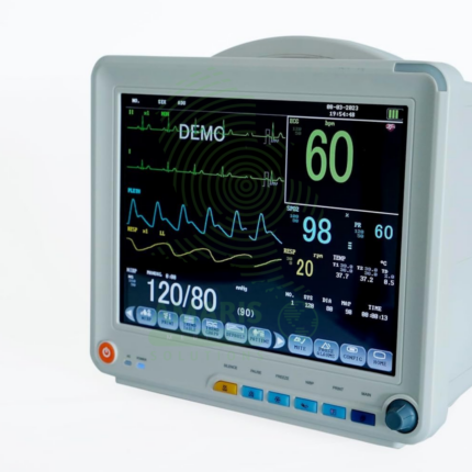
Patient Monitor
The Patient Monitor (6 Parameters BCCMS8000) is a versatile multi-parameter monitor designed for continuous surveillance of core vital signs in various clinical settings. It tracks six essential parameters—ECG, SpO2, Non-Invasive Blood Pressure (NIBP), Respiration, Temperature, and Pulse Rate—providing clinicians with real-time waveforms and numerical data on a clear color display. With its robust alarm system, battery backup for transport, and reliable performance, it is a fundamental tool for ensuring patient safety on general hospital wards, during procedures, and in emergency departments. Its design balances comprehensive monitoring capability with user-friendly operation.
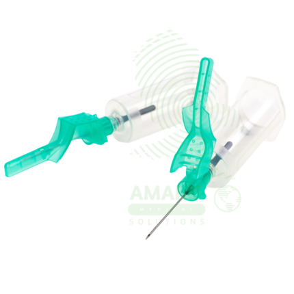
SAFE SERIES Blood Collection Needles
The SAFE SERIES (AM-SAFE) by Beijing Precil (Mindray subsidiary) is a comprehensive line of safety-engineered blood collection devices designed to prevent accidental needlestick injuries in healthcare workers. The series includes venous blood collection needles (AM-SAFE-V), standard safety needles (AM-SAFE1), winged infusion sets/butterfly needles (AM-SAFE2), high-gauge thin-wall needles for fragile veins (AM-SAFE3), premium coated needles for enhanced patient comfort (AM-GOLD), and nurse-friendly one-handed safety variants (AM-NURSE). All devices incorporate integral safety mechanisms that shield the contaminated needle immediately after withdrawal, complying with OSHA Needlestick Safety and Prevention Act requirements. Available in various gauges (18G-25G) and configurations for routine venipuncture, difficult vein access, pediatric/geriatric patients, and frequent blood draws. Sterile, single-use, latex-free, and color-coded for easy gauge identification. Essential safety devices for blood collection in hospitals, clinics, laboratories, and home healthcare settings.
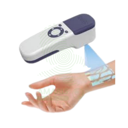
Vein Finder
A Vein Finder is a non-invasive medical imaging device that uses near-infrared light to visualize subcutaneous veins in real time and project a map of the vascular network directly onto the patient's skin. Primarily used to facilitate difficult venipuncture and IV access in challenging patient populations (pediatric, obese, elderly, dark-skinned), it enhances first-stick success rates, improves patient comfort, and reduces procedure time. As a handheld, portable aid, it complements the clinician's skill by providing clear visual guidance, making it a valuable tool in emergency rooms, operating theaters, infusion centers, and phlebotomy services.