Dermatoscope and Magnifiers
 Diagnostic Kits
 Vital Signs Monitors
 Stethoscopes and Accessories
 Otoscopes, Ophthalmoscopes, and Retinoscopes
 Reflex Hammers and Neurological Tools
 Scales and Measuring Devices
 Spirometers and Pulmonary Function Tests
 Electrosurgical Units and Accessories
 Cutting Instruments
 Grasping and Holding Instruments
 Hemostatic Instruments
 Specialized Surgical Sets
 Single-Use Procedure Trays and Packs
 Surgical Drapes, Gowns, and Covers
 Tissue Unifying Instruments
 Radiation Protection
 X-Ray Machines and Accessories
 Ultrasound Systems and Probes
 MRI and CT Scanners
 Radiology Consumables
 Bone Densitometers
 Fluoroscopy Equipment
 Imaging Tables and Positioning Aids
 Microscopes and Accessories
 Centrifuges and Separators
 Analyzers
 Incubators and Ovens
 Pipettes, Dispensers, and Lab Glassware
 Refrigerators, Freezers, and Storage Units
 Lab Consumables
 Sterilizers and Autoclaves for Lab Use
 Multi-Parameter Monitors
 Ventilators and Respiratory Support Devices
 Defibrillators and AEDs
 Infusion Pumps and IV Systems
 Patient Warmers and Cooling Devices
 Central Monitoring Stations
 Accessories
 Anesthesia Machines and Workstations
 Oxygen Concentrators and Delivery Systems
 Nebulizers and Inhalers
 CPAP/BiPAP Machines
 Airway Management
 Anesthesia Masks, Circuits, and Bags
 Humidifiers and Heaters
 Respiratory Therapy Accessories
 First Aid Kits and Cabinets
 Emergency Resuscitation Equipment
 Trauma Supplies
 Emergency Carts and Crash Carts
 Burn Care Products
 Bleeding Control
 Automated External Defibrillators (AEDs)
 Transport and Evacuation
 Wheelchairs and Accessories
 Walkers, Crutches, and Canes
 Prosthetics and Orthotics
 Physical Therapy Equipment
 Transfer Devices
 Bathroom Safety
 Orthopedic Traction and Tables
 Hot/Cold Therapy Packs and Units
 Beds and Mattresses
 Chairs and Stools
 Tables
 Cabinets and Storage
 Privacy Screens & Curtains
 Stands and Racks
 Linens and Textiles
 Lighting
 Autoclaves and Sterilizers
 Ultrasonic Cleaners
 Disinfectant Solutions and Wipes
 Sterilization Pouches, Wraps, and Indicators
 Instrument Trays and Containers
 UV and Ozone Disinfection Devices
 Washer Disinfectors
 Wound Care
 Gloves
 Masks and Respirators
 Catheters and Tubing
 Swabs, Applicators, and Sponges
 Incontinence Products
 Personal Protective Equipment (PPE)
 Dental Chairs and Units
 Handpieces and Burs
 Instruments
 Consumables
 Sterilization for Dental Use
 Orthodontic Supplies
 Endodontic Tools
 Slit Lamps and Tonometers
 Lensometers and Phoropters
 Ophthalmic Surgical Instruments
 Eyewear Frames and Lenses
 Contact Lens Supplies
 Vision Testing Charts and Devices
 Eye Care Consumables
 Laser Systems for Eye Care
 ENT Exam Chairs and Tables
 Endoscopes
 Audiometers and Hearing Tests
 ENT Instruments
 Nasal and Throat Packs
 Hearing Aids and Accessories
 Otology Supplies
 Fetal Dopplers and Monitors
 Delivery Beds and Tables
 Gynecological Instruments
 Neonatal Incubators and Warmers
 Breast Pumps and Accessories
 Contraceptive Devices
 Maternity Supports and Pads
 Neonatal Consumables
 Cystoscopes and Urethroscopes
 Dialysis Machines and Supplies
 Urological Catheters and Bags
 Lithotripters
 Prostate Treatment Devices
 Urinary Incontinence Products
 Kidney Stone Management Tools
 Consumables & Disposables
 EEG and EMG Machines
 Neurosurgical Instruments
 Nerve Stimulators
 Headrests and Positioning Aids
 Lumbar Puncture Kits
 Seizure Monitoring Devices
 Consumables
 Rehabilitation for Neurological Conditions
 ECG Machines and Accessories
 Holter Monitors
 Stress Test Systems
 Pacemakers and Defibrillator Accessories
 Vascular Access Devices
 Cardiac Catheters and Guidewires
 Blood Flow Meters
 Consumables
 Orthopedic Instruments
 Casts, Splints, and Padding
 Joint Replacement Supplies
 Prosthetic Limbs and Components
 Bone Grafts and Substitutes
 Traction Devices
 Orthopedic Braces and Supports
 Rehabilitation Aids for Orthopedics
 Home Oxygen Therapy
 Hospital Beds for Home Use
 Mobility Aids
 Bathroom and Daily Living Aids
 Wound Care for Home
 Monitoring Devices
 Enteral Feeding Pumps and Tubes
 Hand Sanitizers and Dispensers
 Face Shields and Goggles
 Isolation Gowns and Suits
 Biohazard Waste Containers
 Air Purifiers and HEPA Filters
 Surface Disinfectants
 Sharps Containers
 Protective Barriers
 Cardiovascular & Endurance Training
 Strength Training & Weightlifting
 Functional Training & Core Conditioning
 Physical Therapy & Rehabilitation
 Sports & Outdoor Recreation
 Gym Flooring & Facility Equipment
 Fitness Monitoring & Accessories
 Kids & Novelties
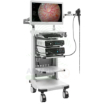
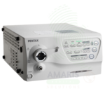
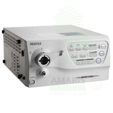
Imaging Processor
WhatsApp Order
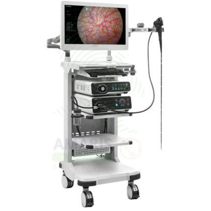
An Imaging Processor is the central processing unit for endoscopic systems, receiving video signals from endoscope cameras and outputting high-definition images for real-time visualization. Incorporating advanced image processing algorithms including narrow band imaging, blue light imaging, and digital contrast enhancement, it enables detection of subtle mucosal abnormalities and early neoplasia. Integrated with hospital information systems and PACS, it provides image storage, documentation, and seamless data transfer for patient records.
Description
Imaging Processor
PRIMARY CLINICAL & DIAGNOSTIC USES
1. Image Acquisition and Processing for Endoscopic Procedures
-
Primary Use: Serves as the central processing unit for endoscopic imaging systems, receiving video signals from the endoscope camera, processing the image data, and outputting high-definition video to monitors for real-time visualization during diagnostic and therapeutic procedures.
-
How it helps: For the endoscopist and surgical team, the imaging processor transforms raw optical signals from the endoscope into high-definition images that guide every aspect of the procedure—providing the clarity and detail needed to identify subtle mucosal abnormalities, navigate through complex anatomy, and perform precise therapeutic interventions. For the patient, superior image processing means more accurate diagnosis, safer procedures, and better outcomes.
2. Image Enhancement and Advanced Visualization
-
Primary Use: Incorporates advanced image processing algorithms including narrow band imaging, blue light imaging, i-scan, and digital contrast enhancement that highlight mucosal and vascular patterns, improving detection of dysplasia and early neoplasia.
-
How it helps: For the gastroenterologist and endoscopist, advanced image processing provides an “optical biopsy” capability—differentiating between benign and malignant tissues with high accuracy, guiding targeted biopsy, and enabling precise delineation of lesion margins. For the patient with Barrett’s esophagus, inflammatory bowel disease, or colorectal polyps, these technologies improve the detection of dysplasia and early cancer.
3. Image Storage and Documentation
-
Primary Use: Captures and stores high-definition still images and video recordings of endoscopic findings, creating a permanent record for patient charts, referral documentation, quality assurance, and medicolegal purposes.
-
How it helps: For the healthcare team and medical records department, stored images provide a permanent record of endoscopic findings that can be shared with referring physicians, reviewed by specialists, and compared with future examinations. For the patient, this documentation ensures continuity of care and facilitates communication between providers.
4. Integration with Hospital Information Systems
-
Primary Use: Connects with electronic medical records, picture archiving and communication systems, and hospital information systems for seamless data transfer, allowing endoscopic images and reports to be integrated into the patient’s medical record.
-
How it helps: For the healthcare facility and information technology team, integrated imaging processors streamline workflow, reduce manual data entry, and ensure that endoscopic findings are immediately available to all members of the patient’s care team. For the patient, this integration ensures that their imaging results are accessible to all providers involved in their care.
5. Digital Signal Processing for Image Optimization
-
Primary Use: Applies digital signal processing algorithms to optimize image quality, including noise reduction, edge enhancement, color correction, and contrast adjustment, ensuring optimal visualization regardless of lighting conditions or tissue characteristics.
-
How it helps: For the endoscopist, digital signal processing ensures consistent, high-quality images throughout the procedure—automatically adjusting to optimize visualization of different tissue types, lighting conditions, and anatomical structures. For the patient, consistent image quality means fewer repeat procedures due to inadequate visualization.
SECONDARY & SUPPORTIVE USES
1. Virtual Chromoendoscopy: Processing algorithms that simulate dye-based chromoendoscopy without the need for physical staining agents.
2. Magnification Processing: Digital zoom and magnification capabilities for detailed assessment of mucosal architecture and pit patterns.
3. Autofluorescence Imaging: Processing of tissue autofluorescence signals to detect changes associated with dysplasia and early cancer.
4. 3D Image Reconstruction: Processing of image data to create three-dimensional reconstructions for surgical planning and navigation.
5. Image Archiving: Long-term storage of endoscopic images and videos for patient records and research.
6. Telemedicine Integration: Compression and transmission of endoscopic images for remote consultation and second opinions.
KEY PRODUCT FEATURES
1. BASIC IDENTIFICATION ATTRIBUTES
-
Device Type: A central processing unit that receives, processes, and outputs video signals from endoscopic camera systems.
-
Designation: Imaging Processor, Endoscopy Processor, Video Processor, Image Processing Unit, Endoscopy System Processor.
-
Key Components:
-
Video Input: Connection ports for endoscope camera heads.
-
Image Processing Unit: Hardware and software for image enhancement.
-
Video Output: High-definition output to monitors and recording devices.
-
Image Storage: Internal or external storage for still images and video.
-
Network Connectivity: Integration with hospital information systems and PACS.
-
User Interface: Touchscreen or control panel for operation.
-
2. TECHNICAL & PERFORMANCE PROPERTIES
-
Resolution: HD (1080p) or 4K (2160p) output.
-
Image Processing: Narrow band imaging, blue light imaging, i-scan, digital contrast enhancement.
-
Frame Rate: High frame rate for smooth real-time visualization.
-
Storage Capacity: Internal storage for thousands of images; expandable options.
-
Connectivity: DICOM, HDMI, SDI, USB, Ethernet.
-
Image Formats: Standard medical image formats for compatibility.
3. PHYSICAL & OPERATIONAL PROPERTIES
-
Construction: Compact, durable housing for clinical environment.
-
User Interface: Intuitive controls for image adjustment and capture.
-
Integration: Compatible with multiple endoscope types and manufacturers.
-
Portability: Cart-mounted or tabletop configurations.
4. SAFETY & COMPLIANCE ATTRIBUTES
-
Regulatory Status: Class II medical device regulated by FDA.
-
Electrical Safety: Compliant with IEC 60601-1.
-
Data Security: Compliant with HIPAA and data protection regulations.
-
EMC Compliance: Electromagnetic compatibility for use in clinical settings.
5. STORAGE & HANDLING ATTRIBUTES
-
Storage: Stored in the procedure room or equipment cart.
-
Cleaning: Wipe with hospital-grade disinfectants.
-
Maintenance: Regular software updates and preventive maintenance.
6. LABORATORY & CLINICAL APPLICATIONS
-
Primary Application: Image processing for endoscopic procedures in gastroenterology, pulmonology, urology, gynecology, and surgery.
-
Clinical Role: Essential component of modern endoscopic systems, enabling high-definition visualization and advanced imaging technologies.
SAFETY HANDLING PRECAUTIONS
1. SAFETY PRECAUTIONS
-
Electrical Safety: Ensure proper grounding; keep away from liquids.
-
Data Security: Follow facility protocols for protected health information.
-
Equipment Compatibility: Ensure compatibility with endoscopes and accessories.
-
Calibration: Regular calibration for optimal image quality.
2. FIRST AID MEASURES
-
Electrical Shock: If shock occurs, disconnect power; seek medical attention if needed.
-
Equipment Failure: If the processor fails, have backup equipment available.
3. FIRE FIGHTING MEASURES
-
Flammability: Plastic components are combustible; electrical components may pose fire risk.
-
Extinguishing Media: For electrical fire, use CO₂ or dry chemical extinguisher.
Related products
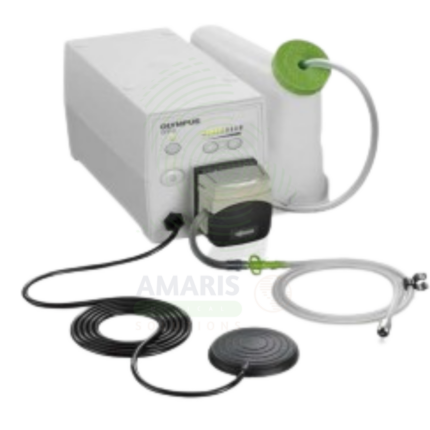
Active Medical Flushing Pump
An Active Medical Flushing Pump is a powered irrigation device used during endoscopic procedures to maintain a clear visual field, control bleeding, and facilitate therapeutic interventions. Providing adjustable flow rates and pressure settings, it enables targeted irrigation, submucosal injection for lesion resection, and delivery of hemostatic agents. Integrated with endoscopic systems, it ensures optimal visualization for accurate diagnosis and precise treatment, improving patient outcomes.
Diagnostic Kit (ENT)
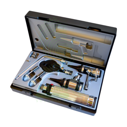
A Diagnostic Kit (ENT) is a comprehensive, portable set of specialized instruments essential for the complete physical examination of the Ear, Nose, and Throat. It typically includes a head mirror/light, otoscope, nasal speculum, laryngeal mirrors, tuning forks, and curettes, housed in a durable carrying case. This kit enables otolaryngologists, general practitioners, and emergency physicians to visually inspect, diagnose, and manage a wide array of conditions from common infections and allergies to anatomical abnormalities. Its effectiveness relies on the clinician's expertise and rigorous adherence to infection control protocols for the cleaning and sterilization of its reusable components.
ENT Endoscopes
ENT Endoscopes are specialized optical instruments, either flexible or rigid, used to diagnose and treat conditions of the ear, nose, throat, and upper airway. Flexible nasopharyngolaryngoscopes enable in-office visualization of the nasal passages and larynx, while rigid telescopes (with various angles like 0°, 30°, 70°) are used for surgical procedures such as sinus surgery and microlaryngoscopy. Their safe use requires meticulous reprocessing—especially high-level disinfection for flexible scopes—gentle technique to avoid mucosal injury or epistaxis, and proper patient preparation. They are indispensable tools in both the otolaryngology clinic and operating room.
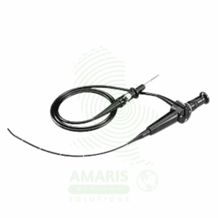
Flexible Fiber Optic Laryngoscope
A Flexible Fiber Optic Laryngoscope is a flexible endoscope (2-5 mm diameter, 30-60 cm working length) with fiber optic image transmission and steerable tip (120-180° angulation) for visualization of the upper airway and facilitation of difficult intubations. Features include control handle with angulation lever, working channel (1-2 mm) for suction or oxygen, external light source (halogen/xenon/LED), and optional camera for video display. Primary clinical applications include awake intubation in difficult airway management (limited mouth opening, cervical spine instability, obstructing pathology), nasotracheal intubation for oral surgery or maxillofacial trauma, intubation with cervical spine precautions (minimal neck movement), diagnostic airway assessment (stridor, hoarseness, vocal cord dysfunction, masses), double-lumen tube placement for thoracic surgery, pediatric difficult airway management, and tracheostomy tube placement guidance. Class II medical device requiring FDA clearance. Critical safety considerations include mandatory leak testing before immersion, antifog preparation, gentle insertion technique, airway maintenance with oxygen, topical anesthesia for patient comfort, suction availability, backup airway device, and strict infection control with validated reprocessing protocols.
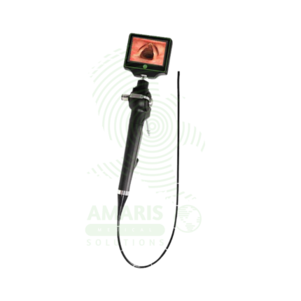
High-Definition Endoscope
A High-Definition Endoscope is an advanced endoscopic system providing superior image resolution, color accuracy, and contrast for diagnostic and therapeutic procedures. Incorporating HD or 4K imaging with advanced visualization technologies such as narrow band imaging, blue light imaging, and autofluorescence, it enables detection of subtle mucosal abnormalities and early neoplasia that may be missed with standard definition systems. Essential for early cancer detection, precise therapeutic intervention, and documentation, it represents the standard of care for advanced endoscopy.
High-Definition Imaging Processor
A High-Definition Imaging Processor is an advanced image processing unit for endoscopic systems, supporting HD and 4K ultra-high-definition visualization. Incorporating technologies such as narrow band imaging, blue light imaging, and digital contrast enhancement, it enables detection of subtle mucosal abnormalities and early neoplasia. With high-resolution image storage and seamless integration with hospital information systems, it provides superior image quality for diagnosis, documentation, and therapeutic precision.
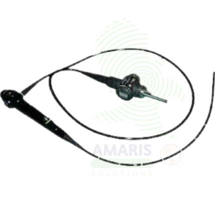
Medical Endoscope
A Medical Endoscope is an optical instrument used for direct visualization of internal organs and body cavities for diagnostic and therapeutic purposes. Flexible endoscopes navigate the gastrointestinal tract, respiratory tract, and urinary tract, while rigid endoscopes are used for laparoscopy, arthroscopy, and cystoscopy. Integrated working channels allow passage of instruments for biopsy, polypectomy, hemostasis, and stent placement. Essential for cancer screening, minimally invasive surgery, and management of bleeding and obstruction, endoscopy has transformed the diagnosis and treatment of diseases across multiple medical specialties.