Dermatoscope and Magnifiers
 Diagnostic Kits
 Vital Signs Monitors
 Stethoscopes and Accessories
 Otoscopes, Ophthalmoscopes, and Retinoscopes
 Reflex Hammers and Neurological Tools
 Scales and Measuring Devices
 Spirometers and Pulmonary Function Tests
 Electrosurgical Units and Accessories
 Cutting Instruments
 Grasping and Holding Instruments
 Hemostatic Instruments
 Specialized Surgical Sets
 Single-Use Procedure Trays and Packs
 Surgical Drapes, Gowns, and Covers
 Tissue Unifying Instruments
 Radiation Protection
 X-Ray Machines and Accessories
 Ultrasound Systems and Probes
 MRI and CT Scanners
 Radiology Consumables
 Bone Densitometers
 Fluoroscopy Equipment
 Imaging Tables and Positioning Aids
 Microscopes and Accessories
 Centrifuges and Separators
 Analyzers
 Incubators and Ovens
 Pipettes, Dispensers, and Lab Glassware
 Refrigerators, Freezers, and Storage Units
 Lab Consumables
 Sterilizers and Autoclaves for Lab Use
 Multi-Parameter Monitors
 Ventilators and Respiratory Support Devices
 Defibrillators and AEDs
 Infusion Pumps and IV Systems
 Patient Warmers and Cooling Devices
 Central Monitoring Stations
 Accessories
 Anesthesia Machines and Workstations
 Oxygen Concentrators and Delivery Systems
 Nebulizers and Inhalers
 CPAP/BiPAP Machines
 Airway Management
 Anesthesia Masks, Circuits, and Bags
 Humidifiers and Heaters
 Respiratory Therapy Accessories
 First Aid Kits and Cabinets
 Emergency Resuscitation Equipment
 Trauma Supplies
 Emergency Carts and Crash Carts
 Burn Care Products
 Bleeding Control
 Automated External Defibrillators (AEDs)
 Transport and Evacuation
 Wheelchairs and Accessories
 Walkers, Crutches, and Canes
 Prosthetics and Orthotics
 Physical Therapy Equipment
 Transfer Devices
 Bathroom Safety
 Orthopedic Traction and Tables
 Hot/Cold Therapy Packs and Units
 Beds and Mattresses
 Chairs and Stools
 Tables
 Cabinets and Storage
 Privacy Screens & Curtains
 Stands and Racks
 Linens and Textiles
 Lighting
 Autoclaves and Sterilizers
 Ultrasonic Cleaners
 Disinfectant Solutions and Wipes
 Sterilization Pouches, Wraps, and Indicators
 Instrument Trays and Containers
 UV and Ozone Disinfection Devices
 Washer Disinfectors
 Wound Care
 Gloves
 Masks and Respirators
 Catheters and Tubing
 Swabs, Applicators, and Sponges
 Incontinence Products
 Personal Protective Equipment (PPE)
 Dental Chairs and Units
 Handpieces and Burs
 Instruments
 Consumables
 Sterilization for Dental Use
 Orthodontic Supplies
 Endodontic Tools
 Slit Lamps and Tonometers
 Lensometers and Phoropters
 Ophthalmic Surgical Instruments
 Eyewear Frames and Lenses
 Contact Lens Supplies
 Vision Testing Charts and Devices
 Eye Care Consumables
 Laser Systems for Eye Care
 ENT Exam Chairs and Tables
 Endoscopes
 Audiometers and Hearing Tests
 ENT Instruments
 Nasal and Throat Packs
 Hearing Aids and Accessories
 Otology Supplies
 Fetal Dopplers and Monitors
 Delivery Beds and Tables
 Gynecological Instruments
 Neonatal Incubators and Warmers
 Breast Pumps and Accessories
 Contraceptive Devices
 Maternity Supports and Pads
 Neonatal Consumables
 Cystoscopes and Urethroscopes
 Dialysis Machines and Supplies
 Urological Catheters and Bags
 Lithotripters
 Prostate Treatment Devices
 Urinary Incontinence Products
 Kidney Stone Management Tools
 Consumables & Disposables
 EEG and EMG Machines
 Neurosurgical Instruments
 Nerve Stimulators
 Headrests and Positioning Aids
 Lumbar Puncture Kits
 Seizure Monitoring Devices
 Consumables
 Rehabilitation for Neurological Conditions
 ECG Machines and Accessories
 Holter Monitors
 Stress Test Systems
 Pacemakers and Defibrillator Accessories
 Vascular Access Devices
 Cardiac Catheters and Guidewires
 Blood Flow Meters
 Consumables
 Orthopedic Instruments
 Casts, Splints, and Padding
 Joint Replacement Supplies
 Prosthetic Limbs and Components
 Bone Grafts and Substitutes
 Traction Devices
 Orthopedic Braces and Supports
 Rehabilitation Aids for Orthopedics
 Home Oxygen Therapy
 Hospital Beds for Home Use
 Mobility Aids
 Bathroom and Daily Living Aids
 Wound Care for Home
 Monitoring Devices
 Enteral Feeding Pumps and Tubes
 Hand Sanitizers and Dispensers
 Face Shields and Goggles
 Isolation Gowns and Suits
 Biohazard Waste Containers
 Air Purifiers and HEPA Filters
 Surface Disinfectants
 Sharps Containers
 Protective Barriers
 Cardiovascular & Endurance Training
 Strength Training & Weightlifting
 Functional Training & Core Conditioning
 Physical Therapy & Rehabilitation
 Sports & Outdoor Recreation
 Gym Flooring & Facility Equipment
 Fitness Monitoring & Accessories
 Kids & Novelties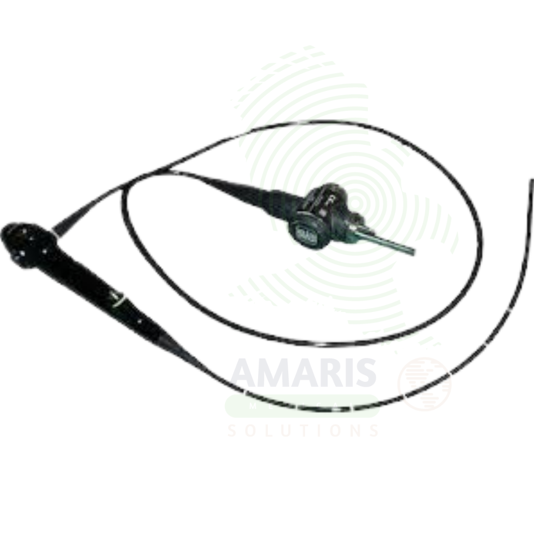
Medical Endoscope
$1.00
WhatsApp Order
A Medical Endoscope is an optical instrument used for direct visualization of internal organs and body cavities for diagnostic and therapeutic purposes. Flexible endoscopes navigate the gastrointestinal tract, respiratory tract, and urinary tract, while rigid endoscopes are used for laparoscopy, arthroscopy, and cystoscopy. Integrated working channels allow passage of instruments for biopsy, polypectomy, hemostasis, and stent placement. Essential for cancer screening, minimally invasive surgery, and management of bleeding and obstruction, endoscopy has transformed the diagnosis and treatment of diseases across multiple medical specialties.
Description
Medical Endoscope
PRIMARY CLINICAL & DIAGNOSTIC USES
1. Visualization and Diagnosis of Hollow Organs and Body Cavities
-
Primary Use: Provides direct visualization of internal organs and structures including the gastrointestinal tract (esophagus, stomach, duodenum, colon), respiratory tract (trachea, bronchi), urinary tract (bladder, ureters), and body cavities (abdomen, thorax, joints). The endoscope transmits high-definition images to a monitor for real-time diagnostic assessment.
-
How it helps: For the gastroenterologist, pulmonologist, urologist, and surgeon, the endoscope transforms internal examination from blind speculation into direct visualization—navigating through the GI tract, airways, or body cavities to see exactly what lies within. For the patient with unexplained bleeding, chronic pain, or suspicious imaging findings, endoscopy offers the chance for a definitive diagnosis without a surgical incision, often as an outpatient procedure with minimal discomfort and rapid recovery.
2. Therapeutic Intervention Through the Working Channel
-
Primary Use: Enables minimally invasive therapeutic procedures through the endoscope’s working channel, including polypectomy, biopsy, foreign body removal, hemostasis (clipping, cautery), stent placement, dilation of strictures, and endoscopic mucosal resection.
-
How it helps: For the interventional endoscopist and surgeon, the endoscope is not just a diagnostic tool but a surgical platform—allowing them to remove polyps before they become cancer, stop bleeding from ulcers, retrieve swallowed objects, and open narrowed passages. For the patient, this means that conditions which once required major surgery can now be treated through natural orifices or tiny incisions, with less pain, shorter hospital stays, and faster return to normal life.
3. Screening and Surveillance for Cancer
-
Primary Use: Essential for cancer screening programs including colonoscopy for colorectal cancer, upper endoscopy for esophageal and gastric cancer, and bronchoscopy for lung cancer. Enables detection and removal of precancerous lesions before they become malignant.
-
How it helps: For the gastroenterologist and preventive medicine specialist, endoscopy provides the opportunity to detect and remove precancerous lesions before they ever become malignant—interrupting the natural history of diseases like colorectal cancer. For the asymptomatic individual undergoing screening, or the patient with Barrett’s esophagus under surveillance, regular endoscopic examinations offer the best chance for cancer prevention through early detection and intervention.
4. Biopsy and Tissue Sampling
-
Primary Use: Allows precise biopsy of suspicious lesions, polyps, and abnormal mucosa for histopathological diagnosis. The working channel enables passage of biopsy forceps, cytology brushes, and fine-needle aspiration devices under direct visualization.
-
How it helps: For the pathologist and clinician, endoscopically guided biopsy provides the tissue diagnosis that guides all subsequent treatment decisions—confirming malignancy, determining tumor type, and guiding surgical planning. For the patient with a suspicious lesion, endoscopic biopsy provides definitive diagnosis without the need for open surgical biopsy.
5. Evaluation of Hemorrhage and Bleeding Control
-
Primary Use: Critical for identifying sources of gastrointestinal bleeding, hemoptysis, and hematuria. Enables therapeutic intervention through the working channel including injection of epinephrine, placement of hemostatic clips, cauterization, and band ligation of varices.
-
How it helps: For the gastroenterologist and pulmonologist managing patients with active bleeding, endoscopy provides both diagnosis and treatment in a single procedure—identifying the bleeding source and delivering targeted therapy to achieve hemostasis. For the patient with gastrointestinal or airway bleeding, prompt endoscopic intervention can stop life-threatening hemorrhage and avoid surgical exploration.
SECONDARY & SUPPORTIVE USES
1. Enteral Access and Feeding Tube Placement: Used to place percutaneous endoscopic gastrostomy (PEG) tubes and nasoenteric feeding tubes for patients unable to take oral nutrition.
2. Foreign Body Removal: Retrieval of swallowed or aspirated foreign bodies including coins, batteries, food impactions, and other objects.
3. Dilation of Strictures: Balloon or bougie dilation of esophageal, pyloric, colonic, and tracheal strictures under direct visualization.
4. Stent Placement: Deployment of self-expanding metal or plastic stents for malignant and benign strictures of the GI tract, biliary tree, and airways.
5. Endoscopic Ultrasound: Specialized endoscopes with ultrasound capability for staging cancers, evaluating submucosal lesions, and guiding fine-needle aspiration.
6. Intraoperative Endoscopy: Used during surgery to evaluate anastomoses, localize tumors, and assess for complications.
KEY PRODUCT FEATURES
1. BASIC IDENTIFICATION ATTRIBUTES
-
Device Type: A flexible or rigid optical instrument with a camera and light source used to visualize internal organs and perform therapeutic interventions.
-
Designation: Medical Endoscope, Endoscopy System, Flexible Endoscope, Rigid Endoscope.
-
Types:
-
Flexible Endoscopes: Used for gastrointestinal, respiratory, and urinary tract; can navigate tortuous anatomy.
-
Rigid Endoscopes: Used for laparoscopy, arthroscopy, cystoscopy; provide stable platforms for surgery.
-
Capsule Endoscopy: Wireless capsule camera for small bowel imaging.
-
Video Endoscopes: Integrated camera chip at the distal tip for high-definition imaging.
-
-
Key Components:
-
Insertion Tube: Flexible or rigid shaft containing optics, light fibers, and working channel.
-
Distal Tip: Contains camera, light source, and instrument channel opening.
-
Control Section: Handpiece with controls for tip deflection, suction, and air/water.
-
Working Channel: Channel for passage of instruments, suction, and irrigation.
-
Light Source: High-intensity light source for illumination.
-
Video Processor: Converts optical signal to high-definition video output.
-
2. TECHNICAL & PERFORMANCE PROPERTIES
-
Image Resolution: High-definition (HD) and 4K resolution for detailed visualization.
-
Field of View: Wide-angle optics for comprehensive visualization.
-
Tip Deflection: Multi-directional tip articulation for navigation and positioning.
-
Working Channel Diameter: Varies by endoscope type (2.0-4.2 mm) for instrument passage.
-
Insertion Tube Diameter: Varies by application; pediatric and adult sizes available.
-
Length: Varies by application (colonoscope: 130-170 cm; gastroscope: 100-110 cm; bronchoscope: 60 cm).
-
Sterilization: High-level disinfection or sterilization depending on endoscope type and clinical application.
3. PHYSICAL & OPERATIONAL PROPERTIES
-
Construction: Durable materials for repeated use; waterproof design for immersion cleaning.
-
Ergonomics: Designed for single-operator use with intuitive controls.
-
Light Source: Xenon, LED, or laser-based illumination.
-
Video Output: Standardized connectors for integration with monitors and recording systems.
4. SAFETY & COMPLIANCE ATTRIBUTES
-
Regulatory Status: Class II medical device regulated by FDA.
-
Electrical Safety: Compliant with medical electrical equipment standards.
-
Biocompatibility: Materials safe for contact with mucous membranes and internal tissues.
-
Sterility: High-level disinfection or sterilization required between patients.
5. STORAGE & HANDLING ATTRIBUTES
-
Storage: Stored in hanging cabinets to prevent damage; protect from moisture.
-
Cleaning: Thorough cleaning immediately after use; enzymatic cleaners for debris removal.
-
Disinfection: High-level disinfection or sterilization per facility protocol.
-
Inspection: Regular inspection for damage, leaks, or wear; leak testing required.
6. LABORATORY & CLINICAL APPLICATIONS
-
Primary Application: Diagnostic and therapeutic procedures in gastroenterology, pulmonology, urology, gynecology, and surgery.
-
Clinical Role: Essential equipment for minimally invasive diagnosis and treatment across multiple specialties.
SAFETY HANDLING PRECAUTIONS
1. SAFETY PRECAUTIONS
-
Leak Testing: Perform leak test before each use to ensure integrity.
-
Sterilization: Follow appropriate disinfection and sterilization protocols.
-
Insertion Technique: Insert with care; use gentle pressure; do not force.
-
Sedation: Monitor patient sedation during procedures.
-
Informed Consent: Explain procedure, risks, and alternatives to patients.
2. FIRST AID MEASURES
-
Perforation: If perforation is suspected, discontinue procedure; obtain surgical consultation.
-
Bleeding: If significant bleeding occurs, attempt endoscopic hemostasis; consider surgical consultation.
-
Equipment Failure: If the endoscope fails during the procedure, have backup equipment available.
3. FIRE FIGHTING MEASURES
-
Flammability: Plastic components are combustible; light sources may generate heat.
-
Extinguishing Media: Use CO₂ or dry chemical extinguisher for electrical fires.
Related products
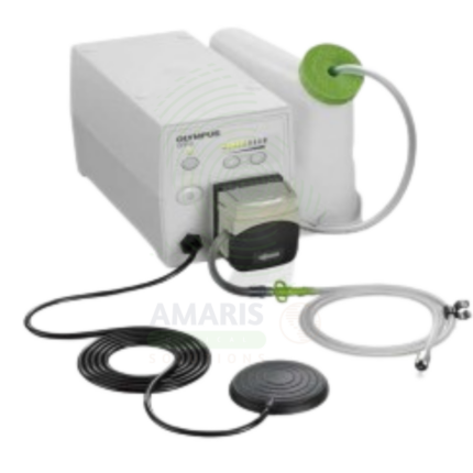
Active Medical Flushing Pump
An Active Medical Flushing Pump is a powered irrigation device used during endoscopic procedures to maintain a clear visual field, control bleeding, and facilitate therapeutic interventions. Providing adjustable flow rates and pressure settings, it enables targeted irrigation, submucosal injection for lesion resection, and delivery of hemostatic agents. Integrated with endoscopic systems, it ensures optimal visualization for accurate diagnosis and precise treatment, improving patient outcomes.
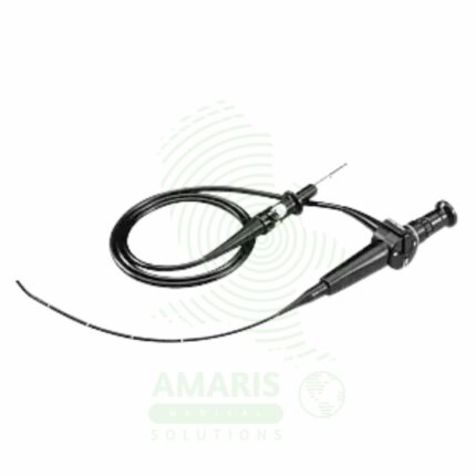
ENT Endoscopes
ENT Endoscopes are specialized optical instruments, either flexible or rigid, used to diagnose and treat conditions of the ear, nose, throat, and upper airway. Flexible nasopharyngolaryngoscopes enable in-office visualization of the nasal passages and larynx, while rigid telescopes (with various angles like 0°, 30°, 70°) are used for surgical procedures such as sinus surgery and microlaryngoscopy. Their safe use requires meticulous reprocessing—especially high-level disinfection for flexible scopes—gentle technique to avoid mucosal injury or epistaxis, and proper patient preparation. They are indispensable tools in both the otolaryngology clinic and operating room.
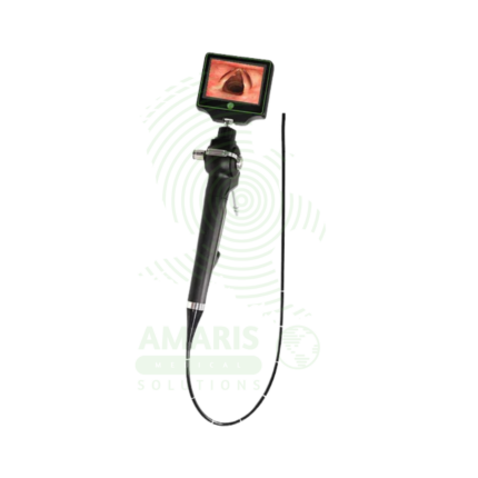
Flexible Fiber Optic Laryngoscope
A Flexible Fiber Optic Laryngoscope is a flexible endoscope (2-5 mm diameter, 30-60 cm working length) with fiber optic image transmission and steerable tip (120-180° angulation) for visualization of the upper airway and facilitation of difficult intubations. Features include control handle with angulation lever, working channel (1-2 mm) for suction or oxygen, external light source (halogen/xenon/LED), and optional camera for video display. Primary clinical applications include awake intubation in difficult airway management (limited mouth opening, cervical spine instability, obstructing pathology), nasotracheal intubation for oral surgery or maxillofacial trauma, intubation with cervical spine precautions (minimal neck movement), diagnostic airway assessment (stridor, hoarseness, vocal cord dysfunction, masses), double-lumen tube placement for thoracic surgery, pediatric difficult airway management, and tracheostomy tube placement guidance. Class II medical device requiring FDA clearance. Critical safety considerations include mandatory leak testing before immersion, antifog preparation, gentle insertion technique, airway maintenance with oxygen, topical anesthesia for patient comfort, suction availability, backup airway device, and strict infection control with validated reprocessing protocols.
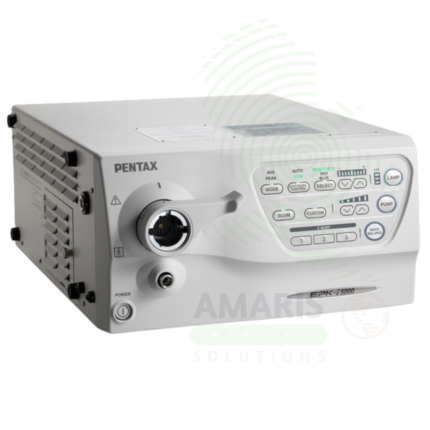
High-Definition Imaging Processor
A High-Definition Imaging Processor is an advanced image processing unit for endoscopic systems, supporting HD and 4K ultra-high-definition visualization. Incorporating technologies such as narrow band imaging, blue light imaging, and digital contrast enhancement, it enables detection of subtle mucosal abnormalities and early neoplasia. With high-resolution image storage and seamless integration with hospital information systems, it provides superior image quality for diagnosis, documentation, and therapeutic precision.
Laparoscope
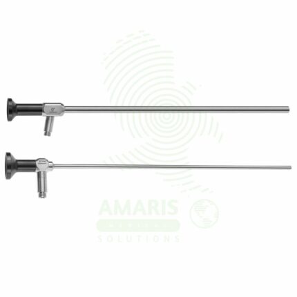
A Laparoscope is a rigid, sterile optical telescope used in minimally invasive surgery to visualize the abdominal and pelvic cavities. Featuring a Hopkins rod-lens system for brilliant, high-resolution images, it comes in standard diameters (5mm, 10mm) and viewing angles (0° for straight-ahead or 30°/45° to look around structures). It connects to a high-intensity light source and a video camera system. Meticulous handling, mandatory pre-sterilization leak testing, and careful cleaning are required to maintain its integrity and prevent infection. As the primary visual conduit for the surgeon, its clarity and reliability are fundamental to the safety and success of any laparoscopic procedure.
Laparoscopic Instrument
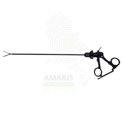
Laparoscopic Instruments are specialized surgical tools designed for use through small incisions during minimally invasive abdominal and pelvic procedures. Available in grasping, cutting, dissecting, hemostatic, and suturing configurations, they enable surgeons to perform complex operations with precision while minimizing patient trauma. With shaft diameters of 3-12 mm and lengths optimized for abdominal access, these instruments allow for the full range of surgical techniques through small incisions, resulting in reduced post-operative pain, shorter hospital stays, and faster recovery.
Metal speculums
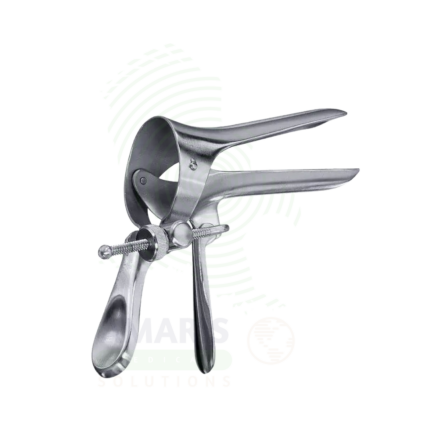
Metal Speculums are reusable, autoclavable instruments designed to gently separate and retract tissue for visualization and access to internal body cavities including the vagina, cervix, nasal passages, and ear canal. Constructed from medical-grade stainless steel for durability and corrosion resistance, these instruments are available in multiple types—including Cusco and Graves vaginal speculums, Vienna nasal speculums, and Brunning ear speculums—with various sizes to accommodate pediatric through adult patients. For the clinician, metal speculums provide reliable, precise tissue retraction that enables accurate diagnostic examination, specimen collection, and therapeutic procedures. For the patient, the use of properly sized, well-maintained metal speculums facilitates timely diagnosis of conditions such as cervical dysplasia, sinusitis, and otitis media, enabling early intervention and optimal outcomes.