Dermatoscope and Magnifiers
 Diagnostic Kits
 Vital Signs Monitors
 Stethoscopes and Accessories
 Otoscopes, Ophthalmoscopes, and Retinoscopes
 Reflex Hammers and Neurological Tools
 Scales and Measuring Devices
 Spirometers and Pulmonary Function Tests
 Electrosurgical Units and Accessories
 Cutting Instruments
 Grasping and Holding Instruments
 Hemostatic Instruments
 Specialized Surgical Sets
 Single-Use Procedure Trays and Packs
 Surgical Drapes, Gowns, and Covers
 Tissue Unifying Instruments
 Radiation Protection
 X-Ray Machines and Accessories
 Ultrasound Systems and Probes
 MRI and CT Scanners
 Radiology Consumables
 Bone Densitometers
 Fluoroscopy Equipment
 Imaging Tables and Positioning Aids
 Microscopes and Accessories
 Centrifuges and Separators
 Analyzers
 Incubators and Ovens
 Pipettes, Dispensers, and Lab Glassware
 Refrigerators, Freezers, and Storage Units
 Lab Consumables
 Sterilizers and Autoclaves for Lab Use
 Multi-Parameter Monitors
 Ventilators and Respiratory Support Devices
 Defibrillators and AEDs
 Infusion Pumps and IV Systems
 Patient Warmers and Cooling Devices
 Central Monitoring Stations
 Accessories
 Anesthesia Machines and Workstations
 Oxygen Concentrators and Delivery Systems
 Nebulizers and Inhalers
 CPAP/BiPAP Machines
 Airway Management
 Anesthesia Masks, Circuits, and Bags
 Humidifiers and Heaters
 Respiratory Therapy Accessories
 First Aid Kits and Cabinets
 Emergency Resuscitation Equipment
 Trauma Supplies
 Emergency Carts and Crash Carts
 Burn Care Products
 Bleeding Control
 Automated External Defibrillators (AEDs)
 Transport and Evacuation
 Wheelchairs and Accessories
 Walkers, Crutches, and Canes
 Prosthetics and Orthotics
 Physical Therapy Equipment
 Transfer Devices
 Bathroom Safety
 Orthopedic Traction and Tables
 Hot/Cold Therapy Packs and Units
 Beds and Mattresses
 Chairs and Stools
 Tables
 Cabinets and Storage
 Privacy Screens & Curtains
 Stands and Racks
 Linens and Textiles
 Lighting
 Autoclaves and Sterilizers
 Ultrasonic Cleaners
 Disinfectant Solutions and Wipes
 Sterilization Pouches, Wraps, and Indicators
 Instrument Trays and Containers
 UV and Ozone Disinfection Devices
 Washer Disinfectors
 Wound Care
 Gloves
 Masks and Respirators
 Catheters and Tubing
 Swabs, Applicators, and Sponges
 Incontinence Products
 Personal Protective Equipment (PPE)
 Dental Chairs and Units
 Handpieces and Burs
 Instruments
 Consumables
 Sterilization for Dental Use
 Orthodontic Supplies
 Endodontic Tools
 Slit Lamps and Tonometers
 Lensometers and Phoropters
 Ophthalmic Surgical Instruments
 Eyewear Frames and Lenses
 Contact Lens Supplies
 Vision Testing Charts and Devices
 Eye Care Consumables
 Laser Systems for Eye Care
 ENT Exam Chairs and Tables
 Endoscopes
 Audiometers and Hearing Tests
 ENT Instruments
 Nasal and Throat Packs
 Hearing Aids and Accessories
 Otology Supplies
 Fetal Dopplers and Monitors
 Delivery Beds and Tables
 Gynecological Instruments
 Neonatal Incubators and Warmers
 Breast Pumps and Accessories
 Contraceptive Devices
 Maternity Supports and Pads
 Neonatal Consumables
 Cystoscopes and Urethroscopes
 Dialysis Machines and Supplies
 Urological Catheters and Bags
 Lithotripters
 Prostate Treatment Devices
 Urinary Incontinence Products
 Kidney Stone Management Tools
 Consumables & Disposables
 EEG and EMG Machines
 Neurosurgical Instruments
 Nerve Stimulators
 Headrests and Positioning Aids
 Lumbar Puncture Kits
 Seizure Monitoring Devices
 Consumables
 Rehabilitation for Neurological Conditions
 ECG Machines and Accessories
 Holter Monitors
 Stress Test Systems
 Pacemakers and Defibrillator Accessories
 Vascular Access Devices
 Cardiac Catheters and Guidewires
 Blood Flow Meters
 Consumables
 Orthopedic Instruments
 Casts, Splints, and Padding
 Joint Replacement Supplies
 Prosthetic Limbs and Components
 Bone Grafts and Substitutes
 Traction Devices
 Orthopedic Braces and Supports
 Rehabilitation Aids for Orthopedics
 Home Oxygen Therapy
 Hospital Beds for Home Use
 Mobility Aids
 Bathroom and Daily Living Aids
 Wound Care for Home
 Monitoring Devices
 Enteral Feeding Pumps and Tubes
 Hand Sanitizers and Dispensers
 Face Shields and Goggles
 Isolation Gowns and Suits
 Biohazard Waste Containers
 Air Purifiers and HEPA Filters
 Surface Disinfectants
 Sharps Containers
 Protective Barriers
 Cardiovascular & Endurance Training
 Strength Training & Weightlifting
 Functional Training & Core Conditioning
 Physical Therapy & Rehabilitation
 Sports & Outdoor Recreation
 Gym Flooring & Facility Equipment
 Fitness Monitoring & Accessories
 Kids & Novelties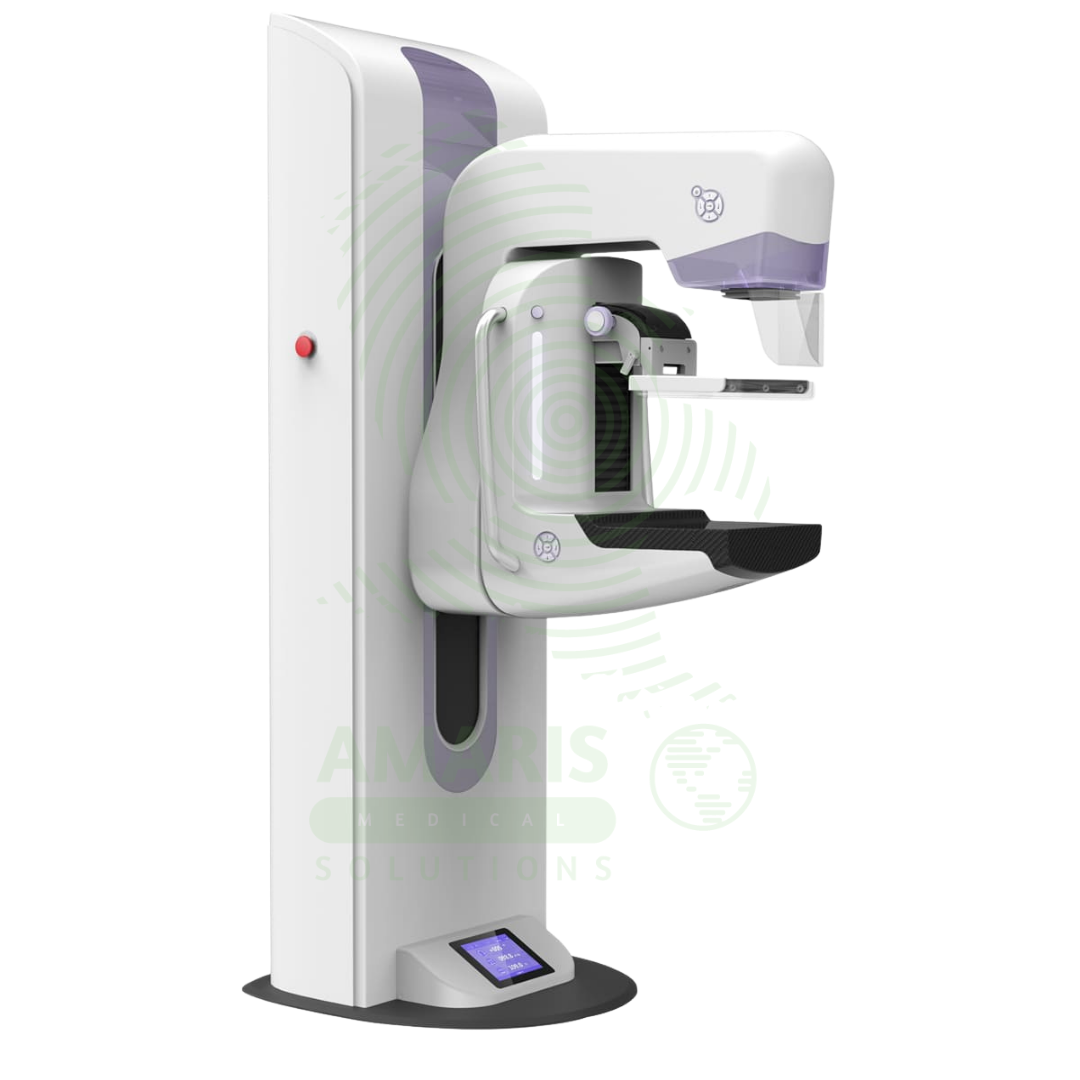

Digital Mammography System
$1.00
WhatsApp Order
A Digital Mammography System is a specialized X-ray imaging system designed for breast cancer screening and diagnosis. Using high-resolution digital detectors, it provides superior image quality with lower radiation dose compared to film mammography. Advanced systems offer tomosynthesis (3D) imaging, which improves cancer detection rates and reduces false positives. Used in breast imaging centers, radiology departments, and women’s health facilities, it is the gold standard for early detection of breast cancer.
Description
Digital Mammography System
PRIMARY CLINICAL & DIAGNOSTIC USES
1. Breast Cancer Screening
-
Primary Use: Provides high-resolution digital X-ray imaging of the breast for routine screening of asymptomatic women to detect early, non-palpable breast cancer. Digital technology offers superior image quality with lower radiation dose compared to film mammography, enabling earlier detection of microcalcifications and subtle masses.
-
How it helps: For the radiologist and breast imaging specialist, digital mammography is the most powerful tool for finding breast cancer at its earliest, most treatable stage—detecting tumors years before they can be felt, identifying microcalcifications that signal early disease, and revealing subtle architectural distortions. For the woman undergoing routine screening, digital mammography means a lower radiation dose, faster examination time, and the best chance for early detection when treatment is most effective and survival rates are highest.
2. Diagnostic Evaluation of Breast Symptoms
-
Primary Use: Investigates clinical signs such as a palpable lump, nipple discharge, skin changes, or persistent breast pain. Diagnostic mammography involves additional views, magnification, and spot compression to characterize abnormalities found on screening or during physical examination.
-
How it helps: For the breast surgeon and radiologist evaluating a patient with a concerning finding, diagnostic digital mammography provides the detailed characterization needed to determine the next steps—distinguishing benign from suspicious findings, mapping the extent of disease, and guiding biopsy decisions. For the patient who has found a lump or noticed a change in her breast, diagnostic imaging provides answers, whether the reassurance of a benign finding or the definitive diagnosis that allows treatment to begin.
3. Tomosynthesis (3D Mammography)
-
Primary Use: Advanced digital mammography systems offer tomosynthesis (3D) imaging, which acquires multiple low-dose images of the breast from different angles and reconstructs them into three-dimensional slices, reducing tissue overlap and improving detection of subtle abnormalities while reducing recall rates.
-
How it helps: For the radiologist, tomosynthesis significantly improves cancer detection rates and reduces false positives by eliminating overlapping tissue that can obscure lesions or mimic abnormalities. For the patient, 3D mammography means fewer callbacks for additional imaging, reduced anxiety, and increased confidence in screening results.
4. Pre-operative Localization and Guidance
-
Primary Use: Used to guide needle localization procedures to mark non-palpable lesions for surgical biopsy or excision, and to guide stereotactic core needle biopsies, allowing for precise tissue sampling of suspicious areas without open surgery.
-
How it helps: For the breast surgeon and interventional radiologist, digital mammography guidance transforms a blind surgical exploration into a precisely targeted procedure—placing localization wires exactly at the lesion site, guiding biopsy needles to the exact area of suspicion, and ensuring that the right tissue is sampled or removed. For the patient with a non-palpable abnormality, image-guided procedures mean a definitive diagnosis can be obtained with minimal discomfort and scarring.
5. Evaluation of Breast Implants
-
Primary Use: Specialized views (Eklund displacement technique) are used to image breast tissue in patients with implants, assessing for rupture and detecting cancers that may be obscured by the implant. Digital technology allows for optimal visualization of breast tissue around implants.
-
How it helps: For the plastic surgeon and breast imager, dedicated implant views displace the implant backward while bringing breast tissue forward, allowing clear visualization of tissue that would otherwise be hidden. For the woman with breast implants, whether for reconstruction after cancer or cosmetic augmentation, specialized digital mammographic techniques ensure that her breast tissue is adequately screened despite the presence of implants.
SECONDARY & SUPPORTIVE USES
1. Assessment of High-Risk Patients: Used as part of a surveillance strategy for women with a strong family history or genetic predisposition (BRCA mutations) to breast cancer, often starting at a younger age.
2. Monitoring Neoadjuvant Chemotherapy Response: Used to assess tumor response to chemotherapy before surgery, helping guide treatment decisions.
3. Post-Operative Surveillance: Used for monitoring patients with a personal history of breast cancer to detect recurrence or new primary cancers.
4. Guiding Other Interventional Procedures: Can be used to guide other percutaneous breast procedures, such as cyst aspiration.
5. Research and Clinical Trials: Used to acquire standardized images for research studies on breast density, cancer risk, and the efficacy of new imaging technologies.
6. Screening in Dense Breast Tissue: Digital mammography, particularly with tomosynthesis, offers improved detection in women with dense breast tissue.
KEY PRODUCT FEATURES
1. BASIC IDENTIFICATION ATTRIBUTES
-
Device Type: A digital X-ray system specifically designed for breast imaging.
-
Designation: Digital Mammography System, Full-Field Digital Mammography (FFDM), 3D Mammography, Tomosynthesis System.
-
Key Components:
-
X-ray Tube: Specialized tube with molybdenum, rhodium, or tungsten target for breast imaging.
-
Digital Detector: Flat panel detector for high-resolution image capture.
-
Compression Paddle: Clear plastic paddle for breast compression and immobilization.
-
Patient Positioning: Dedicated design for patient comfort and optimal imaging.
-
Workstation: High-resolution monitors for image interpretation.
-
Tomosynthesis Capability: Some systems offer 3D imaging.
-
2. TECHNICAL & PERFORMANCE PROPERTIES
-
Detector Type: Direct or indirect conversion flat panel digital detector.
-
Pixel Size: 50-100 microns for high-resolution imaging.
-
Spatial Resolution: High resolution for microcalcification detection.
-
Target Materials: Molybdenum, rhodium, tungsten for optimal X-ray spectrum.
-
Tomosynthesis: 3D imaging with 1-3 mm slice thickness; 15-30 degree sweep.
-
Dose: Lower radiation dose compared to film mammography.
-
CAD (Computer-Aided Detection): Optional software for assisting radiologist interpretation.
3. PHYSICAL & OPERATIONAL PROPERTIES
-
Construction: Dedicated breast imaging system with ergonomic design.
-
Patient Positioning: Designed for comfortable, reproducible positioning.
-
Compression: Automatic compression with manual override; pressure sensing.
-
Workflow: Integrated with PACS and mammography reporting systems.
-
BI-RADS Integration: Compatible with standardized reporting.
4. SAFETY & COMPLIANCE ATTRIBUTES
-
Regulatory Status: Class II medical device regulated by FDA; MQSA compliant.
-
Radiation Safety: AEC (Automatic Exposure Control) for dose optimization.
-
Quality Assurance: MQSA-mandated quality control testing.
-
Breast Density Reporting: Required density assessment for patient notification.
5. STORAGE & HANDLING ATTRIBUTES
-
Storage: Permanent installation in dedicated mammography suite.
-
Room Requirements: Lead-shielded walls, patient privacy area.
-
Maintenance: Regular quality control testing per MQSA requirements.
-
Calibration: Daily, weekly, monthly, and annual quality control tests.
6. LABORATORY & CLINICAL APPLICATIONS
-
Primary Application: Breast cancer screening and diagnosis in asymptomatic and symptomatic women.
-
Clinical Role: Essential equipment in breast imaging centers, radiology departments, and women's health facilities.
SAFETY HANDLING PRECAUTIONS
1. SAFETY PRECAUTIONS
-
Compression: Proper compression essential for image quality and dose reduction; patients may experience discomfort.
-
Pregnancy: Screen for pregnancy; defer screening mammography if pregnant.
-
Breast Implants: Use specialized techniques for patients with implants; ensure adequate visualization.
-
Patient Positioning: Proper positioning essential to visualize all breast tissue.
-
Radiation Dose: Follow ALARA principles; use AEC and appropriate technique factors.
2. FIRST AID MEASURES
-
Patient Syncope: If patient becomes faint during procedure, lower compression, provide support, monitor vital signs.
-
Skin Injury: If compression causes skin injury, assess and treat as needed; document.
3. FIRE FIGHTING MEASURES
-
Flammability: Equipment is non-flammable; fire risk from electrical components.
-
Extinguishing Media: For electrical fire, use CO₂ or dry chemical extinguisher.
Related products
Analogue Fixed X-ray Machine
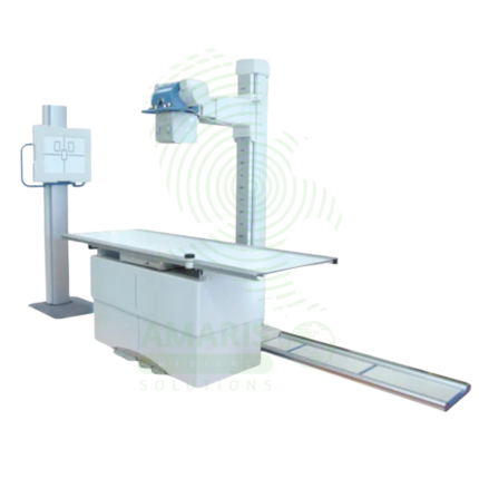
An Analogue Fixed X-ray Machine is a permanent installation X-ray system using traditional film cassettes for general radiography in radiology departments and imaging centers. Featuring ceiling-mounted tube assemblies, tilting tables, and wall stands, it provides essential diagnostic imaging for skeletal, chest, abdominal, and extremity examinations using film technology. Film cassettes are processed in darkroom facilities, producing permanent physical images for patient records and consultation. Used in facilities without digital radiography, as backup for digital systems, and in resource-limited settings.
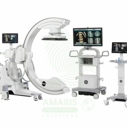
C-Arm Surgical System
A C-Arm Surgical System is a mobile fluoroscopic X-ray imaging device with a distinctive C-shaped arm connecting the X-ray tube and detector. It is an indispensable tool in modern operating rooms and interventional suites, providing real-time live imaging to guide complex procedures in orthopedics, spine surgery, pain management, and vascular interventions. Its mobility allows precise positioning around the patient, while features like pulsed fluoroscopy and dose monitoring are critical for radiation safety. Modern flat-panel systems offer high-resolution imaging and advanced capabilities like 3D Cone-Beam CT. Safe operation demands rigorous adherence to radiation protection protocols (ALARA) for both patients and the surgical team.
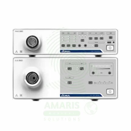
Endoscopy System
The Endoscopy System is a complete video processor and light source stack that forms the core of a modern digital endoscopy suite. It provides high-definition imaging for compatible Fujinon flexible video endoscopes, enabling diagnostic and therapeutic procedures in gastroenterology, pulmonology, and urology. Key features include advanced image processing and enhancement technologies like Narrow Band Imaging (NBI) for improved diagnostic accuracy. As a critical piece of capital equipment, it requires careful handling, the use of compatible accessories, and regular professional maintenance to ensure optimal performance and safety.
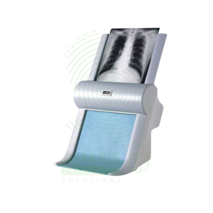
Film Digitizer
A Film Digitizer is a specialized scanner that converts analog radiographic films into high-fidelity digital images (DICOM format). It is the essential tool for migrating historical film archives into a modern digital PACS, enabling filmless workflow, remote access, and long-term preservation of diagnostic records. Its critical performance characteristics are a wide optical density range (to capture all film details) and high spatial resolution. By creating secure, accessible digital copies, it protects against the loss of physical films and integrates past patient history with current digital imaging, supporting comprehensive care and efficient radiology practice.
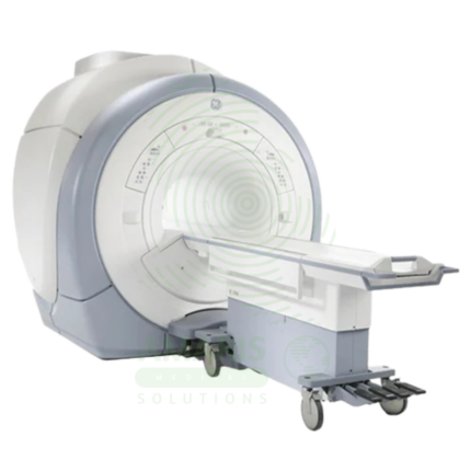
Magnetic Resonance Imaging
Magnetic Resonance Imaging (MRI) is a non-invasive diagnostic imaging modality that uses powerful magnetic fields and radiofrequency waves to produce detailed images of soft tissues, organs, and internal structures without ionizing radiation. It is the gold standard for imaging the brain, spinal cord, joints, muscles, and ligaments, and is essential for neurological, musculoskeletal, oncologic, and cardiovascular diagnosis. MRI provides exceptional soft tissue contrast, enabling precise anatomical characterization, tumor staging, and treatment planning. Strict safety protocols for ferromagnetic screening and contrast administration are essential for patient safety.
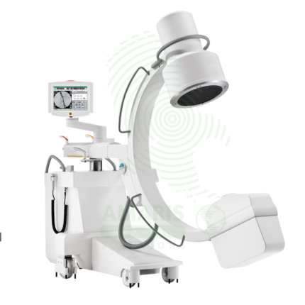
Mobile C-arm Surgical System
A Mobile C-arm Surgical System is a portable fluoroscopic imaging device used for real-time intraoperative guidance during orthopedic, spinal, vascular, and pain management procedures. The C-shaped arm allows flexible positioning around the patient, providing AP, lateral, and oblique views to verify instrument placement, fracture reduction, and device deployment. Essential for minimally invasive surgery, it enables surgeons to achieve precision and accuracy while reducing operative time and improving patient outcomes.
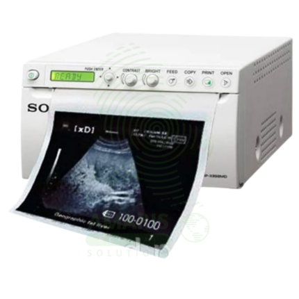
Sony Ultrasound Printer
A Sony Ultrasound Printer is a specialized medical-grade printer that produces high-quality hard copies of ultrasound images. Utilizing dry thermal, laser, or dye-sublimation technology, it creates grayscale or color prints on photographic paper or film, providing a physical record for patient charts, referrals, and consultations. While its role has diminished with the rise of fully digital PACS workflows, it remains essential in settings requiring immediate tangible output, patient education, or where digital infrastructure is limited. Its performance is defined by high spatial and contrast resolution to accurately reproduce the subtleties of sonographic images.