Dermatoscope and Magnifiers
 Diagnostic Kits
 Vital Signs Monitors
 Stethoscopes and Accessories
 Otoscopes, Ophthalmoscopes, and Retinoscopes
 Reflex Hammers and Neurological Tools
 Scales and Measuring Devices
 Spirometers and Pulmonary Function Tests
 Electrosurgical Units and Accessories
 Cutting Instruments
 Grasping and Holding Instruments
 Hemostatic Instruments
 Specialized Surgical Sets
 Single-Use Procedure Trays and Packs
 Surgical Drapes, Gowns, and Covers
 Tissue Unifying Instruments
 Radiation Protection
 X-Ray Machines and Accessories
 Ultrasound Systems and Probes
 MRI and CT Scanners
 Radiology Consumables
 Bone Densitometers
 Fluoroscopy Equipment
 Imaging Tables and Positioning Aids
 Microscopes and Accessories
 Centrifuges and Separators
 Analyzers
 Incubators and Ovens
 Pipettes, Dispensers, and Lab Glassware
 Refrigerators, Freezers, and Storage Units
 Lab Consumables
 Sterilizers and Autoclaves for Lab Use
 Multi-Parameter Monitors
 Ventilators and Respiratory Support Devices
 Defibrillators and AEDs
 Infusion Pumps and IV Systems
 Patient Warmers and Cooling Devices
 Central Monitoring Stations
 Accessories
 Anesthesia Machines and Workstations
 Oxygen Concentrators and Delivery Systems
 Nebulizers and Inhalers
 CPAP/BiPAP Machines
 Airway Management
 Anesthesia Masks, Circuits, and Bags
 Humidifiers and Heaters
 Respiratory Therapy Accessories
 First Aid Kits and Cabinets
 Emergency Resuscitation Equipment
 Trauma Supplies
 Emergency Carts and Crash Carts
 Burn Care Products
 Bleeding Control
 Automated External Defibrillators (AEDs)
 Transport and Evacuation
 Wheelchairs and Accessories
 Walkers, Crutches, and Canes
 Prosthetics and Orthotics
 Physical Therapy Equipment
 Transfer Devices
 Bathroom Safety
 Orthopedic Traction and Tables
 Hot/Cold Therapy Packs and Units
 Beds and Mattresses
 Chairs and Stools
 Tables
 Cabinets and Storage
 Privacy Screens & Curtains
 Stands and Racks
 Linens and Textiles
 Lighting
 Autoclaves and Sterilizers
 Ultrasonic Cleaners
 Disinfectant Solutions and Wipes
 Sterilization Pouches, Wraps, and Indicators
 Instrument Trays and Containers
 UV and Ozone Disinfection Devices
 Washer Disinfectors
 Wound Care
 Gloves
 Masks and Respirators
 Catheters and Tubing
 Swabs, Applicators, and Sponges
 Incontinence Products
 Personal Protective Equipment (PPE)
 Dental Chairs and Units
 Handpieces and Burs
 Instruments
 Consumables
 Sterilization for Dental Use
 Orthodontic Supplies
 Endodontic Tools
 Slit Lamps and Tonometers
 Lensometers and Phoropters
 Ophthalmic Surgical Instruments
 Eyewear Frames and Lenses
 Contact Lens Supplies
 Vision Testing Charts and Devices
 Eye Care Consumables
 Laser Systems for Eye Care
 ENT Exam Chairs and Tables
 Endoscopes
 Audiometers and Hearing Tests
 ENT Instruments
 Nasal and Throat Packs
 Hearing Aids and Accessories
 Otology Supplies
 Fetal Dopplers and Monitors
 Delivery Beds and Tables
 Gynecological Instruments
 Neonatal Incubators and Warmers
 Breast Pumps and Accessories
 Contraceptive Devices
 Maternity Supports and Pads
 Neonatal Consumables
 Cystoscopes and Urethroscopes
 Dialysis Machines and Supplies
 Urological Catheters and Bags
 Lithotripters
 Prostate Treatment Devices
 Urinary Incontinence Products
 Kidney Stone Management Tools
 Consumables & Disposables
 EEG and EMG Machines
 Neurosurgical Instruments
 Nerve Stimulators
 Headrests and Positioning Aids
 Lumbar Puncture Kits
 Seizure Monitoring Devices
 Consumables
 Rehabilitation for Neurological Conditions
 ECG Machines and Accessories
 Holter Monitors
 Stress Test Systems
 Pacemakers and Defibrillator Accessories
 Vascular Access Devices
 Cardiac Catheters and Guidewires
 Blood Flow Meters
 Consumables
 Orthopedic Instruments
 Casts, Splints, and Padding
 Joint Replacement Supplies
 Prosthetic Limbs and Components
 Bone Grafts and Substitutes
 Traction Devices
 Orthopedic Braces and Supports
 Rehabilitation Aids for Orthopedics
 Home Oxygen Therapy
 Hospital Beds for Home Use
 Mobility Aids
 Bathroom and Daily Living Aids
 Wound Care for Home
 Monitoring Devices
 Enteral Feeding Pumps and Tubes
 Hand Sanitizers and Dispensers
 Face Shields and Goggles
 Isolation Gowns and Suits
 Biohazard Waste Containers
 Air Purifiers and HEPA Filters
 Surface Disinfectants
 Sharps Containers
 Protective Barriers
 Cardiovascular & Endurance Training
 Strength Training & Weightlifting
 Functional Training & Core Conditioning
 Physical Therapy & Rehabilitation
 Sports & Outdoor Recreation
 Gym Flooring & Facility Equipment
 Fitness Monitoring & Accessories
 Kids & Novelties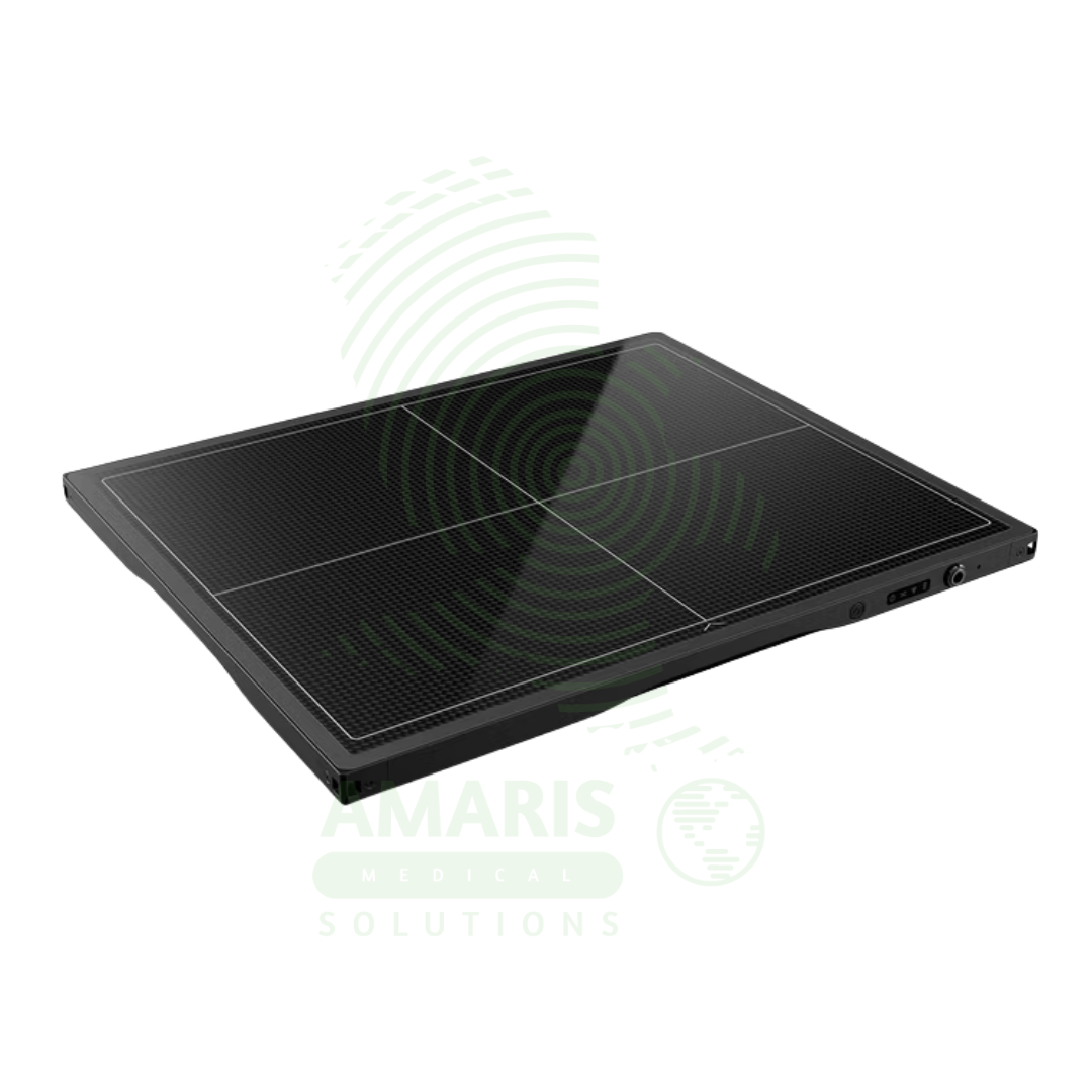
Wire & Wireless Flat Panel Detector
$1.00
WhatsApp Order
A Wire & Wireless Flat Panel Detector is a digital X-ray image receptor that converts X-ray energy into high-quality digital images for general radiography. Available in wired and wireless configurations, these detectors replace traditional film cassettes, providing immediate image preview, eliminating film processing, and enabling seamless digital workflow integration. Wireless detectors offer flexibility for bedside imaging in ICUs, emergency departments, and operating rooms, while wired detectors provide reliable connectivity for fixed systems. High detective quantum efficiency enables dose reduction while maintaining diagnostic image quality.
Description
Wire & Wireless Flat Panel Detector
PRIMARY CLINICAL & DIAGNOSTIC USES
1. Digital X-ray Image Capture for General Radiography
-
Primary Use: Serves as the image receptor in digital radiography systems, converting X-ray energy into high-quality digital images. The flat panel detector replaces traditional film cassettes, providing immediate image preview, eliminating film processing, and enabling electronic image distribution to PACS.
-
How it helps: For the radiologist and radiology technologist, the flat panel detector transforms the imaging workflow from film-based delays to immediate digital capture—providing instant image feedback, eliminating repeat exposures due to processing errors, and enabling rapid transmission to interpreting physicians. For the patient, this means shorter examination times, fewer repeat X-rays, and faster diagnosis.
2. Wireless Flexibility for Mobile and Emergency Imaging
-
Primary Use: Wireless flat panel detectors allow for portable imaging without the constraint of cables, enabling easy positioning for bedside examinations in intensive care units, emergency departments, and operating rooms. The detector can be placed under patients, between mattress and patient, or in the wall stand Bucky tray.
-
How it helps: For the radiology technologist performing bedside imaging, a wireless detector eliminates cable management issues—allowing for flexible positioning around IV lines, monitoring equipment, and ventilators without the risk of tripping or disconnecting cables. For the critically ill patient, wireless imaging means faster, more efficient examinations with less disruption to their care environment.
3. High-Resolution Imaging for Skeletal and Chest Radiography
-
Primary Use: Provides high-resolution digital images for skeletal, chest, and abdominal radiography, capturing fine anatomical details including trabecular bone patterns, pulmonary nodules, and soft tissue interfaces with excellent contrast resolution.
-
How it helps: For the radiologist interpreting images, the high resolution and contrast of flat panel detectors enable visualization of subtle findings—early lung nodules, fine fracture lines, and small calcifications that might be missed with lower resolution systems. For the patient, this means more accurate diagnosis and earlier detection of disease.
4. Dose Efficiency and Image Quality
-
Primary Use: Flat panel detectors offer high detective quantum efficiency, converting a higher percentage of incident X-rays into image signal compared to film or computed radiography systems. This allows for reduced radiation dose while maintaining diagnostic image quality.
-
How it helps: For the medical physicist and radiology department, flat panel detectors enable dose optimization protocols that reduce patient radiation exposure while maintaining image quality—supporting ALARA (As Low As Reasonably Achievable) principles. For the patient, particularly pediatric patients and those requiring frequent imaging, this means lower cumulative radiation exposure over time.
5. Seamless Integration with Digital Workflow
-
Primary Use: Connects wirelessly or via cable to X-ray generators, workstations, and PACS, enabling seamless digital workflow from image acquisition to interpretation. Automatic image processing and DICOM transfer eliminate manual steps and reduce potential for errors.
-
How it helps: For the radiology department, integrated digital workflow reduces examination time, eliminates film and chemical costs, and enables rapid image distribution for remote interpretation and teleradiology. For the patient, this means faster results and coordinated care across multiple facilities.
SECONDARY & SUPPORTIVE USES
1. Pediatric Imaging: High sensitivity allows for reduced dose protocols optimized for pediatric patients.
2. Bariatric Imaging: Large-format detectors accommodate patients of size with full field coverage.
3. Operating Room Use: Wireless detectors can be used for intraoperative imaging without cables crossing the sterile field.
4. Emergency Department: Rapid imaging for trauma patients with wireless detectors placed under stretcher patients.
5. Mobile X-ray: Wireless detectors are essential for mobile X-ray systems, allowing for bedside imaging without cable constraints.
6. Veterinary Imaging: Flat panel detectors used in veterinary radiology for animal imaging.
KEY PRODUCT FEATURES
1. BASIC IDENTIFICATION ATTRIBUTES
-
Device Type: A digital X-ray image receptor using flat panel technology for direct digital capture.
-
Designation: Flat Panel Detector, Digital Radiography Detector, DR Detector, Wireless Detector, Portable Detector.
-
Types:
-
Wired Detector: Connects via cable to workstation and generator.
-
Wireless Detector: Battery-powered with wireless transmission; no cable required.
-
-
Detector Technologies:
-
Direct Conversion: Amorphous selenium (a-Se) converts X-rays directly to electrical signals.
-
Indirect Conversion: Amorphous silicon (a-Si) with a scintillator (CsI or Gd2O2S) converts X-rays to light, then to electrical signals.
-
-
Key Components:
-
Detector Panel: Active matrix array with photodiodes or TFTs.
-
Scintillator: Cesium iodide (CsI) or gadolinium oxysulfide (Gd2O2S) for indirect conversion.
-
Electronic Readout: Converts electrical signal to digital image.
-
Battery (Wireless): Rechargeable battery for portable operation.
-
Wireless Interface: WiFi or Bluetooth for image transmission.
-
2. TECHNICAL & PERFORMANCE PROPERTIES
-
Detector Size: Common sizes: 14x17 inches, 17x17 inches, 10x12 inches; various sizes available.
-
Pixel Matrix: 3-10 megapixels depending on detector size.
-
Pixel Pitch: 100-200 microns for high resolution.
-
Detective Quantum Efficiency: 60-80% for optimal dose efficiency.
-
Dynamic Range: Wide dynamic range for optimal image contrast.
-
Acquisition Time: Immediate preview; full image in seconds.
-
Battery Life (Wireless): 8-12 hours of typical use; rechargeable.
3. PHYSICAL & OPERATIONAL PROPERTIES
-
Construction: Rugged housing for clinical use; impact-resistant.
-
Weight: 8-15 lbs depending on size.
-
Portability: Wireless detectors are portable for bedside and mobile use.
-
Interface: Wireless or wired connection to generator and workstation.
-
Durability: Designed for repeated use; resistant to minor impacts.
4. SAFETY & COMPLIANCE ATTRIBUTES
-
Regulatory Status: Class II medical device regulated by FDA.
-
Radiation Safety: Dose optimization enabled by high DQE; reduced patient exposure.
-
Battery Safety (Wireless): Sealed batteries with safety features.
-
Drop Testing: Designed to withstand minor drops and impacts.
5. STORAGE & HANDLING ATTRIBUTES
-
Storage: Stored in a charging cradle (wireless) or docking station (wired) when not in use.
-
Cleaning: Wipe with hospital-grade disinfectants; follow manufacturer guidelines.
-
Battery Charging (Wireless): Docking station or plug-in charger.
-
Maintenance: Regular calibration and software updates required.
6. LABORATORY & CLINICAL APPLICATIONS
-
Primary Application: Digital X-ray image capture for general radiography, mobile imaging, and bedside examinations.
-
Clinical Role: Essential component of digital radiography systems; enables filmless workflow.
SAFETY HANDLING PRECAUTIONS
1. SAFETY PRECAUTIONS
-
Drop Prevention: Handle with care; avoid dropping detectors.
-
Cable Management (Wired): Manage cables to prevent tripping and disconnection.
-
Battery Management (Wireless): Keep charged for emergency use; monitor battery status.
-
Infection Control: Clean between patients; follow isolation protocols.
-
Dose Management: Use appropriate exposure factors; take advantage of high DQE for dose reduction.
2. FIRST AID MEASURES
-
Detector Drop: If detector is dropped, remove from service; inspect for damage; contact service provider.
-
Battery Failure: If battery fails, use wired connection if available; contact service provider.
3. FIRE FIGHTING MEASURES
-
Flammability: Plastic components are combustible; batteries may pose fire risk.
-
Extinguishing Media: For electrical fire, use CO₂ or dry chemical extinguisher.
Related products
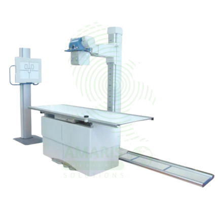
Analogue Fixed X-ray Machine
An Analogue Fixed X-ray Machine is a permanent installation X-ray system using traditional film cassettes for general radiography in radiology departments and imaging centers. Featuring ceiling-mounted tube assemblies, tilting tables, and wall stands, it provides essential diagnostic imaging for skeletal, chest, abdominal, and extremity examinations using film technology. Film cassettes are processed in darkroom facilities, producing permanent physical images for patient records and consultation. Used in facilities without digital radiography, as backup for digital systems, and in resource-limited settings.
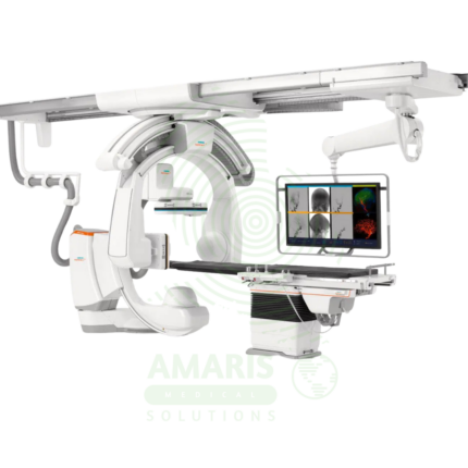
Angiographic Intervention System
An Angiographic Intervention System is a high-end fluoroscopic imaging system designed for guiding minimally invasive vascular and interventional procedures. Essential for cardiac catheterization laboratories, interventional radiology suites, and hybrid operating rooms, it provides real-time, high-resolution visualization of blood vessels, catheters, and devices during coronary interventions, peripheral vascular procedures, neurovascular interventions, and structural heart procedures. Advanced features include rotational angiography for 3D reconstruction, digital subtraction angiography, and dose reduction technologies, enabling precise treatment with minimal radiation exposure.
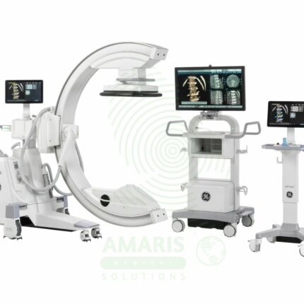
C-Arm Surgical System
A C-Arm Surgical System is a mobile fluoroscopic X-ray imaging device with a distinctive C-shaped arm connecting the X-ray tube and detector. It is an indispensable tool in modern operating rooms and interventional suites, providing real-time live imaging to guide complex procedures in orthopedics, spine surgery, pain management, and vascular interventions. Its mobility allows precise positioning around the patient, while features like pulsed fluoroscopy and dose monitoring are critical for radiation safety. Modern flat-panel systems offer high-resolution imaging and advanced capabilities like 3D Cone-Beam CT. Safe operation demands rigorous adherence to radiation protection protocols (ALARA) for both patients and the surgical team.
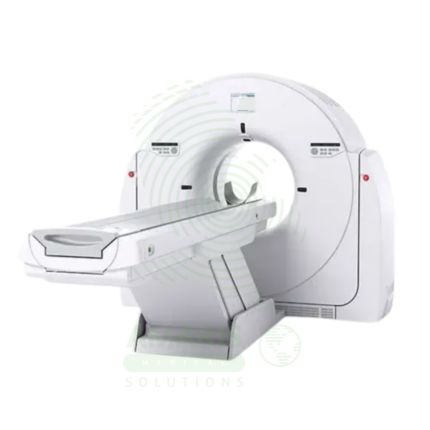
CT Scanner
A CT Scanner is an advanced diagnostic imaging device that uses a rotating X-ray source and detector array to create detailed cross-sectional images ("slices") of the body. By combining these slices, it generates comprehensive 2D and 3D views of bones, organs, and blood vessels, making it indispensable for trauma evaluation, cancer staging, vascular assessment, and guiding complex procedures. While offering unparalleled diagnostic clarity, its operation requires strict adherence to radiation safety principles (ALARA) and protocols for the safe use of contrast agents to maximize patient benefit and minimize risk.
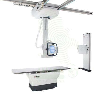
Digital Ceiling X-ray
A Digital Ceiling X-ray is a ceiling-mounted digital radiography system for general diagnostic imaging of the skeletal, chest, abdominal, and extremity anatomy. The ceiling-mounted tube assembly provides full room coverage for flexible patient positioning, while digital flat panel detectors produce immediate high-resolution images for rapid diagnosis. Integrated with PACS and RIS, it supports efficient digital workflow from image acquisition to interpretation. Used in radiology departments, emergency rooms, and outpatient imaging centers.
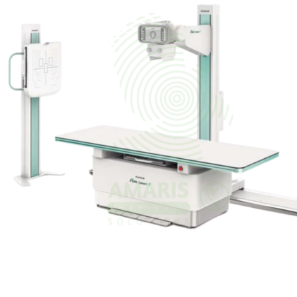
Digital Fixed X-ray
A Digital Fixed X-ray is a permanent installation digital radiography system designed for high-volume general imaging in radiology departments and outpatient imaging centers. Featuring digital flat panel detectors, ceiling-mounted tube assemblies, and tilting tables, it provides high-resolution images for skeletal, chest, abdominal, and extremity examinations. Integrated with PACS and RIS, it supports efficient digital workflow from image acquisition to interpretation, enabling rapid diagnosis and treatment planning.
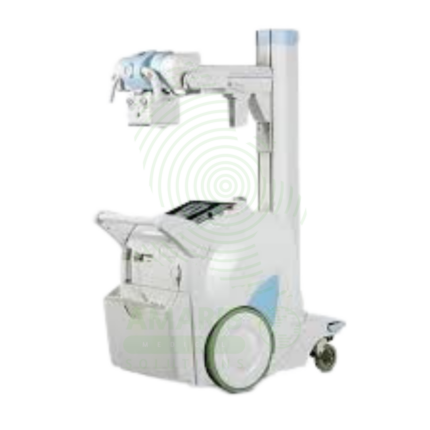
Digital Mobile X-ray
A Digital Mobile X-ray is a battery-powered, portable digital radiography system designed for bedside imaging in intensive care units, neonatal intensive care units, emergency departments, and operating rooms. The mobile unit enables high-quality chest, abdominal, and extremity imaging at the patient's bedside, eliminating the risks associated with transporting critically ill patients. Wireless digital detectors provide immediate image capture and transmission to PACS, supporting rapid diagnosis and treatment decisions.