Dermatoscope and Magnifiers
 Diagnostic Kits
 Vital Signs Monitors
 Stethoscopes and Accessories
 Otoscopes, Ophthalmoscopes, and Retinoscopes
 Reflex Hammers and Neurological Tools
 Scales and Measuring Devices
 Spirometers and Pulmonary Function Tests
 Electrosurgical Units and Accessories
 Cutting Instruments
 Grasping and Holding Instruments
 Hemostatic Instruments
 Specialized Surgical Sets
 Single-Use Procedure Trays and Packs
 Surgical Drapes, Gowns, and Covers
 Tissue Unifying Instruments
 Radiation Protection
 X-Ray Machines and Accessories
 Ultrasound Systems and Probes
 MRI and CT Scanners
 Radiology Consumables
 Bone Densitometers
 Fluoroscopy Equipment
 Imaging Tables and Positioning Aids
 Microscopes and Accessories
 Centrifuges and Separators
 Analyzers
 Incubators and Ovens
 Pipettes, Dispensers, and Lab Glassware
 Refrigerators, Freezers, and Storage Units
 Lab Consumables
 Sterilizers and Autoclaves for Lab Use
 Multi-Parameter Monitors
 Ventilators and Respiratory Support Devices
 Defibrillators and AEDs
 Infusion Pumps and IV Systems
 Patient Warmers and Cooling Devices
 Central Monitoring Stations
 Accessories
 Anesthesia Machines and Workstations
 Oxygen Concentrators and Delivery Systems
 Nebulizers and Inhalers
 CPAP/BiPAP Machines
 Airway Management
 Anesthesia Masks, Circuits, and Bags
 Humidifiers and Heaters
 Respiratory Therapy Accessories
 First Aid Kits and Cabinets
 Emergency Resuscitation Equipment
 Trauma Supplies
 Emergency Carts and Crash Carts
 Burn Care Products
 Bleeding Control
 Automated External Defibrillators (AEDs)
 Transport and Evacuation
 Wheelchairs and Accessories
 Walkers, Crutches, and Canes
 Prosthetics and Orthotics
 Physical Therapy Equipment
 Transfer Devices
 Bathroom Safety
 Orthopedic Traction and Tables
 Hot/Cold Therapy Packs and Units
 Beds and Mattresses
 Chairs and Stools
 Tables
 Cabinets and Storage
 Privacy Screens & Curtains
 Stands and Racks
 Linens and Textiles
 Lighting
 Autoclaves and Sterilizers
 Ultrasonic Cleaners
 Disinfectant Solutions and Wipes
 Sterilization Pouches, Wraps, and Indicators
 Instrument Trays and Containers
 UV and Ozone Disinfection Devices
 Washer Disinfectors
 Wound Care
 Gloves
 Masks and Respirators
 Catheters and Tubing
 Swabs, Applicators, and Sponges
 Incontinence Products
 Personal Protective Equipment (PPE)
 Dental Chairs and Units
 Handpieces and Burs
 Instruments
 Consumables
 Sterilization for Dental Use
 Orthodontic Supplies
 Endodontic Tools
 Slit Lamps and Tonometers
 Lensometers and Phoropters
 Ophthalmic Surgical Instruments
 Eyewear Frames and Lenses
 Contact Lens Supplies
 Vision Testing Charts and Devices
 Eye Care Consumables
 Laser Systems for Eye Care
 ENT Exam Chairs and Tables
 Endoscopes
 Audiometers and Hearing Tests
 ENT Instruments
 Nasal and Throat Packs
 Hearing Aids and Accessories
 Otology Supplies
 Fetal Dopplers and Monitors
 Delivery Beds and Tables
 Gynecological Instruments
 Neonatal Incubators and Warmers
 Breast Pumps and Accessories
 Contraceptive Devices
 Maternity Supports and Pads
 Neonatal Consumables
 Cystoscopes and Urethroscopes
 Dialysis Machines and Supplies
 Urological Catheters and Bags
 Lithotripters
 Prostate Treatment Devices
 Urinary Incontinence Products
 Kidney Stone Management Tools
 Consumables & Disposables
 EEG and EMG Machines
 Neurosurgical Instruments
 Nerve Stimulators
 Headrests and Positioning Aids
 Lumbar Puncture Kits
 Seizure Monitoring Devices
 Consumables
 Rehabilitation for Neurological Conditions
 ECG Machines and Accessories
 Holter Monitors
 Stress Test Systems
 Pacemakers and Defibrillator Accessories
 Vascular Access Devices
 Cardiac Catheters and Guidewires
 Blood Flow Meters
 Consumables
 Orthopedic Instruments
 Casts, Splints, and Padding
 Joint Replacement Supplies
 Prosthetic Limbs and Components
 Bone Grafts and Substitutes
 Traction Devices
 Orthopedic Braces and Supports
 Rehabilitation Aids for Orthopedics
 Home Oxygen Therapy
 Hospital Beds for Home Use
 Mobility Aids
 Bathroom and Daily Living Aids
 Wound Care for Home
 Monitoring Devices
 Enteral Feeding Pumps and Tubes
 Hand Sanitizers and Dispensers
 Face Shields and Goggles
 Isolation Gowns and Suits
 Biohazard Waste Containers
 Air Purifiers and HEPA Filters
 Surface Disinfectants
 Sharps Containers
 Protective Barriers
 Cardiovascular & Endurance Training
 Strength Training & Weightlifting
 Functional Training & Core Conditioning
 Physical Therapy & Rehabilitation
 Sports & Outdoor Recreation
 Gym Flooring & Facility Equipment
 Fitness Monitoring & Accessories
 Kids & Novelties


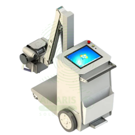
Digital Mobile X-ray
WhatsApp Order
A Digital Mobile X-ray is a battery-powered, portable digital radiography system designed for bedside imaging in intensive care units, neonatal intensive care units, emergency departments, and operating rooms. The mobile unit enables high-quality chest, abdominal, and extremity imaging at the patient’s bedside, eliminating the risks associated with transporting critically ill patients. Wireless digital detectors provide immediate image capture and transmission to PACS, supporting rapid diagnosis and treatment decisions.
Description
Digital Mobile X-ray
PRIMARY CLINICAL & DIAGNOSTIC USES
1. Portable Imaging for Bedside Patient Care
-
Primary Use: Provides high-resolution digital X-ray imaging at the patient’s bedside, eliminating the need to transport critically ill, unstable, or immobilized patients to the radiology department. The mobile unit can be moved to patient rooms, intensive care units, emergency departments, and operating rooms.
-
How it helps: For the intensive care unit team and radiology staff, the digital mobile X-ray brings imaging directly to the patient—eliminating the risks associated with transporting critically ill patients through the facility, reducing the physical strain on staff, and enabling immediate imaging when clinical status changes. For the critically ill or unstable patient, bedside imaging means they receive necessary diagnostic studies without the danger of transport, dislodgement of lines, or interruption of life-support equipment.
2. Intensive Care Unit Imaging
-
Primary Use: Essential for monitoring intubated and ventilated patients, evaluating endotracheal tube and central line placement, detecting pneumothorax, assessing for pulmonary edema, and monitoring progression of pneumonia or acute respiratory distress syndrome (ARDS) in ICU patients.
-
How it helps: For the intensivist and critical care team, mobile X-ray provides the ability to obtain immediate images of the chest and abdomen without moving the patient from the ICU bed—enabling rapid assessment of tube and line placement, detection of complications such as pneumothorax, and monitoring of pulmonary status. For the critically ill patient, this means timely diagnosis and intervention without the risk of transport.
3. Neonatal Intensive Care Unit Imaging
-
Primary Use: Provides safe, portable imaging for premature and critically ill newborns in the NICU, allowing for assessment of chest, abdomen, and skeletal structures without removing the infant from the incubator or warmer.
-
How it helps: For the neonatologist and NICU team, mobile X-ray enables imaging of fragile neonates without disturbing the carefully controlled thermal and respiratory environment of the incubator—minimizing handling, reducing stress, and protecting the infant from temperature fluctuations. For the premature or critically ill infant, bedside imaging means necessary diagnostic studies can be performed safely without disrupting the delicate balance of their care.
4. Operating Room and Interventional Suite Imaging
-
Primary Use: Provides intraoperative imaging for surgical procedures, allowing for immediate assessment of hardware placement, fracture reduction, and post-operative anatomy without moving the patient from the operating table.
-
How it helps: For the orthopedic surgeon, neurosurgeon, and surgical team, mobile X-ray provides immediate feedback during procedures—confirming hardware placement, verifying fracture reduction, and assessing post-operative anatomy before the patient leaves the operating room. For the patient, this means the surgeon can make adjustments immediately if needed, reducing the risk of repeat surgeries and ensuring optimal outcomes.
5. Emergency Department and Trauma Imaging
-
Primary Use: Enables rapid imaging of trauma patients in the emergency department, including chest, abdomen, and extremity X-rays, without moving the patient from the stretcher or compromising spinal precautions.
-
How it helps: For the emergency physician and trauma team, mobile X-ray provides rapid imaging at the bedside—allowing for assessment of chest and abdominal injuries, fracture evaluation, and detection of free air or obstruction without moving the patient from the trauma bay. For the trauma patient, this means faster diagnosis and treatment without the risk of moving unstable patients.
SECONDARY & SUPPORTIVE USES
1. Post-Operative Imaging: Used for immediate post-operative assessment of hardware placement and surgical results.
2. Home Healthcare: Some mobile X-ray services provide portable imaging for homebound patients.
3. Long-Term Care Facilities: Used for imaging residents who cannot be transported to imaging centers.
4. Bariatric Patient Imaging: Mobile units can accommodate patients of size at bedside.
5. Isolation Room Imaging: Reduces risk of pathogen transmission by bringing imaging to isolation rooms.
6. Radiography and Fluoroscopy: Some mobile units offer both digital radiography and fluoroscopy capabilities.
KEY PRODUCT FEATURES
1. BASIC IDENTIFICATION ATTRIBUTES
-
Device Type: A battery-powered, mobile digital X-ray system designed for bedside imaging.
-
Designation: Digital Mobile X-ray, Portable X-ray, Mobile DR System, Mobile Radiography Unit.
-
Key Components:
-
Mobile Cart: Battery-powered drive system for maneuverability.
-
X-ray Tube: Mounted on an articulating arm for flexible positioning.
-
Digital Detector: Wireless flat panel detector for image capture.
-
Generator: High-frequency generator for X-ray production.
-
Control Console: Touchscreen interface for image acquisition.
-
Collimator: Adjustable beam restriction.
-
2. TECHNICAL & PERFORMANCE PROPERTIES
-
Detector Type: Wireless flat panel digital detector (amorphous silicon or CMOS).
-
Detector Size: Typically 14x17 inches for full chest and abdominal imaging.
-
Generator Power: Typically 10-32 kW depending on model.
-
Battery Life: Sufficient for full day of bedside imaging; rechargeable.
-
Drive System: Power-assisted or motorized for ease of movement.
-
Wireless Connectivity: DICOM transmission to PACS.
3. PHYSICAL & OPERATIONAL PROPERTIES
-
Weight: 400-800 lbs depending on model.
-
Dimensions: Compact for maneuverability in patient rooms and tight spaces.
-
Tube Arm: Articulating arm for positioning around patient beds and equipment.
-
Detector: Wireless; can be placed under patient or in Bucky tray.
-
Operation: Single operator can position, expose, and acquire images.
4. SAFETY & COMPLIANCE ATTRIBUTES
-
Regulatory Status: Class II medical device regulated by FDA.
-
Radiation Safety: AEC (Automatic Exposure Control); collimation for beam restriction.
-
Battery Safety: Sealed lead-acid or lithium-ion batteries with safety features.
-
Collision Avoidance: Sensors to prevent equipment collision with patient and surroundings.
5. STORAGE & HANDLING ATTRIBUTES
-
Storage: Stored in designated charging areas when not in use.
-
Battery Charging: Docking station for overnight charging.
-
Cleaning: Wipe with hospital-grade disinfectants between patient uses.
-
Maintenance: Regular calibration and battery maintenance required.
6. LABORATORY & CLINICAL APPLICATIONS
-
Primary Application: Bedside imaging for ICU, NICU, emergency, and operating room patients.
-
Clinical Role: Essential equipment for hospitals with critically ill, unstable, or immobile patients.
SAFETY HANDLING PRECAUTIONS
1. SAFETY PRECAUTIONS
-
Collision Avoidance: Ensure clear path before moving; use caution around patient beds and equipment.
-
Radiation Safety: Use proper collimation and AEC; maintain distance during exposure.
-
Infection Control: Clean unit between patients; follow isolation protocols.
-
Battery Management: Keep unit charged for emergency use; monitor battery status.
-
Detector Care: Handle wireless detectors carefully; avoid drops and impact.
2. FIRST AID MEASURES
-
Collision: If unit collides with patient or equipment, stop movement; assess for injury; inspect unit for damage.
-
Battery Failure: If battery fails, plug into the wall outlet for operation; contact the service provider.
3. FIRE FIGHTING MEASURES
-
Flammability: Equipment is non-flammable; fire risk from electrical components and batteries.
-
Extinguishing Media: For electrical fire, use CO₂ or dry chemical extinguisher.
Related products
Angiographic Intervention System
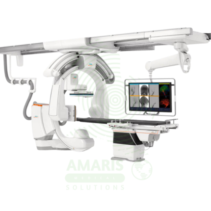
An Angiographic Intervention System is a high-end fluoroscopic imaging system designed for guiding minimally invasive vascular and interventional procedures. Essential for cardiac catheterization laboratories, interventional radiology suites, and hybrid operating rooms, it provides real-time, high-resolution visualization of blood vessels, catheters, and devices during coronary interventions, peripheral vascular procedures, neurovascular interventions, and structural heart procedures. Advanced features include rotational angiography for 3D reconstruction, digital subtraction angiography, and dose reduction technologies, enabling precise treatment with minimal radiation exposure.
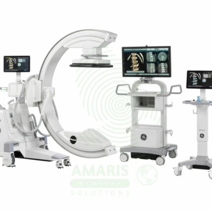
C-Arm Surgical System
A C-Arm Surgical System is a mobile fluoroscopic X-ray imaging device with a distinctive C-shaped arm connecting the X-ray tube and detector. It is an indispensable tool in modern operating rooms and interventional suites, providing real-time live imaging to guide complex procedures in orthopedics, spine surgery, pain management, and vascular interventions. Its mobility allows precise positioning around the patient, while features like pulsed fluoroscopy and dose monitoring are critical for radiation safety. Modern flat-panel systems offer high-resolution imaging and advanced capabilities like 3D Cone-Beam CT. Safe operation demands rigorous adherence to radiation protection protocols (ALARA) for both patients and the surgical team.
Digital & Analog X-ray Machine
A Digital & Analog X-ray Machine is a fundamental medical imaging device that uses a controlled beam of ionizing radiation to produce static or real-time images of the body's internal structures. It is indispensable for diagnosing fractures, lung diseases, dental issues, and many abdominal conditions. The transition from Analog (film-based) to Digital (CR or DR) technology has revolutionized the field, offering faster results, superior image manipulation, improved dose efficiency, and seamless integration into digital healthcare networks. Its operation demands strict adherence to radiation safety protocols (ALARA) to protect patients and staff, making it a cornerstone of safe, effective diagnostic medicine.
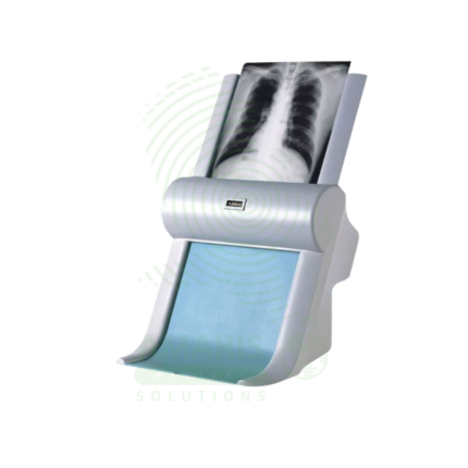
Film Digitizer
A Film Digitizer is a specialized scanner that converts analog radiographic films into high-fidelity digital images (DICOM format). It is the essential tool for migrating historical film archives into a modern digital PACS, enabling filmless workflow, remote access, and long-term preservation of diagnostic records. Its critical performance characteristics are a wide optical density range (to capture all film details) and high spatial resolution. By creating secure, accessible digital copies, it protects against the loss of physical films and integrates past patient history with current digital imaging, supporting comprehensive care and efficient radiology practice.
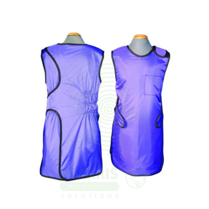
Lead Apron
A Lead Apron is a protective garment worn by healthcare workers to shield against scatter radiation during fluoroscopic procedures, X-ray examinations, and interventional radiology. Made of lead-impregnated material, it attenuates scatter radiation to the thyroid, chest, and reproductive organs, ensuring occupational radiation exposure remains within safe limits. Available in frontal, wrap-around, and two-piece designs with lead equivalence ranging from 0.25 mm to 0.5 mm, proper storage, annual inspection, and use of thyroid shields are essential for effective radiation protection.
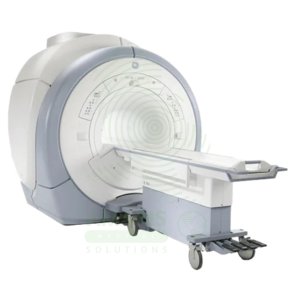
Magnetic Resonance Imaging
Magnetic Resonance Imaging (MRI) is a non-invasive diagnostic imaging modality that uses powerful magnetic fields and radiofrequency waves to produce detailed images of soft tissues, organs, and internal structures without ionizing radiation. It is the gold standard for imaging the brain, spinal cord, joints, muscles, and ligaments, and is essential for neurological, musculoskeletal, oncologic, and cardiovascular diagnosis. MRI provides exceptional soft tissue contrast, enabling precise anatomical characterization, tumor staging, and treatment planning. Strict safety protocols for ferromagnetic screening and contrast administration are essential for patient safety.
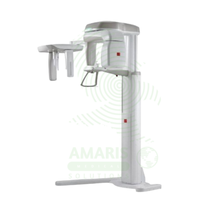
Panoramic X-ray Dental Machine
A Panoramic X-ray Dental Machine is a rotating extraoral radiographic system that produces a single, broad 2D image of the entire jaws, teeth, TMJs, and sinuses. By focusing on a curved "focal trough," it provides an efficient screening tool for wisdom teeth evaluation, orthodontic planning, and detecting large jaw pathologies. While offering a valuable overview with a relatively low radiation dose, its diagnostic utility is entirely dependent on precise patient positioning to avoid blurring and distortion. It is a cornerstone of modern dental diagnostic imaging, serving as a crucial first step in comprehensive oral assessment and treatment planning.