Dermatoscope and Magnifiers
 Diagnostic Kits
 Vital Signs Monitors
 Stethoscopes and Accessories
 Otoscopes, Ophthalmoscopes, and Retinoscopes
 Reflex Hammers and Neurological Tools
 Scales and Measuring Devices
 Spirometers and Pulmonary Function Tests
 Electrosurgical Units and Accessories
 Cutting Instruments
 Grasping and Holding Instruments
 Hemostatic Instruments
 Specialized Surgical Sets
 Single-Use Procedure Trays and Packs
 Surgical Drapes, Gowns, and Covers
 Tissue Unifying Instruments
 Radiation Protection
 X-Ray Machines and Accessories
 Ultrasound Systems and Probes
 MRI and CT Scanners
 Radiology Consumables
 Bone Densitometers
 Fluoroscopy Equipment
 Imaging Tables and Positioning Aids
 Microscopes and Accessories
 Centrifuges and Separators
 Analyzers
 Incubators and Ovens
 Pipettes, Dispensers, and Lab Glassware
 Refrigerators, Freezers, and Storage Units
 Lab Consumables
 Sterilizers and Autoclaves for Lab Use
 Multi-Parameter Monitors
 Ventilators and Respiratory Support Devices
 Defibrillators and AEDs
 Infusion Pumps and IV Systems
 Patient Warmers and Cooling Devices
 Central Monitoring Stations
 Accessories
 Anesthesia Machines and Workstations
 Oxygen Concentrators and Delivery Systems
 Nebulizers and Inhalers
 CPAP/BiPAP Machines
 Airway Management
 Anesthesia Masks, Circuits, and Bags
 Humidifiers and Heaters
 Respiratory Therapy Accessories
 First Aid Kits and Cabinets
 Emergency Resuscitation Equipment
 Trauma Supplies
 Emergency Carts and Crash Carts
 Burn Care Products
 Bleeding Control
 Automated External Defibrillators (AEDs)
 Transport and Evacuation
 Wheelchairs and Accessories
 Walkers, Crutches, and Canes
 Prosthetics and Orthotics
 Physical Therapy Equipment
 Transfer Devices
 Bathroom Safety
 Orthopedic Traction and Tables
 Hot/Cold Therapy Packs and Units
 Beds and Mattresses
 Chairs and Stools
 Tables
 Cabinets and Storage
 Privacy Screens & Curtains
 Stands and Racks
 Linens and Textiles
 Lighting
 Autoclaves and Sterilizers
 Ultrasonic Cleaners
 Disinfectant Solutions and Wipes
 Sterilization Pouches, Wraps, and Indicators
 Instrument Trays and Containers
 UV and Ozone Disinfection Devices
 Washer Disinfectors
 Wound Care
 Gloves
 Masks and Respirators
 Catheters and Tubing
 Swabs, Applicators, and Sponges
 Incontinence Products
 Personal Protective Equipment (PPE)
 Dental Chairs and Units
 Handpieces and Burs
 Instruments
 Consumables
 Sterilization for Dental Use
 Orthodontic Supplies
 Endodontic Tools
 Slit Lamps and Tonometers
 Lensometers and Phoropters
 Ophthalmic Surgical Instruments
 Eyewear Frames and Lenses
 Contact Lens Supplies
 Vision Testing Charts and Devices
 Eye Care Consumables
 Laser Systems for Eye Care
 ENT Exam Chairs and Tables
 Endoscopes
 Audiometers and Hearing Tests
 ENT Instruments
 Nasal and Throat Packs
 Hearing Aids and Accessories
 Otology Supplies
 Fetal Dopplers and Monitors
 Delivery Beds and Tables
 Gynecological Instruments
 Neonatal Incubators and Warmers
 Breast Pumps and Accessories
 Contraceptive Devices
 Maternity Supports and Pads
 Neonatal Consumables
 Cystoscopes and Urethroscopes
 Dialysis Machines and Supplies
 Urological Catheters and Bags
 Lithotripters
 Prostate Treatment Devices
 Urinary Incontinence Products
 Kidney Stone Management Tools
 Consumables & Disposables
 EEG and EMG Machines
 Neurosurgical Instruments
 Nerve Stimulators
 Headrests and Positioning Aids
 Lumbar Puncture Kits
 Seizure Monitoring Devices
 Consumables
 Rehabilitation for Neurological Conditions
 ECG Machines and Accessories
 Holter Monitors
 Stress Test Systems
 Pacemakers and Defibrillator Accessories
 Vascular Access Devices
 Cardiac Catheters and Guidewires
 Blood Flow Meters
 Consumables
 Orthopedic Instruments
 Casts, Splints, and Padding
 Joint Replacement Supplies
 Prosthetic Limbs and Components
 Bone Grafts and Substitutes
 Traction Devices
 Orthopedic Braces and Supports
 Rehabilitation Aids for Orthopedics
 Home Oxygen Therapy
 Hospital Beds for Home Use
 Mobility Aids
 Bathroom and Daily Living Aids
 Wound Care for Home
 Monitoring Devices
 Enteral Feeding Pumps and Tubes
 Hand Sanitizers and Dispensers
 Face Shields and Goggles
 Isolation Gowns and Suits
 Biohazard Waste Containers
 Air Purifiers and HEPA Filters
 Surface Disinfectants
 Sharps Containers
 Protective Barriers
 Cardiovascular & Endurance Training
 Strength Training & Weightlifting
 Functional Training & Core Conditioning
 Physical Therapy & Rehabilitation
 Sports & Outdoor Recreation
 Gym Flooring & Facility Equipment
 Fitness Monitoring & Accessories
 Kids & Novelties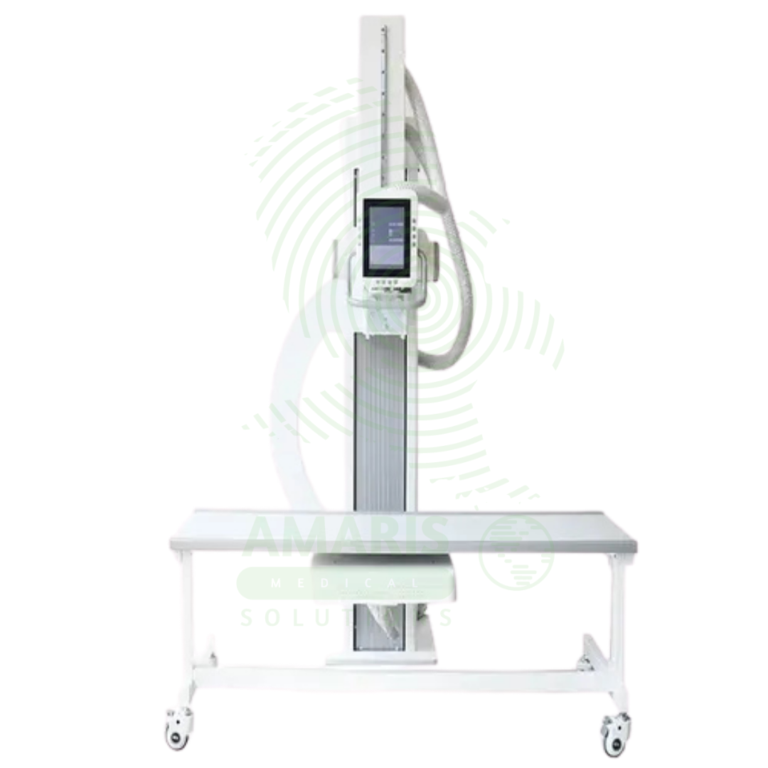
Digital U-arm X-ray
$1.00
WhatsApp Order
A Digital U-arm X-ray is a versatile digital radiography system designed for emergency departments, urgent care centers, and outpatient clinics. The U-arm configuration provides flexible positioning for chest, abdominal, skeletal, and extremity imaging with easy patient access for stretcher and wheelchair patients. Digital detectors produce immediate high-resolution images for rapid diagnosis, while the compact footprint allows installation in space-constrained settings. Essential for rapid, high-quality imaging in emergency and ambulatory care environments.
Description
Digital U-arm X-ray
PRIMARY CLINICAL & DIAGNOSTIC USES
1. Versatile Imaging for Multiple Clinical Settings
-
Primary Use: Provides high-resolution digital X-ray imaging with a U-arm configuration that offers flexible positioning for a wide range of examinations including chest, abdomen, skeletal, and extremity imaging. The U-arm design allows for easy patient access and positioning flexibility in compact spaces.
-
How it helps: For the radiologist and imaging technologist, the U-arm configuration provides the flexibility needed for diverse imaging examinations in a compact footprint—allowing for upright, supine, and lateral positioning without moving the patient. For the patient, a U-arm system means faster, more comfortable examinations with less repositioning.
2. Emergency Department Imaging
-
Primary Use: Ideal for emergency departments where rapid, versatile imaging is required for trauma patients, fracture assessment, and acute chest and abdominal conditions. The U-arm design allows for imaging of patients on stretchers without transfer to a dedicated X-ray table.
-
How it helps: For the emergency physician and trauma team, the U-arm X-ray provides rapid imaging capabilities at the bedside—allowing for assessment of chest, abdominal, and extremity injuries without moving the patient from the stretcher. For the trauma patient, this means faster diagnosis and treatment without the pain and risk of multiple transfers.
3. Orthopedic and Extremity Imaging
-
Primary Use: Provides detailed imaging of extremities, joints, and skeletal structures for fracture assessment, arthritis evaluation, and post-operative follow-up. The U-arm design allows for precise positioning for weight-bearing and non-weight-bearing views.
-
How it helps: For the orthopedic surgeon and sports medicine physician, the U-arm X-ray provides the detailed images needed to assess fractures, joint alignment, and hardware placement—with the flexibility to obtain weight-bearing views for functional assessment. For the patient with orthopedic concerns, this means accurate diagnosis and appropriate treatment planning.
4. Chest and Abdominal Imaging
-
Primary Use: Performs high-quality chest X-rays for pneumonia, lung cancer, heart failure, and pleural effusion assessment, as well as abdominal X-rays for obstruction, free air, and calcifications. The U-arm allows for upright, supine, and lateral views.
-
How it helps: For the pulmonologist, cardiologist, and emergency physician, the U-arm X-ray provides essential chest and abdominal imaging with flexible positioning—allowing for upright views for pleural effusion and free air detection, and supine views for trauma patients. For the patient, this flexibility means the appropriate view can be obtained without repositioning or transferring the patient.
5. Digital Imaging for Rapid Diagnosis
-
Primary Use: Digital detectors produce immediate, high-resolution images that can be viewed, enhanced, and transmitted electronically to PACS for interpretation. The integrated digital workflow eliminates film processing and reduces examination time.
-
How it helps: For the radiology team and referring physicians, digital X-ray provides immediate image availability—eliminating film processing delays, allowing for image enhancement to improve visualization, and enabling rapid transmission to specialists. For the patient, faster results mean quicker diagnosis and treatment decisions.
SECONDARY & SUPPORTIVE USES
1. Point-of-Care Imaging: Compact design allows placement in emergency departments, urgent care centers, and outpatient clinics for rapid, on-site imaging.
2. Bariatric Patient Imaging: U-arm design accommodates patients of size with easy access and flexible positioning.
3. Pediatric Imaging: Quick, efficient imaging minimizes patient handling and examination time for pediatric patients.
4. Portable and Fixed Applications: Some U-arm systems are mobile; others are fixed for dedicated rooms.
5. Rheumatology Assessment: Provides imaging for arthritis and joint disease assessment.
6. Pre-Operative Planning: Used for surgical planning and post-operative hardware assessment.
KEY PRODUCT FEATURES
1. BASIC IDENTIFICATION ATTRIBUTES
-
Device Type: A U-arm configured digital X-ray system for general radiography in emergency and outpatient settings.
-
Designation: Digital U-arm X-ray, U-Arm X-ray System, Digital Radiography U-Arm, U-Arm DR System.
-
Key Components:
-
U-Arm Assembly: C-shaped or U-shaped arm with X-ray tube and detector at opposite ends.
-
Digital Detector: Flat panel detector for image capture.
-
Generator: High-frequency generator.
-
Control Console: Workstation for image acquisition and processing.
-
Patient Support: Stretcher-compatible design for emergency use.
-
2. TECHNICAL & PERFORMANCE PROPERTIES
-
Detector Type: Flat panel digital detector (amorphous silicon or CMOS).
-
Detector Size: Typically 14x17 inches or 17x17 inches.
-
Generator Power: Typically 32-50 kW.
-
U-Arm Rotation: Motorized positioning for various views.
-
Height Adjustment: Motorized vertical travel for patient positioning.
-
Digital Workflow: Integrated with RIS and PACS.
3. PHYSICAL & OPERATIONAL PROPERTIES
-
Configuration: U-arm design with X-ray tube and detector mounted on opposing arms.
-
Space Requirements: Compact footprint suitable for emergency departments and clinics.
-
Mobility: Some models mobile; others fixed installation.
-
Patient Access: Open design for stretcher and wheelchair access.
4. SAFETY & COMPLIANCE ATTRIBUTES
-
Regulatory Status: Class II medical device regulated by FDA.
-
Radiation Safety: AEC (Automatic Exposure Control); collimation for beam restriction.
-
Digital Detector: Lower dose compared to film-based systems.
-
Pediatric Protocols: Age-appropriate exposure settings available.
5. STORAGE & HANDLING ATTRIBUTES
-
Storage: Fixed installation in designated room; mobile units stored in charging area.
-
Room Requirements: Standard X-ray room with shielding.
-
Maintenance: Regular quality control testing and calibration.
6. LABORATORY & CLINICAL APPLICATIONS
-
Primary Application: Emergency department imaging, orthopedic imaging, chest and abdominal radiography.
-
Clinical Role: Essential equipment in emergency departments, urgent care centers, orthopedic clinics, and outpatient imaging.
SAFETY HANDLING PRECAUTIONS
1. SAFETY PRECAUTIONS
-
Radiation Dose: Follow ALARA principles; use AEC and proper collimation.
-
Patient Positioning: Ensure proper positioning to minimize repeat exposures.
-
Stretcher Compatibility: Confirm stretcher compatibility before use.
-
Mobile Operation: Lock wheels when stationary; ensure stability during imaging.
2. FIRST AID MEASURES
-
Patient Fall: If a patient falls during positioning, assess for injury; seek medical attention if needed.
-
Equipment Malfunction: If equipment fails, remove the patient; contact the service provider.
3. FIRE FIGHTING MEASURES
-
Flammability: Equipment is non-flammable; fire risk from electrical components.
-
Extinguishing Media: Use CO₂ or dry chemical extinguisher for electrical fires.
Related products
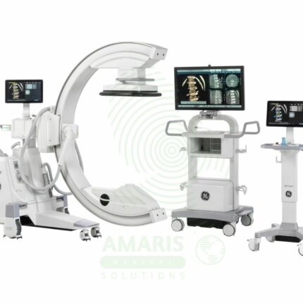
C-Arm Surgical System
A C-Arm Surgical System is a mobile fluoroscopic X-ray imaging device with a distinctive C-shaped arm connecting the X-ray tube and detector. It is an indispensable tool in modern operating rooms and interventional suites, providing real-time live imaging to guide complex procedures in orthopedics, spine surgery, pain management, and vascular interventions. Its mobility allows precise positioning around the patient, while features like pulsed fluoroscopy and dose monitoring are critical for radiation safety. Modern flat-panel systems offer high-resolution imaging and advanced capabilities like 3D Cone-Beam CT. Safe operation demands rigorous adherence to radiation protection protocols (ALARA) for both patients and the surgical team.
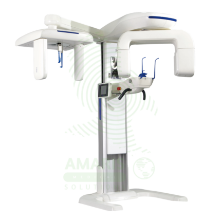
Dental X-ray Machine
A Dental X-ray Machine is a specialized radiographic system designed for imaging teeth, jaws, and facial structures. It encompasses intraoral units for detailed tooth-specific views, panoramic machines for wide screening shots, and advanced Cone Beam CT (CBCT) scanners for 3D surgical planning. Utilizing low-dose radiation and digital imaging technology, it is indispensable for diagnosing cavities, gum disease, infections, and planning treatments like implants, orthodontics, and oral surgery. Its safe operation requires strict adherence to radiation protection protocols, including the use of lead aprons, proper collimation, and operator training to ensure patient and staff safety while obtaining critical diagnostic information.
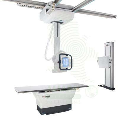
Digital Ceiling X-ray
A Digital Ceiling X-ray is a ceiling-mounted digital radiography system for general diagnostic imaging of the skeletal, chest, abdominal, and extremity anatomy. The ceiling-mounted tube assembly provides full room coverage for flexible patient positioning, while digital flat panel detectors produce immediate high-resolution images for rapid diagnosis. Integrated with PACS and RIS, it supports efficient digital workflow from image acquisition to interpretation. Used in radiology departments, emergency rooms, and outpatient imaging centers.
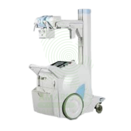
Digital Mobile X-ray
A Digital Mobile X-ray is a battery-powered, portable digital radiography system designed for bedside imaging in intensive care units, neonatal intensive care units, emergency departments, and operating rooms. The mobile unit enables high-quality chest, abdominal, and extremity imaging at the patient's bedside, eliminating the risks associated with transporting critically ill patients. Wireless digital detectors provide immediate image capture and transmission to PACS, supporting rapid diagnosis and treatment decisions.
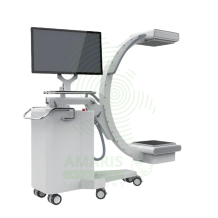
Mobile Film
Mobile Film is a battery-powered, portable X-ray system using traditional film cassettes for bedside imaging in intensive care units, neonatal intensive care units, emergency departments, and operating rooms. The mobile unit enables chest, abdominal, and extremity imaging at the patient's bedside, eliminating the risks associated with transporting critically ill patients. Film cassettes are processed in darkroom or daylight processors for image development. Used in hospitals without digital radiography capabilities or as backup for digital systems.
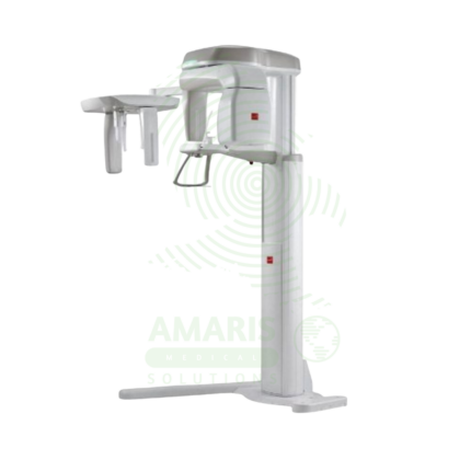
Panoramic X-ray Dental Machine
A Panoramic X-ray Dental Machine is a rotating extraoral radiographic system that produces a single, broad 2D image of the entire jaws, teeth, TMJs, and sinuses. By focusing on a curved "focal trough," it provides an efficient screening tool for wisdom teeth evaluation, orthodontic planning, and detecting large jaw pathologies. While offering a valuable overview with a relatively low radiation dose, its diagnostic utility is entirely dependent on precise patient positioning to avoid blurring and distortion. It is a cornerstone of modern dental diagnostic imaging, serving as a crucial first step in comprehensive oral assessment and treatment planning.
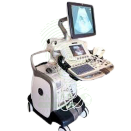
Ultrasound Machine
The Ultrasound Machine is a diagnostic imaging system that uses high-frequency sound waves to produce real-time, dynamic images of internal body structures, including soft tissues, organs, blood vessels, and developing fetuses. It provides non-invasive, radiation-free visualization using multiple transducer probes for abdominal, cardiac, obstetric, gynecological, vascular, and musculoskeletal examinations. With core features like B-mode grayscale imaging, M-mode for motion assessment, and comprehensive Doppler capabilities (Color, Power, Spectral), it offers essential diagnostic functionality for evaluating anatomy, physiology, and blood flow. Its portability, digital image storage, and user-friendly interface make it a practical tool for hospitals, clinics, emergency departments, and point-of-care settings across virtually all medical specialties, requiring proper operator training and adherence to probe disinfection protocols for safe and effective use.