Dermatoscope and Magnifiers
 Diagnostic Kits
 Vital Signs Monitors
 Stethoscopes and Accessories
 Otoscopes, Ophthalmoscopes, and Retinoscopes
 Reflex Hammers and Neurological Tools
 Scales and Measuring Devices
 Spirometers and Pulmonary Function Tests
 Electrosurgical Units and Accessories
 Cutting Instruments
 Grasping and Holding Instruments
 Hemostatic Instruments
 Specialized Surgical Sets
 Single-Use Procedure Trays and Packs
 Surgical Drapes, Gowns, and Covers
 Tissue Unifying Instruments
 Radiation Protection
 X-Ray Machines and Accessories
 Ultrasound Systems and Probes
 MRI and CT Scanners
 Radiology Consumables
 Bone Densitometers
 Fluoroscopy Equipment
 Imaging Tables and Positioning Aids
 Microscopes and Accessories
 Centrifuges and Separators
 Analyzers
 Incubators and Ovens
 Pipettes, Dispensers, and Lab Glassware
 Refrigerators, Freezers, and Storage Units
 Lab Consumables
 Sterilizers and Autoclaves for Lab Use
 Multi-Parameter Monitors
 Ventilators and Respiratory Support Devices
 Defibrillators and AEDs
 Infusion Pumps and IV Systems
 Patient Warmers and Cooling Devices
 Central Monitoring Stations
 Accessories
 Anesthesia Machines and Workstations
 Oxygen Concentrators and Delivery Systems
 Nebulizers and Inhalers
 CPAP/BiPAP Machines
 Airway Management
 Anesthesia Masks, Circuits, and Bags
 Humidifiers and Heaters
 Respiratory Therapy Accessories
 First Aid Kits and Cabinets
 Emergency Resuscitation Equipment
 Trauma Supplies
 Emergency Carts and Crash Carts
 Burn Care Products
 Bleeding Control
 Automated External Defibrillators (AEDs)
 Transport and Evacuation
 Wheelchairs and Accessories
 Walkers, Crutches, and Canes
 Prosthetics and Orthotics
 Physical Therapy Equipment
 Transfer Devices
 Bathroom Safety
 Orthopedic Traction and Tables
 Hot/Cold Therapy Packs and Units
 Beds and Mattresses
 Chairs and Stools
 Tables
 Cabinets and Storage
 Privacy Screens & Curtains
 Stands and Racks
 Linens and Textiles
 Lighting
 Autoclaves and Sterilizers
 Ultrasonic Cleaners
 Disinfectant Solutions and Wipes
 Sterilization Pouches, Wraps, and Indicators
 Instrument Trays and Containers
 UV and Ozone Disinfection Devices
 Washer Disinfectors
 Wound Care
 Gloves
 Masks and Respirators
 Catheters and Tubing
 Swabs, Applicators, and Sponges
 Incontinence Products
 Personal Protective Equipment (PPE)
 Dental Chairs and Units
 Handpieces and Burs
 Instruments
 Consumables
 Sterilization for Dental Use
 Orthodontic Supplies
 Endodontic Tools
 Slit Lamps and Tonometers
 Lensometers and Phoropters
 Ophthalmic Surgical Instruments
 Eyewear Frames and Lenses
 Contact Lens Supplies
 Vision Testing Charts and Devices
 Eye Care Consumables
 Laser Systems for Eye Care
 ENT Exam Chairs and Tables
 Endoscopes
 Audiometers and Hearing Tests
 ENT Instruments
 Nasal and Throat Packs
 Hearing Aids and Accessories
 Otology Supplies
 Fetal Dopplers and Monitors
 Delivery Beds and Tables
 Gynecological Instruments
 Neonatal Incubators and Warmers
 Breast Pumps and Accessories
 Contraceptive Devices
 Maternity Supports and Pads
 Neonatal Consumables
 Cystoscopes and Urethroscopes
 Dialysis Machines and Supplies
 Urological Catheters and Bags
 Lithotripters
 Prostate Treatment Devices
 Urinary Incontinence Products
 Kidney Stone Management Tools
 Consumables & Disposables
 EEG and EMG Machines
 Neurosurgical Instruments
 Nerve Stimulators
 Headrests and Positioning Aids
 Lumbar Puncture Kits
 Seizure Monitoring Devices
 Consumables
 Rehabilitation for Neurological Conditions
 ECG Machines and Accessories
 Holter Monitors
 Stress Test Systems
 Pacemakers and Defibrillator Accessories
 Vascular Access Devices
 Cardiac Catheters and Guidewires
 Blood Flow Meters
 Consumables
 Orthopedic Instruments
 Casts, Splints, and Padding
 Joint Replacement Supplies
 Prosthetic Limbs and Components
 Bone Grafts and Substitutes
 Traction Devices
 Orthopedic Braces and Supports
 Rehabilitation Aids for Orthopedics
 Home Oxygen Therapy
 Hospital Beds for Home Use
 Mobility Aids
 Bathroom and Daily Living Aids
 Wound Care for Home
 Monitoring Devices
 Enteral Feeding Pumps and Tubes
 Hand Sanitizers and Dispensers
 Face Shields and Goggles
 Isolation Gowns and Suits
 Biohazard Waste Containers
 Air Purifiers and HEPA Filters
 Surface Disinfectants
 Sharps Containers
 Protective Barriers
 Cardiovascular & Endurance Training
 Strength Training & Weightlifting
 Functional Training & Core Conditioning
 Physical Therapy & Rehabilitation
 Sports & Outdoor Recreation
 Gym Flooring & Facility Equipment
 Fitness Monitoring & Accessories
 Kids & Novelties
ECG Machine
$1.00
WhatsApp Order
An ECG Machine is a Class II medical device that records and displays the electrical activity of the heart through surface electrodes, producing an electrocardiogram for diagnosis of cardiac conditions. Standard diagnostic machines record 12 simultaneous leads (3 limb, 6 precordial, 1 ground) with frequency response 0.05-150 Hz, sampling rate 500-1,000 Hz, and high-resolution (5-10 µV) signal acquisition. Features include color touchscreen display, thermal array printer, computerized interpretation algorithms, internal memory (50-500+ ECGs), and network connectivity for EMR integration. Lead wires (AHA or IEC color coding) connect to disposable adhesive electrodes. Portable models (5-15 kg) with rechargeable batteries enable bedside and mobile use; cart-mounted units provide full diagnostic capability. Primary clinical applications include diagnosis of arrhythmias (AF, VT, bradycardia), detection of myocardial ischemia/infarction (STEMI, NSTEMI), evaluation of chest pain, preoperative cardiac risk assessment, monitoring electrolyte imbalances, assessment of chamber enlargement, and drug effect/toxicity monitoring. Essential diagnostic equipment in emergency departments, cardiology clinics, ICUs, operating rooms, and primary care settings worldwide.
Description
ECG Machine
PRIMARY CLINICAL & DIAGNOSTIC USES
1. Diagnosis of Cardiac Arrhythmias:
-
Primary Use: Records the electrical activity of the heart to identify and diagnose various arrhythmias including atrial fibrillation, atrial flutter, ventricular tachycardia, bradyarrhythmia’s, and premature beats.
-
How it helps: Enables physicians to prescribe targeted antiarrhythmic therapy, recommend pacemaker implantation, or perform cardioversion with confidence by pinpointing the exact rhythm disorder.
2. Detection of Myocardial Ischemia and Infarction:
-
Primary Use: Essential for identifying ST-segment elevations or depressions, T-wave inversions, and pathological Q-waves indicative of acute coronary syndromes (STEMI, NSTEMI) or prior myocardial infarction.
-
How it helps: Guides emergent interventions, such as activating the cath lab for immediate angioplasty or administering thrombolytics, significantly reducing heart muscle damage and improving survival rates.
3. Evaluation of Chest Pain in Emergency Settings:
-
Primary Use: Rapid ECG acquisition in patients presenting with chest pain helps differentiate cardiac from non-cardiac causes.
-
How it helps: Determines the need for urgent, life-saving treatments versus further cardiac workup, ensuring critical cases are prioritized immediately.
4. Preoperative Cardiac Risk Assessment:
-
Primary Use: Routine ECG screening before surgical procedures identify undiagnosed cardiac conditions, arrhythmias, or ischemic changes.
-
How it helps: Allows the medical team to optimize the patient’s condition beforehand, adjust the anesthetic plan, or cancel elective surgery if the risk is too high.
5. Monitoring Electrolyte Imbalances:
-
Primary Use: ECG changes (peaked T-waves, prolonged QT interval, U-waves) provide clues to electrolyte abnormalities including hyperkalemia, hypokalemia, hypercalcemia, and hypocalcemia.
-
How it helps: Guides urgent correction of electrolyte disturbances before lab results are available, acting as an early warning system to prevent life-threatening cardiac arrhythmias.
6. Assessment of Chamber Enlargement:
-
Primary Use: Identifies criteria for atrial enlargement and ventricular hypertrophy in patients with hypertension, valvular heart disease, or cardiomyopathies.
-
How it helps: Provides critical information about the heart’s structural adaptation to stress, guiding long-term management to prevent heart failure.
7. Drug Effect and Toxicity Monitoring:
-
Primary Use: Serial ECGs monitor for QT prolongation from antiarrhythmic medications, antipsychotics, and other drugs, as well as characteristic changes from digoxin toxicity.
-
How it helps: Guides medication adjustments to maintain therapeutic levels while avoiding dangerous side effects, ensuring patient safety during treatment.
SECONDARY & SUPPORTIVE USES
1. Pre-Participation Sports Screening: ECG screening for athletes to detect underlying cardiac conditions (hypertrophic cardiomyopathy, long QT syndrome) that increase risk of sudden cardiac death.
2. Pre-Employment and Occupational Health: Baseline ECG for safety-sensitive positions (firefighters, pilots, commercial drivers) to identify cardiac conditions that could pose safety risks.
3. Research and Clinical Trials: Used in cardiovascular research studies to assess drug effects, device efficacy, and disease progression.
4. Telemedicine and Remote Monitoring: Portable ECG devices enable remote transmission of ECGs for specialist interpretation in underserved areas or during emergency transport.
5. Teaching and Training: Essential for teaching cardiac electrophysiology and ECG interpretation to medical students, residents, and healthcare providers.
6. Wellness and Preventive Health: Used in health screening programs to detect silent cardiac conditions in asymptomatic individuals.
7. Veterinary Medicine: Adapted for cardiac evaluation in animals, particularly dogs and cats with suspected heart disease.
KEY PRODUCT FEATURES
1. BASIC IDENTIFICATION ATTRIBUTES
-
Product Type: Medical device that records and displays the electrical activity of the heart over time, producing an electrocardiogram (ECG or EKG).
-
Common Names: ECG Machine, EKG Machine, Electrocardiograph, 12-Lead ECG, Holter Monitor (ambulatory), Event Monitor, Telemetry System.
-
Number of Leads:
-
3-Lead: Basic monitoring (telemetry, ambulatory); views three electrical vectors.
-
5-Lead: Enhanced monitoring (ICU, telemetry); provides limb leads and one precordial lead.
-
6-Lead: Limited diagnostic capability (some portable devices).
-
12-Lead: Full diagnostic standard; records 12 different views of cardiac electrical activity.
-
15-Lead: Adds right-sided and posterior leads for enhanced infarction detection.
-
18-Lead: Extended views for comprehensive cardiac assessment.
-
-
ECG Acquisition Types:
-
Resting ECG: Performed with patient supine and at rest (standard diagnostic ECG).
-
Stress ECG: Recorded during exercise (treadmill or bicycle) to detect ischemia.
-
Ambulatory ECG: Continuous recording over 24-48 hours (Holter monitor) to detect intermittent arrhythmias.
-
Event Monitoring: Patient-activated recording during symptoms.
-
-
Display: High-resolution color LCD or LED screen showing real-time ECG waveforms.
-
Printing: Thermal array printer for producing ECG tracings on grid paper.
-
Interpretation: Built-in computerized ECG analysis algorithms with preliminary interpretation.
-
Storage: Internal memory for storing ECGs (50-500+ records); USB, SD card, or network export.
-
Connectivity: USB, Ethernet, Wi-Fi, Bluetooth for EMR integration and remote transmission.
2. TECHNICAL & PERFORMANCE PROPERTIES
-
Frequency Response: 0.05-150 Hz (diagnostic); meets AHA/ACC standards.
-
Sampling Rate: 500-1,000 samples/second per channel for high-fidelity recording.
-
Resolution: 5-10 µV for accurate signal detection.
-
Common Mode Rejection Ratio (CMRR): >100 dB to reject interference.
-
Input Impedance: >10 MΩ for signal fidelity.
-
Lead-off Detection: Automatic detection of loose or disconnected leads.
-
Filters: Adjustable low-pass, high-pass, and notch filters (50/60 Hz) to reduce artifacts.
-
ACquisition Modes: Automatic (10-second standard), manual, and arrhythmia detection.
-
Report Generation: Automated measurement of intervals (PR, QRS, QT, QTc), axes, and amplitudes.
-
Interpretation Algorithm: Proven algorithms (e.g., Glasgow, Marquette) with physician overread capability.
-
Battery Life: 2-8 hours for portable models; rechargeable lithium-ion batteries.
-
Calibration: Internal 1 mV calibration signal; periodic external calibration recommended.
3. PHYSICAL & OPERATIONAL PROPERTIES
-
Dimensions: 30-40 cm × 30-40 cm × 10-20 cm (cart-mounted or portable).
-
Weight: 5-15 kg (portable); 20-40 kg (cart-mounted with accessories).
-
Display: 10-15 inch color LCD touchscreen.
-
Printer: Built-in thermal array printer (8.5 × 11 inch or A4 paper).
-
Controls: Touchscreen, membrane keypad, or rotary knob.
-
Lead Wires: Patient cable with color-coded lead wires (AHA or IEC color coding).
-
Electrodes: Disposable adhesive electrodes (various sizes for adult/pediatric).
-
Power Supply: AC power (100-240 V) with rechargeable battery backup.
-
Cart: Optional mobile cart for transport within facility.
-
Accessories: Electrodes, paper rolls, lead wires, carrying case (portable models).
4. SAFETY & COMPLIANCE ATTRIBUTES
-
Regulatory Status: Class II medical device requiring FDA 510(k) clearance; CE marked.
-
Clinical Standards: Complies with IEC 60601-2-25 (electrocardiographic monitoring equipment), AHA/ACC guidelines, and ISO 13485.
-
Electrical Safety: Compliant with IEC 60601-1 for medical electrical equipment; Type CF applied parts (defibrillator-proof).
-
Defibrillator Protection: Input circuitry withstands defibrillator discharge without damage.
-
Pacemaker Detection: Algorithms to detect pacemaker spikes and pacemaker function.
-
Lead-off Detection: Automatic detection of disconnected leads to prevent misdiagnosis.
-
Artifact Rejection: Filters to reduce muscle tremor, baseline wander, and AC interference.
-
Data Security: HIPAA-compliant data storage and transmission options.
-
Quality Management: Manufactured under ISO 13485 certified processes.
-
Warranty: Typically 1-5 years depending on manufacturer and components.
5. STORAGE & HANDLING ATTRIBUTES
-
Storage: Store in clean, dry environment at room temperature (15-35°C); protect from dust, moisture, and extreme temperatures.
-
Battery Maintenance: Keep batteries charged; replace every 2-3 years or per manufacturer.
-
Lead Wire Care: Inspect for cracks or broken wires; clean with mild detergent; replace as needed.
-
Electrodes: Store in sealed packages at room temperature; check expiration dates.
-
Paper Replacement: Use manufacturer-specified thermal paper; store paper in cool, dry place.
-
Cleaning: Wipe exterior with EPA-registered hospital disinfectant; clean screen with soft, lint-free cloth; do not immerse.
-
Calibration: Annual calibration verification recommended; return to manufacturer or qualified service center.
-
Software Updates: Install manufacturer updates for interpretation algorithms and features.
-
Inspection: Before each use, check lead wires, printer paper, battery status, and calibration signal.
6. LABORATORY & CLINICAL APPLICATIONS
-
Primary Application: Recording and interpretation of cardiac electrical activity for diagnosis of heart conditions.
-
Standard 12-Lead ECG Placement (AHA Guidelines):
-
Limb Leads: RA (right arm), LA (left arm), RL (right leg - ground), LL (left leg).
-
Precordial Leads: V1 (4th intercostal space, right sternal border), V2 (4th intercostal space, left sternal border), V3 (midway between V2 and V4), V4 (5th intercostal space, midclavicular line), V5 (anterior axillary line at V4 level), V6 (midaxillary line at V4 level).
-
-
ECG Interpretation Parameters:
-
Rate: Atrial and ventricular rates (normal 60-100 bpm).
-
Rhythm: Regularity and origin of impulses (sinus, atrial, junctional, ventricular).
-
Intervals: PR (120-200 ms), QRS (<120 ms), QT (corrected for rate).
-
Axis: Mean electrical axis in frontal plane (normal -30° to +90°).
-
Waveforms: P-wave morphology, QRS complexes, ST segments, T-waves.
-
-
Clinical Indications for ECG:
-
Chest pain, palpitations, syncope, dyspnea.
-
Preoperative evaluation.
-
Hypertension, diabetes, known heart disease.
-
Medication monitoring (antiarrhythmics, QT-prolonging drugs).
-
Electrolyte abnormalities.
-
Pre-participation sports screening.
-
-
Pediatric ECG Considerations:
-
Smaller electrodes and lead placement adjustments.
-
Age-specific normal values (rates, intervals, axes differ from adults).
-
Indications: congenital heart disease, Kawasaki disease, syncope, chest pain.
-
-
Stress ECG:
-
Exercise protocols (Bruce, Modified Bruce, Naughton).
-
Monitoring for ischemia, arrhythmias, hemodynamic response.
-
Indications: suspected coronary artery disease, risk stratification.
-
SAFETY HANDLING PRECAUTIONS
1. SAFETY PRECAUTIONS
-
Electrical Safety: Ensure machines are properly grounded; use only hospital-grade outlets; avoid liquid exposure.
-
Defibrillation Precautions: Do not touch patient or ECG leads during defibrillation; machine is defibrillator-protected but operator is not.
-
Lead Placement Accuracy: Correct lead placement essential for accurate interpretation; misplacement leads to misdiagnosis (e.g., dextrocardia, infarction patterns).
-
Electrode Skin Preparation: Clean skin; remove oils and dead skin for good signal; avoid hairy areas; use fresh electrodes.
-
Motion Artifact: Instruct patients to lie still, relax, and not talk during recording.
-
Electromagnetic Interference: Keep away from other electrical devices (infusion pumps, ventilators) that may cause interference.
-
Patient Comfort: Ensure patients are warm and comfortable; cold patients may have tremor artefacts.
-
Infection Control: Clean lead wires between patients; use single-patient electrodes.
-
Pacemaker Patients: Ensure machine detects pacemaker spikes; annotate tracing with pacemaker presence.
-
Quality Control: Regular calibration and maintenance; verify interpretation algorithm accuracy.
2. FIRST AID MEASURES
-
Patient with Chest Pain: Acquire ECG immediately; if STEMI identified, activate cath lab per protocol.
-
Patient with Syncope: ECG during symptoms if possible; if not, continuous monitoring for arrhythmias.
-
Cardiac Arrest: ECG during resuscitation; analyze rhythm for shockable rhythms (VF, pulseless VT).
-
Lead Wire Breakage: Replace damaged lead wire; use backup if available.
-
Artifact Interference: Identify source (muscle tremor, AC interference, loose lead); correct and repeat.
3. FIRE FIGHTING MEASURES
-
Flammability: Plastic components and thermal paper are combustible.
-
Extinguishing Media: For electrical fire, use CO₂ or dry chemical (Class C) extinguisher.
-
Power Off: Disconnect power if safe to do so.
Related products
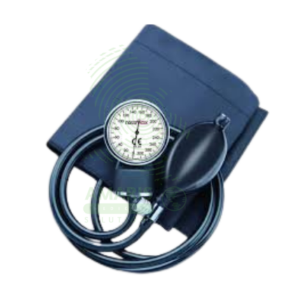
Aneroid Blood Pressure Monitor
The Aneroid Blood Pressure Monitor is a manual, mechanical sphygmomanometer used for the accurate, non-invasive measurement of arterial blood pressure. Operating on the auscultatory method, it consists of an inflatable cuff, a pump bulb with a release valve, and a calibrated dial gauge. Renowned for its reliability, durability, and independence from electrical power, it serves as a clinical gold standard and essential tool in physician offices, hospitals, and for guided home monitoring. Its accuracy is dependent on proper technique, correct cuff size, and regular calibration, making it a fundamental device for diagnosing and managing hypertension and assessing cardiovascular health.
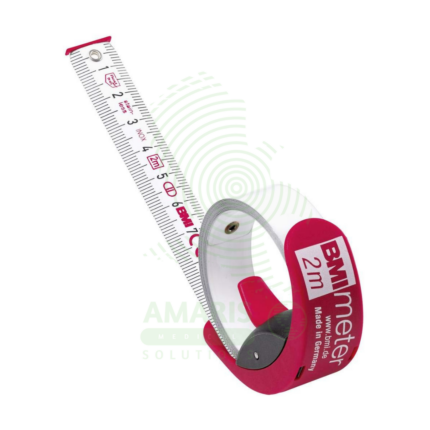
BMI Tape
A BMI Tape is a rapid screening tool that estimates an individual's Body Mass Index (BMI) category by measuring body circumference, most commonly the waist. It features a color-coded scale that directly displays BMI ranges (Underweight, Normal, Overweight, Obese), eliminating the need for separate height and weight measurements and manual calculation. While highly useful for community health screenings, wellness programs, and patient education due to its portability and simplicity, it is an approximate method. Its accuracy is limited compared to BMI calculated from precise clinical measurements, and it should be used as an initial screening indicator rather than a definitive diagnostic tool.
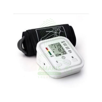
Digital Blood Pressure Monitor
A Digital Blood Pressure Monitor is an automatic electronic device that measures and displays systolic pressure, diastolic pressure, and pulse rate using the oscillometric method. Designed primarily for home and clinical use, it offers a user-friendly alternative to manual aneroid devices, requiring minimal training. Key features often include irregular heartbeat detection, multi-user memory, and connectivity for data tracking. Its accuracy, when clinically validated and used with correct technique, makes it an indispensable tool for the diagnosis and management of hypertension, empowering patients and supporting healthcare providers with reliable ambulatory data.
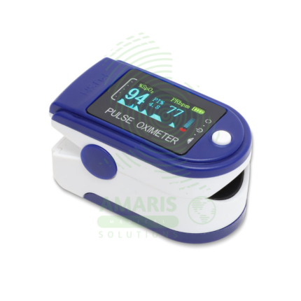
Fingertip Pulse Oximeter
A Fingertip Pulse Oximeter is a compact, non-invasive medical device that clips onto a finger to measure peripheral arterial oxygen saturation (SpO2) and pulse rate in seconds. Utilizing light-based technology, it provides critical, real-time data on respiratory and circulatory status. Its portability and ease of use make it indispensable in hospitals (from ORs to general wards), for patients managing chronic lung conditions at home, and for situational monitoring in sports or high-altitude environments. While an extremely valuable screening tool, readings must be interpreted in context, considering factors like patient movement, perfusion, and clinical symptoms.
Handheld Pulse Oximeter
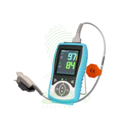
A Handheld Pulse Oximeter is a professional-grade, portable medical device used by healthcare providers for accurate and reliable monitoring of blood oxygen saturation (SpO2) and pulse rate. It consists of a durable, feature-rich monitor unit and separate, interchangeable sensors for use on fingers, toes, or ears. Superior to basic fingertip models, it offers advanced functions like configurable alarms, detailed plethysmograph display, trend memory, and data connectivity. Its robust design, clinical accuracy under challenging conditions (motion, low perfusion), and versatility make it an essential tool in hospitals, clinics, ambulances, and home health for patient assessment and monitoring.

Littman Stethoscope (Classic 4 Cardiology)
A Littman Stethoscope (Classic 4 Cardiology) represents the pinnacle of acoustic auscultation technology. Designed for clinicians who require the highest level of diagnostic accuracy, it features dual tunable diaphragms and dual-lumen tubing to provide exceptional acoustic sensitivity and ambient noise reduction. Its innovative design allows the user to hear both high and low-frequency sounds by simply adjusting pressure on either side of the chestpiece, making it the instrument of choice for cardiologists, intensivists, and specialists who cannot afford to miss a subtle murmur, rub, or breath sound. It combines superior performance with the durability and comfort expected from the Littmann brand.
Littmann Stethoscope (Classic II)
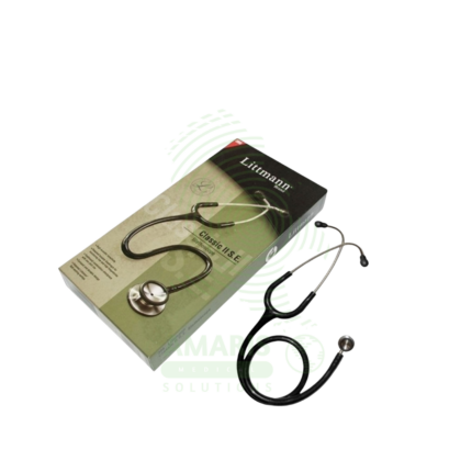
A Littmann Stethoscope (Classic II) is a premium, acoustic dual-head stethoscope renowned for its diagnostic clarity, durability, and innovative tunable diaphragm technology. It allows healthcare professionals to hear both high and low-frequency sounds by simply adjusting pressure on the single-sided chestpiece, eliminating the need to rotate it. With its superior acoustics, comfortable fit, and wide range of colors, it is the instrument of choice for medical students, nurses, and physicians across all specialties who require reliable performance for physical assessment, auscultation of heart and lung sounds, and manual blood pressure measurement.