Dermatoscope and Magnifiers
 Diagnostic Kits
 Vital Signs Monitors
 Stethoscopes and Accessories
 Otoscopes, Ophthalmoscopes, and Retinoscopes
 Reflex Hammers and Neurological Tools
 Scales and Measuring Devices
 Spirometers and Pulmonary Function Tests
 Electrosurgical Units and Accessories
 Cutting Instruments
 Grasping and Holding Instruments
 Hemostatic Instruments
 Specialized Surgical Sets
 Single-Use Procedure Trays and Packs
 Surgical Drapes, Gowns, and Covers
 Tissue Unifying Instruments
 Radiation Protection
 X-Ray Machines and Accessories
 Ultrasound Systems and Probes
 MRI and CT Scanners
 Radiology Consumables
 Bone Densitometers
 Fluoroscopy Equipment
 Imaging Tables and Positioning Aids
 Microscopes and Accessories
 Centrifuges and Separators
 Analyzers
 Incubators and Ovens
 Pipettes, Dispensers, and Lab Glassware
 Refrigerators, Freezers, and Storage Units
 Lab Consumables
 Sterilizers and Autoclaves for Lab Use
 Multi-Parameter Monitors
 Ventilators and Respiratory Support Devices
 Defibrillators and AEDs
 Infusion Pumps and IV Systems
 Patient Warmers and Cooling Devices
 Central Monitoring Stations
 Accessories
 Anesthesia Machines and Workstations
 Oxygen Concentrators and Delivery Systems
 Nebulizers and Inhalers
 CPAP/BiPAP Machines
 Airway Management
 Anesthesia Masks, Circuits, and Bags
 Humidifiers and Heaters
 Respiratory Therapy Accessories
 First Aid Kits and Cabinets
 Emergency Resuscitation Equipment
 Trauma Supplies
 Emergency Carts and Crash Carts
 Burn Care Products
 Bleeding Control
 Automated External Defibrillators (AEDs)
 Transport and Evacuation
 Wheelchairs and Accessories
 Walkers, Crutches, and Canes
 Prosthetics and Orthotics
 Physical Therapy Equipment
 Transfer Devices
 Bathroom Safety
 Orthopedic Traction and Tables
 Hot/Cold Therapy Packs and Units
 Beds and Mattresses
 Chairs and Stools
 Tables
 Cabinets and Storage
 Privacy Screens & Curtains
 Stands and Racks
 Linens and Textiles
 Lighting
 Autoclaves and Sterilizers
 Ultrasonic Cleaners
 Disinfectant Solutions and Wipes
 Sterilization Pouches, Wraps, and Indicators
 Instrument Trays and Containers
 UV and Ozone Disinfection Devices
 Washer Disinfectors
 Wound Care
 Gloves
 Masks and Respirators
 Catheters and Tubing
 Swabs, Applicators, and Sponges
 Incontinence Products
 Personal Protective Equipment (PPE)
 Dental Chairs and Units
 Handpieces and Burs
 Instruments
 Consumables
 Sterilization for Dental Use
 Orthodontic Supplies
 Endodontic Tools
 Slit Lamps and Tonometers
 Lensometers and Phoropters
 Ophthalmic Surgical Instruments
 Eyewear Frames and Lenses
 Contact Lens Supplies
 Vision Testing Charts and Devices
 Eye Care Consumables
 Laser Systems for Eye Care
 ENT Exam Chairs and Tables
 Endoscopes
 Audiometers and Hearing Tests
 ENT Instruments
 Nasal and Throat Packs
 Hearing Aids and Accessories
 Otology Supplies
 Fetal Dopplers and Monitors
 Delivery Beds and Tables
 Gynecological Instruments
 Neonatal Incubators and Warmers
 Breast Pumps and Accessories
 Contraceptive Devices
 Maternity Supports and Pads
 Neonatal Consumables
 Cystoscopes and Urethroscopes
 Dialysis Machines and Supplies
 Urological Catheters and Bags
 Lithotripters
 Prostate Treatment Devices
 Urinary Incontinence Products
 Kidney Stone Management Tools
 Consumables & Disposables
 EEG and EMG Machines
 Neurosurgical Instruments
 Nerve Stimulators
 Headrests and Positioning Aids
 Lumbar Puncture Kits
 Seizure Monitoring Devices
 Consumables
 Rehabilitation for Neurological Conditions
 ECG Machines and Accessories
 Holter Monitors
 Stress Test Systems
 Pacemakers and Defibrillator Accessories
 Vascular Access Devices
 Cardiac Catheters and Guidewires
 Blood Flow Meters
 Consumables
 Orthopedic Instruments
 Casts, Splints, and Padding
 Joint Replacement Supplies
 Prosthetic Limbs and Components
 Bone Grafts and Substitutes
 Traction Devices
 Orthopedic Braces and Supports
 Rehabilitation Aids for Orthopedics
 Home Oxygen Therapy
 Hospital Beds for Home Use
 Mobility Aids
 Bathroom and Daily Living Aids
 Wound Care for Home
 Monitoring Devices
 Enteral Feeding Pumps and Tubes
 Hand Sanitizers and Dispensers
 Face Shields and Goggles
 Isolation Gowns and Suits
 Biohazard Waste Containers
 Air Purifiers and HEPA Filters
 Surface Disinfectants
 Sharps Containers
 Protective Barriers
 Cardiovascular & Endurance Training
 Strength Training & Weightlifting
 Functional Training & Core Conditioning
 Physical Therapy & Rehabilitation
 Sports & Outdoor Recreation
 Gym Flooring & Facility Equipment
 Fitness Monitoring & Accessories
 Kids & Novelties
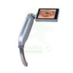
Flexible Fiber Optic Laryngoscope
WhatsApp Order
A Flexible Fiber Optic Laryngoscope is a flexible endoscope (2-5 mm diameter, 30-60 cm working length) with fiber optic image transmission and steerable tip (120-180° angulation) for visualization of the upper airway and facilitation of difficult intubations. Features include control handle with angulation lever, working channel (1-2 mm) for suction or oxygen, external light source (halogen/xenon/LED), and optional camera for video display. Primary clinical applications include awake intubation in difficult airway management (limited mouth opening, cervical spine instability, obstructing pathology), nasotracheal intubation for oral surgery or maxillofacial trauma, intubation with cervical spine precautions (minimal neck movement), diagnostic airway assessment (stridor, hoarseness, vocal cord dysfunction, masses), double-lumen tube placement for thoracic surgery, pediatric difficult airway management, and tracheostomy tube placement guidance. Class II medical device requiring FDA clearance. Critical safety considerations include mandatory leak testing before immersion, antifog preparation, gentle insertion technique, airway maintenance with oxygen, topical anesthesia for patient comfort, suction availability, backup airway device, and strict infection control with validated reprocessing protocols.
Description
Flexible Fiber Optic Laryngoscope
DIAGNOSTIC UPRIMARY CLINICAL &SES
1. Awake Intubation in Difficult Airway Management:
-
Primary Use: Enables tracheal intubation in awake, spontaneously breathing patients with known or predicted difficult airways, including those with limited mouth opening, cervical spine instability, or obstructing airway pathology. The flexible tip can be maneuvered around anatomical obstacles without requiring sedation or paralysis.
-
How it helps: Provides a safe pathway to secure the airway in patients where traditional intubation would be dangerous or impossible, allowing patients to maintain their own breathing while doctors carefully navigate around obstacles.
2. Nasotracheal Intubation:
-
Primary Use: Facilitates nasotracheal intubation for oral surgery, maxillofacial trauma, or patients requiring prolonged intubation where oral access is limited or contraindicated.
-
How it helps: Offers an alternative route to secure the airway when the mouth cannot be used due to surgery, injury, or other conditions, ensuring patients still receive the breathing support they need.
3. Cervical Spine Precautions:
-
Primary Use: Allows intubation with minimal cervical spine movement in patients with suspected or confirmed cervical spine injuries, as the flexible scope can navigate the airway without head extension or manipulation.
-
How it helps: Protects patients with neck injuries from further spinal cord damage during intubation, ensuring their airway is secured without moving their vulnerable cervical spine.
4. Airway Assessment and Diagnosis:
-
Primary Use: Provides direct visualization of the upper airway including nasal passages, pharynx, larynx, and vocal cords for diagnostic evaluation of stridor, hoarseness, vocal cord dysfunction, airway masses, or suspected aspiration.
-
How it helps: Gives specialists a clear view inside the airway to diagnose problems affecting breathing and voice, providing answers for patients suffering from chronic cough, hoarseness, or difficulty swallowing.
5. Double-Lumen Tube Placement:
-
Primary Use: Facilitates accurate placement of double-lumen endotracheal tubes for lung isolation procedures in thoracic surgery.
-
How it helps: Ensures precise positioning of specialized breathing tubes that allow surgeons to operate on one lung while the other continues to ventilate, making complex chest surgeries safer and more effective.
6. Pediatric Difficult Airway:
-
Primary Use: Smaller-diameter flexible scopes allow visualization and intubation in neonates, infants, and children with difficult airways where rigid laryngoscopy may be challenging or impossible.
-
How it helps: Gives anesthesiologists and pediatric specialists the tools they need to secure the airways of the smallest patients, ensuring even babies with complex airway anatomy receive safe, effective care.
7. Tracheostomy Tube Placement:
-
Primary Use: Assists in guiding tracheostomy tube placement and confirming proper position within the trachea.
-
How it helps: Provides visual confirmation that a tracheostomy tube is correctly positioned before it is secured, preventing complications that could arise from improper placement.
SECONDARY & SUPPORTIVE USES
1. Bronchoscopy Assistance: Can be used to guide bronchoscope placement in combined procedures, helping specialists examine deeper airways.
2. Endotracheal Tube Position Confirmation: Verifies correct depth and position of endotracheal tubes, ensuring they are properly placed for effective ventilation.
3. Foreign Body Removal: Assists in visualization and removal of foreign bodies from the airway, helping retrieve objects that have been accidentally inhaled.
4. Teaching and Training: Allows instructors to view airway anatomy during procedures for education, helping train the next generation of airway specialists.
5. Research and Documentation: Records airway findings for research studies and medical records, contributing to scientific knowledge and ensuring accurate documentation.
KEY PRODUCT FEATURES
1. BASIC IDENTIFICATION ATTRIBUTES
-
Device Type: Flexible endoscope with fiber optic image transmission for visualization of the upper airway and facilitation of intubation.
-
Common Names: Flexible Laryngoscope, Fiber Optic Laryngoscope, Flexible Intubation Scope, Nasopharyngoscope.
-
Components:
-
Control Handle: Contains eyepiece or camera connection, angulation controls, and working channel port.
-
Insertion Tube: Flexible shaft containing fiber optic bundles, angulation wires, and working channel.
-
Distal Tip: Steerable tip with objective lens and light source.
-
Light Source: External or integrated LED/halogen light source connected via light cable.
-
Camera System: Optional camera head for video display and recording.
-
-
Insertion Tube Diameter: 2-5 mm; smaller diameters for pediatric/nasal use; larger for adult oral use.
-
Working Length: 30-60 cm; sufficient to reach carina for intubation confirmation.
-
Working Channel: 1-2 mm channel for suction, oxygen delivery, or medication administration.
2. TECHNICAL & PERFORMANCE PROPERTIES
-
Image Transmission: Fiber optic bundle (10,000-30,000 pixels) or digital sensor at tip with video transmission.
-
Resolution: 10-30 line pairs/mm for fiber optic; up to 1920×1080 for digital video scopes.
-
Field of View: 60-120 degrees; wider field improves orientation and navigation.
-
Depth of Field: 3-50 mm; adjustable focus on some models.
-
Tip Angulation: 120-180 degrees up/down; allows navigation through complex airway anatomy.
-
Angulation Control: Lever or knob on handle for precise tip steering.
-
Light Source: Halogen, xenon, or LED; light intensity adjustable; fiber optic light cable transmission.
-
Working Channel: Allows passage of suction catheter, biopsy forceps, or oxygen catheter.
-
Sterilization Method: High-level disinfection (HLD) with chemical solutions or ethylene oxide (EtO) sterilization.
3. PHYSICAL & OPERATIONAL PROPERTIES
-
Handle Material: Medical-grade ABS plastic or anodized aluminum; ergonomic grip.
-
Insertion Tube Material: Polyurethane or silicone jacket with stainless steel braid for durability and flexibility.
-
Weight: 8-24 ounces depending on configuration.
-
Water Resistance: Fully immersible for cleaning and disinfection.
-
Eyepiece: Adjustable diopter for user vision correction.
-
Camera Connection: C-mount or proprietary connector for video camera attachment.
-
Light Post: Standard ACMI or proprietary connector for light cable.
-
Storage: Hanging storage cabinet to maintain straight configuration; foam-lined carrying case.
4. SAFETY & COMPLIANCE ATTRIBUTES
-
Regulatory Status: Class II medical device requiring FDA 510(k) clearance.
-
Intended Use: Indicated for visualization of the upper airway and facilitation of tracheal intubation.
-
Electrical Safety: Compliant with IEC 60601-1 for medical electrical equipment; Type BF applied part.
-
Biocompatibility: Insertion tube materials must be biocompatible for airway contact (ISO 10993).
-
Sterilization Validation: Must have validated high-level disinfection or sterilization protocol per manufacturer.
-
Leak Testing: Must pass leak test before immersion to prevent fluid damage to internal components.
-
Light Source Safety: Automatic shutoff if light cable disconnected to prevent thermal injury.
-
Working Channel: Must maintain patency and seal integrity after repeated use.
5. STORAGE & HANDLING ATTRIBUTES
-
Storage: Hang vertically in clean, dry cabinet to maintain straight configuration; protect from impact and crushing.
-
Cleaning/Disinfection: Manual cleaning followed by high-level disinfection (glutaraldehyde, orthophthalaldehyde, or peracetic acid) or ethylene oxide sterilization per manufacturer instructions.
-
Leak Testing: Perform leak test before each immersion; do not immerse if leak detected.
-
Inspection: Check fiber optic image for broken fibers (black dots); verify tip angulation range; test suction channel patency.
-
Light Source: Inspect light cable for broken fibers; verify light intensity.
-
Reprocessing Records: Maintain logs of HLD/sterilization cycles per facility policy.
-
Replacement: Replace when image quality degrades significantly, when angulation becomes limited, or when channel integrity compromised.
6. LABORATORY & CLINICAL APPLICATIONS
-
Primary Application: Facilitates tracheal intubation in awake patients with difficult airways, nasotracheal intubation, cervical spine precautions, and diagnostic evaluation of upper airway pathology.
-
Limitation: Requires skilled operator with specific training; image quality inferior to video laryngoscopy; fragile and expensive equipment requiring careful handling.
SAFETY HANDLING PRECAUTIONS
1. SAFETY PRECAUTIONS
-
Leak Testing: Always perform leak test before immersion; fluid damage is expensive to repair and compromises patient safety.
-
Antifog Preparation: Apply antifog solution to distal lens or warm scope to body temperature to prevent fogging.
-
Lubrication: Use water-soluble lubricant on insertion tube; avoid petroleum-based products that damage materials.
-
Gentle Insertion: Never force scope against resistance; use angulation to navigate around obstacles.
-
Airway Maintenance: Administer oxygen via working channel or nasal cannula during awake intubation.
-
Topical Anesthesia: Apply topical anesthesia to airway before procedure to suppress gag reflex and improve patient tolerance.
-
Suction Availability: Have suction available via working channel to clear secretions obscuring view.
-
Backup Plan: Always have alternative airway device available in case of scope failure or inability to intubate.
-
Infection Control: Follow strict reprocessing protocols; inadequate disinfection has been linked to patient-to-patient transmission of pathogens.
2. FIRST AID MEASURES
-
Lens Fogging: Remove scope; reapply antifog; warm scope to body temperature.
-
Image Loss (Blackout): Check light source connection; verify light source power; have backup scope available.
-
Patient Desaturation: Stop procedure; ventilate with bag-mask; reassess oxygenation before continuing.
-
Equipment Malfunction: Discontinue use; switch to alternative airway device; document malfunction.
3. FIRE FIGHTING MEASURES
-
Flammability: Plastic components are combustible; fiber optic bundles non-combustible.
-
Extinguishing Media: For electrical fire, use CO₂ or dry chemical (Class C) extinguisher.
-
Light Source Fire Risk: High-intensity light sources can ignite drapes or materials if not properly managed.
Related products
Anti-Streptol Olysin O Titer (ASOT)
Anti-Streptol Olysin O Titer (ASOT) is a quantitative or semi-quantitative serological test (latex agglutination, turbidimetry, nephelometry, or ELISA) for detecting antibodies against streptolysin O, an exotoxin produced by Group A Streptococcus. Elevated or rising titers indicate recent streptococcal infection and are essential for diagnosing post-streptococcal sequelae including acute rheumatic fever (Jones criteria) and post-streptococcal glomerulonephritis. The test requires serum samples; acute and convalescent (2-4 weeks apart) with fourfold rise confirms recent infection. Reference range typically <200-250 Todd units/mL (adults), varies by age and population. Primary clinical applications include diagnosis of Group A streptococcal infections, acute rheumatic fever evaluation, post-streptococcal glomerulonephritis diagnosis, differentiation of acute vs. past infection, evaluation of unexplained arthritis or carditis, pediatric inflammatory conditions (PANDAS), and monitoring disease activity in rheumatic fever. Critical safety precautions include proper timing of acute and convalescent samples, awareness of false negatives/positives, clinical correlation for diagnosis, and standard biohazard precautions. Essential test for rheumatology, nephrology, cardiology, and infectious disease practice.
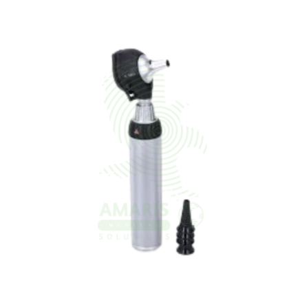
LED Otoscope
An LED Otoscope is a handheld medical device used for illuminating and magnifying the external ear canal and tympanic membrane. Featuring a bright, cool, and long-lasting LED light source, it provides superior visualization for diagnosing common conditions like ear infections, wax impaction, and perforations. Its design, incorporating a reusable handle/head and disposable specula, ensures hygiene and patient safety. As an essential tool in family medicine, pediatrics, and ENT, it enables clinicians to perform accurate otoscopic examinations and, when equipped with an insufflator, crucial pneumatic otoscopy to assess middle ear function.
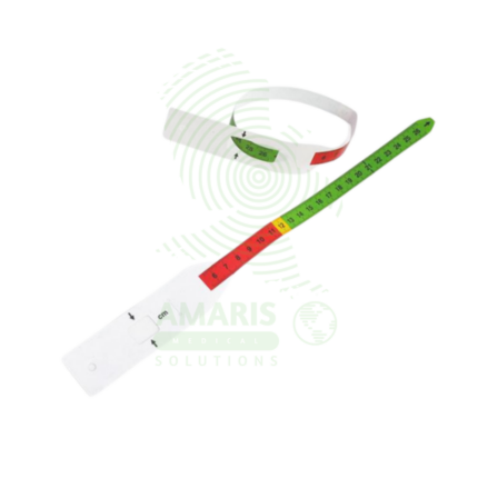
MUAC Tape
A MUAC Tape (Mid-Upper Arm Circumference) is a specialized, color-coded measuring tape used globally as the primary field tool for screening children aged 6-59 months for acute malnutrition. Its simple design—featuring red, yellow, and green zones corresponding to Severe Acute Malnutrition, Moderate Acute Malnutrition, and normal nutritional status—allows for instant, non-invasive assessment without the need for scales, calculation, or literacy. Durable, portable, and inexpensive, it is the cornerstone of community-based nutrition programs, emergency relief efforts, and public health surveillance, enabling early detection and life-saving intervention for malnourished children in even the most resource-constrained settings.
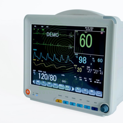
Patient Monitor
The Patient Monitor (6 Parameters BCCMS8000) is a versatile multi-parameter monitor designed for continuous surveillance of core vital signs in various clinical settings. It tracks six essential parameters—ECG, SpO2, Non-Invasive Blood Pressure (NIBP), Respiration, Temperature, and Pulse Rate—providing clinicians with real-time waveforms and numerical data on a clear color display. With its robust alarm system, battery backup for transport, and reliable performance, it is a fundamental tool for ensuring patient safety on general hospital wards, during procedures, and in emergency departments. Its design balances comprehensive monitoring capability with user-friendly operation.
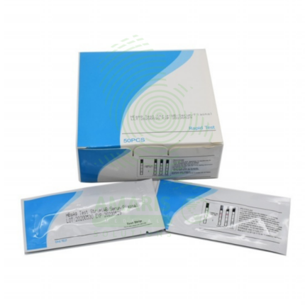
Rapid Test Kits (HIV, HBV, HCV, Malaria)
Rapid Test Kits for HIV, HBV, HCV, and Malaria are single-use, immunochromatographic devices designed for the quick, preliminary screening of these critical infectious diseases at the point of care. By detecting specific antibodies or antigens in a small blood sample (or oral fluid for some HIV tests), they provide a visual result within 15-30 minutes without the need for laboratory equipment. Their primary role is to expand access to testing in community and resource-limited settings, enabling immediate counseling, triage, and referral for confirmatory testing and treatment. As essential tools in public health, they are characterized by their simplicity, speed, and critical importance in early diagnosis and outbreak response.
SAFE SERIES Blood Collection Needles
The SAFE SERIES (AM-SAFE) by Beijing Precil (Mindray subsidiary) is a comprehensive line of safety-engineered blood collection devices designed to prevent accidental needlestick injuries in healthcare workers. The series includes venous blood collection needles (AM-SAFE-V), standard safety needles (AM-SAFE1), winged infusion sets/butterfly needles (AM-SAFE2), high-gauge thin-wall needles for fragile veins (AM-SAFE3), premium coated needles for enhanced patient comfort (AM-GOLD), and nurse-friendly one-handed safety variants (AM-NURSE). All devices incorporate integral safety mechanisms that shield the contaminated needle immediately after withdrawal, complying with OSHA Needlestick Safety and Prevention Act requirements. Available in various gauges (18G-25G) and configurations for routine venipuncture, difficult vein access, pediatric/geriatric patients, and frequent blood draws. Sterile, single-use, latex-free, and color-coded for easy gauge identification. Essential safety devices for blood collection in hospitals, clinics, laboratories, and home healthcare settings.
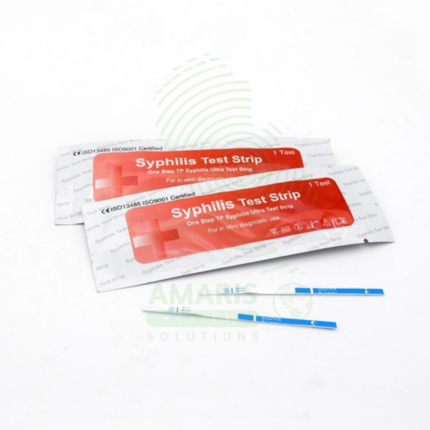
VDRL Test For Syphilis
The VDRL Test For Syphilis (Venereal Disease Research Laboratory) is a Class II medical device (FDA-cleared) non-treponemal flocculation test for qualitative and quantitative detection of reaginic antibodies in serum or cerebrospinal fluid, used for syphilis screening, diagnosis, and treatment monitoring. The test uses an antigen mixture (cardiolipin, cholesterol, lecithin) that reacts with antibodies, forming visible clumping (flocculation) in positive samples. Results are reported as reactive/non-reactive with quantitative titers (e.g., 1:2 to 1:256). Primary clinical applications include initial screening for syphilis infection, confirmation of active syphilis (with treponemal tests), monitoring treatment response (fourfold titer decrease indicates cure), diagnosis of neurosyphilis (CSF testing), prenatal screening for congenital syphilis prevention, screening in high-risk populations (MSM, HIV-positive, multiple partners), and blood donor screening. Critical safety precautions include confirmation of all reactive results with treponemal tests (TPPA, FTA-ABS), awareness of false positives (pregnancy, autoimmune disease, viral infections), proper specimen handling (universal precautions), refrigerated storage of reagents (2-8°C), and mandatory reporting of reactive results to public health authorities. Essential test for syphilis control and congenital syphilis prevention worldwide.