Dermatoscope and Magnifiers
 Diagnostic Kits
 Vital Signs Monitors
 Stethoscopes and Accessories
 Otoscopes, Ophthalmoscopes, and Retinoscopes
 Reflex Hammers and Neurological Tools
 Scales and Measuring Devices
 Spirometers and Pulmonary Function Tests
 Electrosurgical Units and Accessories
 Cutting Instruments
 Grasping and Holding Instruments
 Hemostatic Instruments
 Specialized Surgical Sets
 Single-Use Procedure Trays and Packs
 Surgical Drapes, Gowns, and Covers
 Tissue Unifying Instruments
 Radiation Protection
 X-Ray Machines and Accessories
 Ultrasound Systems and Probes
 MRI and CT Scanners
 Radiology Consumables
 Bone Densitometers
 Fluoroscopy Equipment
 Imaging Tables and Positioning Aids
 Microscopes and Accessories
 Centrifuges and Separators
 Analyzers
 Incubators and Ovens
 Pipettes, Dispensers, and Lab Glassware
 Refrigerators, Freezers, and Storage Units
 Lab Consumables
 Sterilizers and Autoclaves for Lab Use
 Multi-Parameter Monitors
 Ventilators and Respiratory Support Devices
 Defibrillators and AEDs
 Infusion Pumps and IV Systems
 Patient Warmers and Cooling Devices
 Central Monitoring Stations
 Accessories
 Anesthesia Machines and Workstations
 Oxygen Concentrators and Delivery Systems
 Nebulizers and Inhalers
 CPAP/BiPAP Machines
 Airway Management
 Anesthesia Masks, Circuits, and Bags
 Humidifiers and Heaters
 Respiratory Therapy Accessories
 First Aid Kits and Cabinets
 Emergency Resuscitation Equipment
 Trauma Supplies
 Emergency Carts and Crash Carts
 Burn Care Products
 Bleeding Control
 Automated External Defibrillators (AEDs)
 Transport and Evacuation
 Wheelchairs and Accessories
 Walkers, Crutches, and Canes
 Prosthetics and Orthotics
 Physical Therapy Equipment
 Transfer Devices
 Bathroom Safety
 Orthopedic Traction and Tables
 Hot/Cold Therapy Packs and Units
 Beds and Mattresses
 Chairs and Stools
 Tables
 Cabinets and Storage
 Privacy Screens & Curtains
 Stands and Racks
 Linens and Textiles
 Lighting
 Autoclaves and Sterilizers
 Ultrasonic Cleaners
 Disinfectant Solutions and Wipes
 Sterilization Pouches, Wraps, and Indicators
 Instrument Trays and Containers
 UV and Ozone Disinfection Devices
 Washer Disinfectors
 Wound Care
 Gloves
 Masks and Respirators
 Catheters and Tubing
 Swabs, Applicators, and Sponges
 Incontinence Products
 Personal Protective Equipment (PPE)
 Dental Chairs and Units
 Handpieces and Burs
 Instruments
 Consumables
 Sterilization for Dental Use
 Orthodontic Supplies
 Endodontic Tools
 Slit Lamps and Tonometers
 Lensometers and Phoropters
 Ophthalmic Surgical Instruments
 Eyewear Frames and Lenses
 Contact Lens Supplies
 Vision Testing Charts and Devices
 Eye Care Consumables
 Laser Systems for Eye Care
 ENT Exam Chairs and Tables
 Endoscopes
 Audiometers and Hearing Tests
 ENT Instruments
 Nasal and Throat Packs
 Hearing Aids and Accessories
 Otology Supplies
 Fetal Dopplers and Monitors
 Delivery Beds and Tables
 Gynecological Instruments
 Neonatal Incubators and Warmers
 Breast Pumps and Accessories
 Contraceptive Devices
 Maternity Supports and Pads
 Neonatal Consumables
 Cystoscopes and Urethroscopes
 Dialysis Machines and Supplies
 Urological Catheters and Bags
 Lithotripters
 Prostate Treatment Devices
 Urinary Incontinence Products
 Kidney Stone Management Tools
 Consumables & Disposables
 EEG and EMG Machines
 Neurosurgical Instruments
 Nerve Stimulators
 Headrests and Positioning Aids
 Lumbar Puncture Kits
 Seizure Monitoring Devices
 Consumables
 Rehabilitation for Neurological Conditions
 ECG Machines and Accessories
 Holter Monitors
 Stress Test Systems
 Pacemakers and Defibrillator Accessories
 Vascular Access Devices
 Cardiac Catheters and Guidewires
 Blood Flow Meters
 Consumables
 Orthopedic Instruments
 Casts, Splints, and Padding
 Joint Replacement Supplies
 Prosthetic Limbs and Components
 Bone Grafts and Substitutes
 Traction Devices
 Orthopedic Braces and Supports
 Rehabilitation Aids for Orthopedics
 Home Oxygen Therapy
 Hospital Beds for Home Use
 Mobility Aids
 Bathroom and Daily Living Aids
 Wound Care for Home
 Monitoring Devices
 Enteral Feeding Pumps and Tubes
 Hand Sanitizers and Dispensers
 Face Shields and Goggles
 Isolation Gowns and Suits
 Biohazard Waste Containers
 Air Purifiers and HEPA Filters
 Surface Disinfectants
 Sharps Containers
 Protective Barriers
 Cardiovascular & Endurance Training
 Strength Training & Weightlifting
 Functional Training & Core Conditioning
 Physical Therapy & Rehabilitation
 Sports & Outdoor Recreation
 Gym Flooring & Facility Equipment
 Fitness Monitoring & Accessories
 Kids & Novelties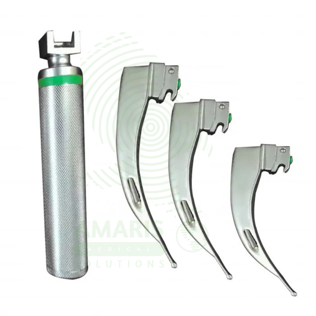
Macintosh Fiber Optic Laryngoscope
$1.00
WhatsApp Order
A Macintosh Fiber Optic Laryngoscope is a rigid laryngoscope with curved Macintosh blade (sizes 0-4, 70-160 mm) incorporating an integrated fiber optic light bundle that transmits bright, focused illumination (2,000-10,000+ Lux) from a handle-mounted LED or xenon bulb to the blade tip for enhanced visualization during tracheal intubation. The curved blade design allows indirect epiglottis elevation by placing the tip in the vallecula, requiring less force and neck extension than straight blades. Features stainless steel reusable blades, ergonomic handles with knurled grip, ISO standard hook-on fittings, and steam autoclave compatibility. Primary clinical applications include routine and emergency tracheal intubation during general anesthesia, difficult airway management with improved illumination, cervical spine precautions (minimal neck movement), teaching and training (enhanced visualization for instructors), rapid sequence intubation, neonatal and pediatric intubation (sizes 0-2), and oral/maxillofacial surgery. Class II medical device requiring FDA clearance. Critical safety considerations include pre-use light check (dark spots indicate broken fibers), appropriate blade size selection, proper lifting technique (not levering on teeth), battery verification, fiber optic care (avoid sharp bending), and backup device availability.
Description
Macintosh Fiber Optic Laryngoscope
PRIMARY CLINICAL & DIAGNOSTIC USES
1. Routine and Emergency Tracheal Intubation:
-
Primary Use: Provides direct visualization of the glottis for tracheal intubation during general anesthesia, emergency resuscitation, and critical care. The Macintosh curved blade design allows elevation of the epiglottis indirectly by placing the tip in the vallecula, providing a clear view of the vocal cords for tube passage.
-
How it helps: Gives anesthesiologists and emergency physicians a time-tested tool with enhanced illumination to secure a patient’s airway, ensuring oxygen can reach the lungs when patients cannot breathe on their own.
2. Difficult Airway Management with Fiber Optic Illumination:
-
Primary Use: The integrated fiber optic light bundle delivers bright, focused illumination to the blade tip, improving visualization in patients with poor lighting conditions, dark oral cavities, or when additional light is needed for difficult airway anatomy.
-
How it helps: Cuts through the darkness of the most challenging airways, providing brilliant illumination that reveals anatomy hidden by blood, secretions, or abnormal tissue.
3. Cervical Spine Precautions:
-
Primary Use: Allows intubation with manual in-line stabilization in patients with suspected or confirmed cervical spine injuries. The curved blade design requires less force and neck extension compared to straight blades, reducing cervical spine movement during intubation.
-
How it helps: Protects patients with neck injuries from further spinal cord damage during intubation, ensuring their airway is secured without compromising their injured cervical spine.
4. Teaching and Training:
-
Primary Use: The bright fiber optic illumination improves visualization for instructors teaching laryngoscopy technique to trainees, allowing better observation of anatomy and blade placement.
-
How it helps: Transforms airway education by allowing instructors to see exactly what their students see, providing targeted feedback that accelerates learning and builds competence.
5. Rapid Sequence Intubation:
-
Primary Use: Used in emergency settings for rapid sequence intubation where speed and first-pass success are critical. The familiar Macintosh design combined with superior illumination enhances success rates.
-
How it helps: Gives emergency physicians every advantage when seconds count, combining a trusted design with superior lighting to maximize first-attempt success.
6. Neonatal and Pediatric Intubation:
-
Primary Use: Smaller Macintosh blades (size 0-2) with fiber optic illumination allow visualization of the pediatric airway in neonates, infants, and children requiring intubation.
-
How it helps: Provides pediatric specialists with the appropriately sized tools and enhanced illumination needed to secure airways in the smallest patients, from premature infants to young children.
7. Oral and Maxillofacial Surgery:
-
Primary Use: Facilitates intubation in patients undergoing oral surgery where access may be limited and optimal lighting is essential for successful airway management.
-
How it helps: Supports complex surgical procedures by ensuring the airway is secured even when the surgical field limits access, allowing surgeons to focus on their work.
SECONDARY & SUPPORTIVE USES
1. Airway Assessment: Provides visualization of the oropharynx and glottis for preoperative airway evaluation, helping anesthesiologists plan their approach before surgery.
2. Foreign Body Removal: Assists in visualization and removal of foreign bodies from the upper airway, helping retrieve objects that are blocking breathing.
3. Endotracheal Tube Position Confirmation: Allows direct visualization of tube passage through vocal cords, ensuring the breathing tube is correctly placed before ventilation begins.
4. Suctioning Under Direct Vision: Enables targeted suctioning of secretions under direct visualization, clearing the airway of blood or mucus that could interfere with intubation.
5. Research and Documentation: Can be used with camera attachments for recording intubation procedures, contributing to education, research, and quality improvement.
KEY PRODUCT FEATURES
1. BASIC IDENTIFICATION ATTRIBUTES
-
Device Type: Rigid laryngoscope with Macintosh curved blade incorporating fiber optic light transmission for tracheal intubation.
-
Common Names: Macintosh Laryngoscope, Fiber Optic Laryngoscope, Rigid Laryngoscope, Curved Blade Laryngoscope.
-
Components:
-
Handle: Contains batteries and provides grip; standard or stubby sizes; accepts fiber optic blade.
-
Blade: Macintosh curved blade with integrated fiber optic light carrier; detachable from handle.
-
Light Source: Fiber optic bundle transmitting light from handle bulb to blade tip.
-
Bulb: LED or xenon bulb in handle (not at blade tip) for fiber optic illumination.
-
-
Blade Sizes: 0 (neonatal), 1 (infant), 2 (child/small adult), 3 (adult), 4 (large adult).
-
Blade Length: 70-160 mm depending on size.
-
Blade Material: Stainless steel with fiber optic bundle embedded along blade.
2. TECHNICAL & PERFORMANCE PROPERTIES
-
Fiber Optic Illumination: 2,000-10,000+ Lux at blade tip; bright, focused light without shadowing from bulb at tip.
-
Light Source: LED (longer life, brighter, cooler) or xenon (traditional, reliable) bulb in handle.
-
Fiber Optic Bundle: 2-3 mm diameter; glass or acrylic fibers; transmits light from handle to blade tip.
-
Light Intensity: Adjustable on some models; constant high output on others.
-
Blade Curvature: Radius of curvature 30-45 mm; designed to fit vallecula and lift epiglottis indirectly.
-
Blade Flange: Height 10-20 mm; provides channel for tube passage and displaces tongue.
-
Blade Tip: Rounded, blunt tip designed for vallecula placement without tissue trauma.
-
Battery Type: C, AA, or proprietary rechargeable lithium-ion; 2-4 hours continuous use.
-
Autoclavable: Blades and handles (with battery removed) are steam autoclavable.
3. PHYSICAL & OPERATIONAL PROPERTIES
-
Handle Material: Stainless steel, anodized aluminum, or chrome-plated brass; knurled or textured grip.
-
Blade Material: Stainless steel (reusable) or medical-grade plastic (disposable).
-
Fiber Optic Jacketing: Stainless steel or polymer protection along blade.
-
Weight: 8-16 ounces complete (handle + blade).
-
Connection: International Standard (ISO 7376) hook-on fitting; compatible across manufacturers.
-
Battery Access: Screw-on or snap-off cap; O-ring sealed for autoclaving.
-
Light Activation: Automatic when blade is deployed (some models) or manual switch.
-
Cleaning Compatibility: Fully immersible for cleaning; autoclavable (with battery removed).
4. SAFETY & COMPLIANCE ATTRIBUTES
-
Regulatory Status: Class II medical device requiring FDA 510(k) clearance.
-
Intended Use: Indicated for oral intubation providing direct visualization of the glottis.
-
Electrical Safety: Compliant with IEC 60601-1 for medical electrical equipment; battery-operated.
-
Biocompatibility: Blade materials must be biocompatible for oral contact (ISO 10993).
-
Light Source Safety: LED bulbs produce minimal heat; xenon bulbs warm but within safe limits.
-
Autoclave Compatibility: Must withstand repeated steam sterilization cycles without degradation.
-
Fiber Optic Integrity: Fiber bundle must maintain light transmission after repeated sterilization.
-
ISO Compliance: Meets ISO 7376 for handle/blade compatibility and ISO 7376/1 for fiber optic laryngoscopes.
5. STORAGE & HANDLING ATTRIBUTES
-
Storage: Store blades and handles in a clean, dry environment; blade storage rack or case; protect from impact.
-
Cleaning: Manual cleaning with enzymatic detergent; rinse thoroughly; dry before sterilization.
-
Sterilization: Steam autoclave (gravity or prevacuum) at 132-135°C for 3-4 minutes (wrapped). Remove batteries before autoclaving.
-
Battery Maintenance: Use only specified batteries; remove if storing long-term; recharge rechargeables per manufacturer.
-
Inspection: Check fiber optic illumination for dark spots (broken fibers); verify blade locking mechanism; test light activation.
-
Replacement: Replace when a fiber optic bundle has >20% broken fibers (significant light loss), when blade bent or damaged, or when locking mechanism fails.
6. LABORATORY & CLINICAL APPLICATIONS
-
Primary Application: Provides direct visualization for tracheal intubation during general anesthesia, emergency resuscitation, and critical care using the familiar Macintosh curved blade design with enhanced fiber optic illumination.
-
Limitation: Requires direct line-of-sight to glottis; may be difficult in patients with limited mouth opening, large tongue, or anterior airway.
SAFETY HANDLING PRECAUTIONS
1. SAFETY PRECAUTIONS
-
Light Check Before Use: Verify bright, even illumination before each intubation attempt; dark spots indicate broken fibers and reduced light output.
-
Blade Selection: Choose appropriate blade size for patient anatomy; too small fails to lift epiglottis; too large causes trauma.
-
Technique: Place blade tip in vallecula; lift upward at 45-degree angle (not levering on teeth). Fiber optic illumination improves visualization but does not replace proper technique.
-
Tooth Protection: Avoid using blade as lever on upper teeth; use lifting motion to prevent dental trauma.
-
Sterility: Use sterile blade for surgical cases; high-level disinfection for non-sterile procedures.
-
Battery Check: Verify adequate battery charge before procedure; have backup handle available.
-
Fiber Optic Care: Do not bend blade sharply; fiber bundle may break. Clean gently to avoid damaging fibers.
-
Alternative Available: Always have backup laryngoscope (conventional or video) available.
-
Infection Control: Clean and sterilize between patients per facility protocol; prion diseases require special processing.
2. FIRST AID MEASURES
-
Light Failure: Check battery; replace handle; have backup device available.
-
Blade Lock Failure: Remove blade; use backup device; do not attempt intubation with unstable blade.
-
Patient Trauma: If dental or soft tissue injury occurs, document and manage per protocol.
-
Equipment Malfunction: Discontinue use; switch to alternative device; document malfunction.
3. FIRE FIGHTING MEASURES
-
Flammability: Metal components non-combustible; plastic handles combustible.
-
Extinguishing Media: For electrical fire, use CO₂ or dry chemical (Class C) extinguisher.
Related products
Fingertip Pulse Oximeter
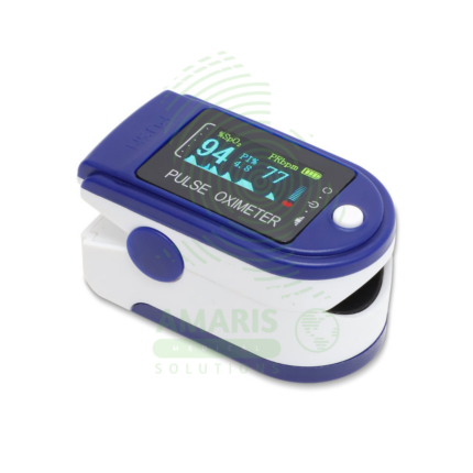
A Fingertip Pulse Oximeter is a compact, non-invasive medical device that clips onto a finger to measure peripheral arterial oxygen saturation (SpO2) and pulse rate in seconds. Utilizing light-based technology, it provides critical, real-time data on respiratory and circulatory status. Its portability and ease of use make it indispensable in hospitals (from ORs to general wards), for patients managing chronic lung conditions at home, and for situational monitoring in sports or high-altitude environments. While an extremely valuable screening tool, readings must be interpreted in context, considering factors like patient movement, perfusion, and clinical symptoms.
Handheld Pulse Oximeter
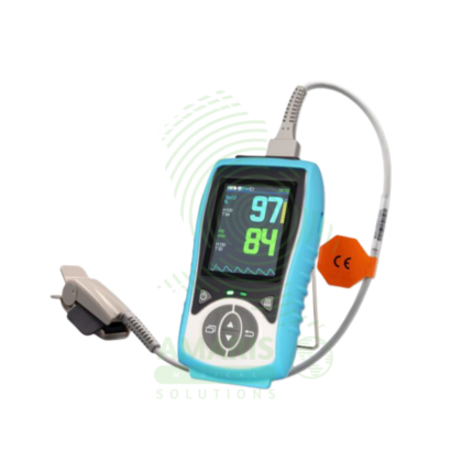
A Handheld Pulse Oximeter is a professional-grade, portable medical device used by healthcare providers for accurate and reliable monitoring of blood oxygen saturation (SpO2) and pulse rate. It consists of a durable, feature-rich monitor unit and separate, interchangeable sensors for use on fingers, toes, or ears. Superior to basic fingertip models, it offers advanced functions like configurable alarms, detailed plethysmograph display, trend memory, and data connectivity. Its robust design, clinical accuracy under challenging conditions (motion, low perfusion), and versatility make it an essential tool in hospitals, clinics, ambulances, and home health for patient assessment and monitoring.

Littman Stethoscope (Classic 4 Cardiology)
A Littman Stethoscope (Classic 4 Cardiology) represents the pinnacle of acoustic auscultation technology. Designed for clinicians who require the highest level of diagnostic accuracy, it features dual tunable diaphragms and dual-lumen tubing to provide exceptional acoustic sensitivity and ambient noise reduction. Its innovative design allows the user to hear both high and low-frequency sounds by simply adjusting pressure on either side of the chestpiece, making it the instrument of choice for cardiologists, intensivists, and specialists who cannot afford to miss a subtle murmur, rub, or breath sound. It combines superior performance with the durability and comfort expected from the Littmann brand.
Littmann Stethoscope (Classic II)
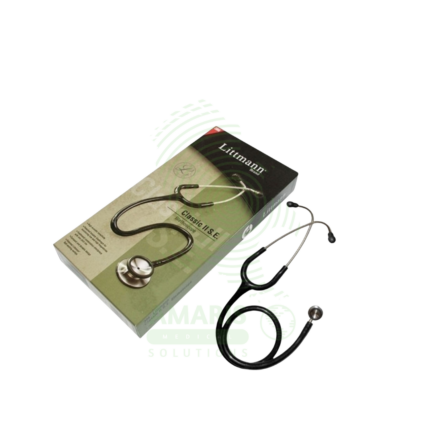
A Littmann Stethoscope (Classic II) is a premium, acoustic dual-head stethoscope renowned for its diagnostic clarity, durability, and innovative tunable diaphragm technology. It allows healthcare professionals to hear both high and low-frequency sounds by simply adjusting pressure on the single-sided chestpiece, eliminating the need to rotate it. With its superior acoustics, comfortable fit, and wide range of colors, it is the instrument of choice for medical students, nurses, and physicians across all specialties who require reliable performance for physical assessment, auscultation of heart and lung sounds, and manual blood pressure measurement.
MUAC Tape
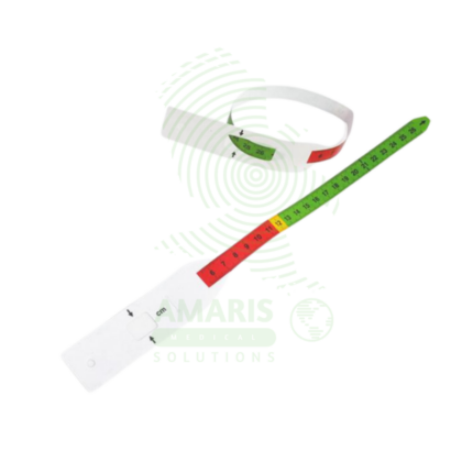
A MUAC Tape (Mid-Upper Arm Circumference) is a specialized, color-coded measuring tape used globally as the primary field tool for screening children aged 6-59 months for acute malnutrition. Its simple design—featuring red, yellow, and green zones corresponding to Severe Acute Malnutrition, Moderate Acute Malnutrition, and normal nutritional status—allows for instant, non-invasive assessment without the need for scales, calculation, or literacy. Durable, portable, and inexpensive, it is the cornerstone of community-based nutrition programs, emergency relief efforts, and public health surveillance, enabling early detection and life-saving intervention for malnourished children in even the most resource-constrained settings.
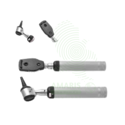
Ophthalmoscope
An Ophthalmoscope is a handheld diagnostic instrument used to examine the interior of the eye, specifically the fundus (retina, optic disc, blood vessels, and macula). By providing illuminated magnification, it serves as a vital screening tool for detecting ocular diseases like glaucoma and retinal disorders, and more importantly, for identifying signs of systemic conditions such as diabetes, hypertension, and increased intracranial pressure. Its operation involves selecting appropriate apertures and compensating lenses to obtain a clear view. While offering a detailed but narrow field of vision, mastery of the direct ophthalmoscope remains a cornerstone skill in general medicine, neurology, and ophthalmology for assessing vascular and neurological health.
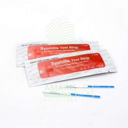
VDRL Test For Syphilis
The VDRL Test For Syphilis (Venereal Disease Research Laboratory) is a Class II medical device (FDA-cleared) non-treponemal flocculation test for qualitative and quantitative detection of reaginic antibodies in serum or cerebrospinal fluid, used for syphilis screening, diagnosis, and treatment monitoring. The test uses an antigen mixture (cardiolipin, cholesterol, lecithin) that reacts with antibodies, forming visible clumping (flocculation) in positive samples. Results are reported as reactive/non-reactive with quantitative titers (e.g., 1:2 to 1:256). Primary clinical applications include initial screening for syphilis infection, confirmation of active syphilis (with treponemal tests), monitoring treatment response (fourfold titer decrease indicates cure), diagnosis of neurosyphilis (CSF testing), prenatal screening for congenital syphilis prevention, screening in high-risk populations (MSM, HIV-positive, multiple partners), and blood donor screening. Critical safety precautions include confirmation of all reactive results with treponemal tests (TPPA, FTA-ABS), awareness of false positives (pregnancy, autoimmune disease, viral infections), proper specimen handling (universal precautions), refrigerated storage of reagents (2-8°C), and mandatory reporting of reactive results to public health authorities. Essential test for syphilis control and congenital syphilis prevention worldwide.