Dermatoscope and Magnifiers
 Diagnostic Kits
 Vital Signs Monitors
 Stethoscopes and Accessories
 Otoscopes, Ophthalmoscopes, and Retinoscopes
 Reflex Hammers and Neurological Tools
 Scales and Measuring Devices
 Spirometers and Pulmonary Function Tests
 Electrosurgical Units and Accessories
 Cutting Instruments
 Grasping and Holding Instruments
 Hemostatic Instruments
 Specialized Surgical Sets
 Single-Use Procedure Trays and Packs
 Surgical Drapes, Gowns, and Covers
 Tissue Unifying Instruments
 Radiation Protection
 X-Ray Machines and Accessories
 Ultrasound Systems and Probes
 MRI and CT Scanners
 Radiology Consumables
 Bone Densitometers
 Fluoroscopy Equipment
 Imaging Tables and Positioning Aids
 Microscopes and Accessories
 Centrifuges and Separators
 Analyzers
 Incubators and Ovens
 Pipettes, Dispensers, and Lab Glassware
 Refrigerators, Freezers, and Storage Units
 Lab Consumables
 Sterilizers and Autoclaves for Lab Use
 Multi-Parameter Monitors
 Ventilators and Respiratory Support Devices
 Defibrillators and AEDs
 Infusion Pumps and IV Systems
 Patient Warmers and Cooling Devices
 Central Monitoring Stations
 Accessories
 Anesthesia Machines and Workstations
 Oxygen Concentrators and Delivery Systems
 Nebulizers and Inhalers
 CPAP/BiPAP Machines
 Airway Management
 Anesthesia Masks, Circuits, and Bags
 Humidifiers and Heaters
 Respiratory Therapy Accessories
 First Aid Kits and Cabinets
 Emergency Resuscitation Equipment
 Trauma Supplies
 Emergency Carts and Crash Carts
 Burn Care Products
 Bleeding Control
 Automated External Defibrillators (AEDs)
 Transport and Evacuation
 Wheelchairs and Accessories
 Walkers, Crutches, and Canes
 Prosthetics and Orthotics
 Physical Therapy Equipment
 Transfer Devices
 Bathroom Safety
 Orthopedic Traction and Tables
 Hot/Cold Therapy Packs and Units
 Beds and Mattresses
 Chairs and Stools
 Tables
 Cabinets and Storage
 Privacy Screens & Curtains
 Stands and Racks
 Linens and Textiles
 Lighting
 Autoclaves and Sterilizers
 Ultrasonic Cleaners
 Disinfectant Solutions and Wipes
 Sterilization Pouches, Wraps, and Indicators
 Instrument Trays and Containers
 UV and Ozone Disinfection Devices
 Washer Disinfectors
 Wound Care
 Gloves
 Masks and Respirators
 Catheters and Tubing
 Swabs, Applicators, and Sponges
 Incontinence Products
 Personal Protective Equipment (PPE)
 Dental Chairs and Units
 Handpieces and Burs
 Instruments
 Consumables
 Sterilization for Dental Use
 Orthodontic Supplies
 Endodontic Tools
 Slit Lamps and Tonometers
 Lensometers and Phoropters
 Ophthalmic Surgical Instruments
 Eyewear Frames and Lenses
 Contact Lens Supplies
 Vision Testing Charts and Devices
 Eye Care Consumables
 Laser Systems for Eye Care
 ENT Exam Chairs and Tables
 Endoscopes
 Audiometers and Hearing Tests
 ENT Instruments
 Nasal and Throat Packs
 Hearing Aids and Accessories
 Otology Supplies
 Fetal Dopplers and Monitors
 Delivery Beds and Tables
 Gynecological Instruments
 Neonatal Incubators and Warmers
 Breast Pumps and Accessories
 Contraceptive Devices
 Maternity Supports and Pads
 Neonatal Consumables
 Cystoscopes and Urethroscopes
 Dialysis Machines and Supplies
 Urological Catheters and Bags
 Lithotripters
 Prostate Treatment Devices
 Urinary Incontinence Products
 Kidney Stone Management Tools
 Consumables & Disposables
 EEG and EMG Machines
 Neurosurgical Instruments
 Nerve Stimulators
 Headrests and Positioning Aids
 Lumbar Puncture Kits
 Seizure Monitoring Devices
 Consumables
 Rehabilitation for Neurological Conditions
 ECG Machines and Accessories
 Holter Monitors
 Stress Test Systems
 Pacemakers and Defibrillator Accessories
 Vascular Access Devices
 Cardiac Catheters and Guidewires
 Blood Flow Meters
 Consumables
 Orthopedic Instruments
 Casts, Splints, and Padding
 Joint Replacement Supplies
 Prosthetic Limbs and Components
 Bone Grafts and Substitutes
 Traction Devices
 Orthopedic Braces and Supports
 Rehabilitation Aids for Orthopedics
 Home Oxygen Therapy
 Hospital Beds for Home Use
 Mobility Aids
 Bathroom and Daily Living Aids
 Wound Care for Home
 Monitoring Devices
 Enteral Feeding Pumps and Tubes
 Hand Sanitizers and Dispensers
 Face Shields and Goggles
 Isolation Gowns and Suits
 Biohazard Waste Containers
 Air Purifiers and HEPA Filters
 Surface Disinfectants
 Sharps Containers
 Protective Barriers
 Cardiovascular & Endurance Training
 Strength Training & Weightlifting
 Functional Training & Core Conditioning
 Physical Therapy & Rehabilitation
 Sports & Outdoor Recreation
 Gym Flooring & Facility Equipment
 Fitness Monitoring & Accessories
 Kids & Novelties
 Amaris Medical")
LED Otoscope
WhatsApp Order
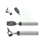
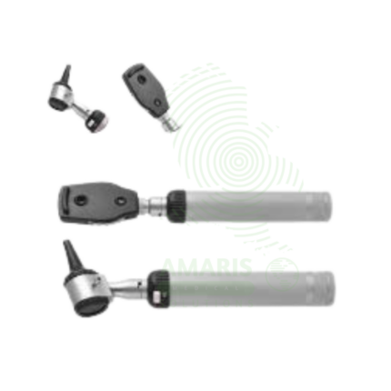
An LED Otoscope is a handheld medical device used for illuminating and magnifying the external ear canal and tympanic membrane. Featuring a bright, cool, and long-lasting LED light source, it provides superior visualization for diagnosing common conditions like ear infections, wax impaction, and perforations. Its design, incorporating a reusable handle/head and disposable specula, ensures hygiene and patient safety. As an essential tool in family medicine, pediatrics, and ENT, it enables clinicians to perform accurate otoscopic examinations and, when equipped with an insufflator, crucial pneumatic otoscopy to assess middle ear function.
Description
LED Otoscope
PRIMARY CLINICAL & DIAGNOSTIC USES
1. Examination of the External Auditory Canal and Tympanic Membrane:
-
Primary Use: The primary and definitive use is for the visual inspection of the outer ear canal and eardrum (tympanic membrane) to assess for abnormalities, inflammation, infection, or obstruction.
-
How it helps: Gives doctors a clear view inside the ear, allowing them to see what patients cannot describe—whether it’s a bulging eardrum, a pocket of fluid, or a foreign object causing discomfort.
2. Diagnosis of Otitis Media and Externa:
-
Primary Use: Essential for diagnosing acute otitis media (middle ear infection, characterized by a bulging, red, immobile tympanic membrane) and otitis externa (swimmer’s ear, characterized by canal edema, redness, and discharge).
-
How it helps: Provides the visual evidence needed to distinguish between different types of ear infections, ensuring patients receive the right treatment—antibiotics for middle ear infections, antibiotic drops for swimmer’s ear, or simple pain relief for viral causes.
3. Evaluation of Ear Pain (Otalgia), Hearing Loss, and Tinnitus:
-
Primary Use: A first-line diagnostic tool for patients presenting with ear-related symptoms to identify potential causes such as cerumen (earwax) impaction, foreign bodies, perforations, cholesteatoma, or signs of barotrauma.
-
How it helps: Takes the guesswork out of ear complaints, quickly revealing whether hearing loss is caused by a simple wax blockage requiring removal or something more serious requiring specialist referral.
4. Routine Pediatric and Adult Wellness Exams:
-
Primary Use: A standard component of the physical examination in both pediatric and adult medicine to screen for asymptomatic conditions and establish a baseline.
-
How it helps: Catches silent problems during routine check-ups, from fluid behind a child’s eardrum that could affect speech development to chronic changes in an adult’s ear that might signal underlying health issues.
5. Removal of Cerumen or Foreign Bodies:
-
Primary Use: Under direct visualization provided by the otoscope, a clinician can safely perform or guide the removal of obstructive earwax or small foreign objects from the ear canal using specialized instruments.
-
How it helps: Turns a blind procedure into a guided one, allowing doctors to see exactly what they are doing as they remove wax or retrieve objects, preventing injury to the delicate ear canal and eardrum.
SECONDARY & SUPPORTIVE USES
1. Pre- and Post-Operative Assessment: Used in ENT (Ear, Nose, and Throat) and audiology practices to examine ears before procedures (e.g., tympanostomy tube placement) and to monitor healing and tube function post-operatively.
2. Pneumatic Otoscopy: When equipped with a sealed speculum and insufflator bulb, it allows for pneumatic otoscopy—the assessment of tympanic membrane mobility by applying gentle positive and negative air pressure. This is critical for detecting middle ear effusion (fluid behind the eardrum).
3. Teaching Aid: A fundamental tool for instructing medical students, nursing students, and residents in otologic examination, training the next generation of healthcare providers to recognize the visual signs of ear disease.
KEY PRODUCT FEATURES
1. BASIC IDENTIFICATION ATTRIBUTES
-
Device Type: Handheld, diagnostic optical instrument for otoscopy.
-
Light Source: LED (Light Emitting Diode) illumination. This is a key advancement, replacing older halogen bulbs.
-
Components: Consists of a handle (containing power source), a head (containing the light source, lens, and speculum attachment point), and disposable plastic specula of various sizes.
-
Optics: Provides a magnified, illuminated view. May be direct viewing (looking straight through a lens) or fiber optic (where light is transmitted via fibers around a viewing path).
2. TECHNICAL & PERFORMANCE PROPERTIES
-
LED Advantages:
-
Cool Light: Emits minimal heat, preventing patient discomfort and reducing the risk of burns.
-
Bright, White Light: Provides excellent color rendering and illumination of the ear canal and tympanic membrane.
-
Long Lifespan & Low Power Consumption: LEDs last for tens of thousands of hours and are highly efficient, leading to longer battery life.
-
-
Magnification: Typically provides low-power magnification (e.g., 3x to 8x) sufficient for detailed examination.
-
Field of View: Determined by the speculum size and optical design. A wider view is helpful for examining the entire canal.
-
Pneumatic Capability: Some models have a built-in or attachable insufflator port to connect a rubber bulb for performing pneumatic otoscopy.
3. PHYSICAL & OPERATIONAL PROPERTIES
-
Power Source: Powered by standard batteries (e.g., AA or rechargeable packs) housed in the handle.
-
Durability: The head and handle are typically made of durable, medical-grade metal (e.g., stainless steel, aluminum) or high-impact plastic.
-
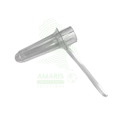
Speculum Sizes: Comes with a set of disposable, single-use plastic specula in sizes from 2 mm to 5 mm to fit different patient ages and canal sizes (pediatric to adult).
4. SAFETY & COMPLIANCE ATTRIBUTES
-
Regulatory Status: Classified as a Class I medical device in most jurisdictions (low-risk, non-invasive diagnostic tool). Requires CE Marking (EU) and may be subject to FDA General Controls (US).
-
Biocompatibility: The specula that contact the patient are made of medical-grade, non-toxic plastic.
-
Electrical Safety: For rechargeable models, complies with relevant electrical safety standards.
5. STORAGE & HANDLING ATTRIBUTES
-
Storage: Store in a clean, dry place. Often kept in a wall charger or protective case. Store specula in their original packaging.
-
Cleaning and Disinfection:
-
Handle and Head: Wipe down thoroughly with a hospital-grade disinfectant wipe (e.g., 70% isopropyl alcohol, quaternary ammonium) after each patient use. Do not immerse in liquid or autoclave unless explicitly stated by the manufacturer.
-
Specula: Disposable and single-use only. Discard after each patient in appropriate medical waste.
-
-
Battery Maintenance: Replace batteries or recharge as needed. For rechargeable models, follow the manufacturer's charging instructions to preserve battery life.
6. LABORATORY & CLINICAL APPLICATIONS
-
Primary Application: The standard instrument for conducting a visual examination of the ear in any clinical setting, from primary care offices to emergency departments and specialist ENT practices.
-
Skill-Dependent: Diagnostic accuracy depends heavily on the clinician's technique (pulling the pinna correctly to straighten the canal) and experience in interpreting the findings.
SAFETY HANDLING PRECAUTIONS
1. SAFETY PRECAUTIONS
-
Infection Control: Use a new, disposable speculum for each patient. Clean the handle and head between patients.
-
Patient Comfort and Safety: Choose the appropriate speculum size. Insert the speculum gently only into the outer third of the ear canal. Never force the otoscope. Exercise extreme caution in patients with pain, suspected foreign body, or active infection.
-
Contraindication: Do not use if there is active bleeding from the ear or a suspected cerebrospinal fluid (CSF) leak, as this may indicate a skull base fracture.
2. FIRST AID MEASURES
-
Trauma: If the otoscope causes accidental trauma to the ear canal (abrasion, perforation), stop the procedure. Apply no treatment to the canal itself. Refer the patient to an ENT specialist for evaluation.
-
Electrical Issue: If a rechargeable unit malfunctions, overheats, or emits smoke, disconnect from power immediately and discontinue use.
3. FIRE FIGHTING MEASURES
-
Flammability: Plastic components and the battery are combustible.
-
Extinguishing Media: For electrical fires, use a CO₂ or dry chemical extinguisher.
Related products
Digital Blood Pressure Monitor
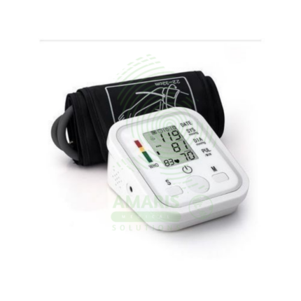
A Digital Blood Pressure Monitor is an automatic electronic device that measures and displays systolic pressure, diastolic pressure, and pulse rate using the oscillometric method. Designed primarily for home and clinical use, it offers a user-friendly alternative to manual aneroid devices, requiring minimal training. Key features often include irregular heartbeat detection, multi-user memory, and connectivity for data tracking. Its accuracy, when clinically validated and used with correct technique, makes it an indispensable tool for the diagnosis and management of hypertension, empowering patients and supporting healthcare providers with reliable ambulatory data.
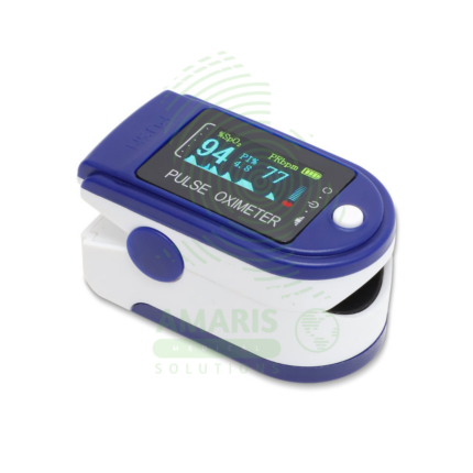
Fingertip Pulse Oximeter
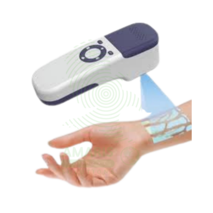
A Fingertip Pulse Oximeter is a compact, non-invasive medical device that clips onto a finger to measure peripheral arterial oxygen saturation (SpO2) and pulse rate in seconds. Utilizing light-based technology, it provides critical, real-time data on respiratory and circulatory status. Its portability and ease of use make it indispensable in hospitals (from ORs to general wards), for patients managing chronic lung conditions at home, and for situational monitoring in sports or high-altitude environments. While an extremely valuable screening tool, readings must be interpreted in context, considering factors like patient movement, perfusion, and clinical symptoms.
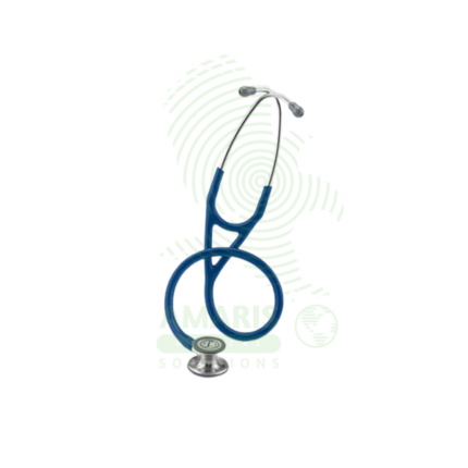
Littman Stethoscope (Classic 4 Cardiology)
A Littman Stethoscope (Classic 4 Cardiology) represents the pinnacle of acoustic auscultation technology. Designed for clinicians who require the highest level of diagnostic accuracy, it features dual tunable diaphragms and dual-lumen tubing to provide exceptional acoustic sensitivity and ambient noise reduction. Its innovative design allows the user to hear both high and low-frequency sounds by simply adjusting pressure on either side of the chestpiece, making it the instrument of choice for cardiologists, intensivists, and specialists who cannot afford to miss a subtle murmur, rub, or breath sound. It combines superior performance with the durability and comfort expected from the Littmann brand.
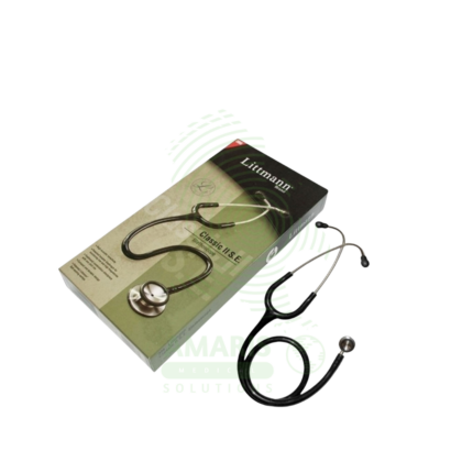
Littmann Stethoscope (Classic II)
A Littmann Stethoscope (Classic II) is a premium, acoustic dual-head stethoscope renowned for its diagnostic clarity, durability, and innovative tunable diaphragm technology. It allows healthcare professionals to hear both high and low-frequency sounds by simply adjusting pressure on the single-sided chestpiece, eliminating the need to rotate it. With its superior acoustics, comfortable fit, and wide range of colors, it is the instrument of choice for medical students, nurses, and physicians across all specialties who require reliable performance for physical assessment, auscultation of heart and lung sounds, and manual blood pressure measurement.
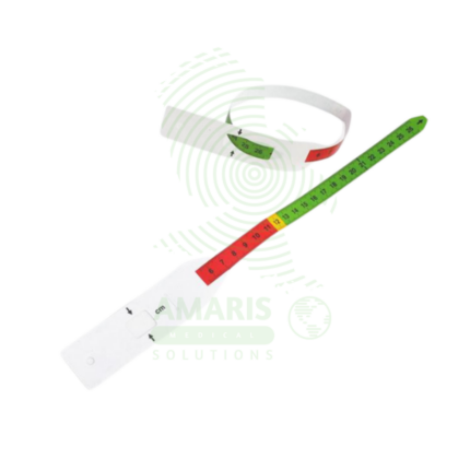
MUAC Tape
A MUAC Tape (Mid-Upper Arm Circumference) is a specialized, color-coded measuring tape used globally as the primary field tool for screening children aged 6-59 months for acute malnutrition. Its simple design—featuring red, yellow, and green zones corresponding to Severe Acute Malnutrition, Moderate Acute Malnutrition, and normal nutritional status—allows for instant, non-invasive assessment without the need for scales, calculation, or literacy. Durable, portable, and inexpensive, it is the cornerstone of community-based nutrition programs, emergency relief efforts, and public health surveillance, enabling early detection and life-saving intervention for malnourished children in even the most resource-constrained settings.
Ophthalmoscope
An Ophthalmoscope is a handheld diagnostic instrument used to examine the interior of the eye, specifically the fundus (retina, optic disc, blood vessels, and macula). By providing illuminated magnification, it serves as a vital screening tool for detecting ocular diseases like glaucoma and retinal disorders, and more importantly, for identifying signs of systemic conditions such as diabetes, hypertension, and increased intracranial pressure. Its operation involves selecting appropriate apertures and compensating lenses to obtain a clear view. While offering a detailed but narrow field of vision, mastery of the direct ophthalmoscope remains a cornerstone skill in general medicine, neurology, and ophthalmology for assessing vascular and neurological health.
Proctoscope
A Proctoscope is a rigid, straight-tube endoscope specifically designed for the examination and treatment of the anal canal and distal rectum. It enables direct visualization to diagnose conditions like hemorrhoids, fissures, and proctitis, and serves as a conduit for therapeutic procedures such as band ligation. Available in reusable (autoclavable metal) or disposable (single-use plastic) formats, it is a fundamental tool in colorectal practice. Its effective use requires proper patient preparation, gentle insertion technique, and stringent adherence to sterilization protocols to ensure patient safety and diagnostic accuracy.