Dermatoscope and Magnifiers
 Diagnostic Kits
 Vital Signs Monitors
 Stethoscopes and Accessories
 Otoscopes, Ophthalmoscopes, and Retinoscopes
 Reflex Hammers and Neurological Tools
 Scales and Measuring Devices
 Spirometers and Pulmonary Function Tests
 Electrosurgical Units and Accessories
 Cutting Instruments
 Grasping and Holding Instruments
 Hemostatic Instruments
 Specialized Surgical Sets
 Single-Use Procedure Trays and Packs
 Surgical Drapes, Gowns, and Covers
 Tissue Unifying Instruments
 Radiation Protection
 X-Ray Machines and Accessories
 Ultrasound Systems and Probes
 MRI and CT Scanners
 Radiology Consumables
 Bone Densitometers
 Fluoroscopy Equipment
 Imaging Tables and Positioning Aids
 Microscopes and Accessories
 Centrifuges and Separators
 Analyzers
 Incubators and Ovens
 Pipettes, Dispensers, and Lab Glassware
 Refrigerators, Freezers, and Storage Units
 Lab Consumables
 Sterilizers and Autoclaves for Lab Use
 Multi-Parameter Monitors
 Ventilators and Respiratory Support Devices
 Defibrillators and AEDs
 Infusion Pumps and IV Systems
 Patient Warmers and Cooling Devices
 Central Monitoring Stations
 Accessories
 Anesthesia Machines and Workstations
 Oxygen Concentrators and Delivery Systems
 Nebulizers and Inhalers
 CPAP/BiPAP Machines
 Airway Management
 Anesthesia Masks, Circuits, and Bags
 Humidifiers and Heaters
 Respiratory Therapy Accessories
 First Aid Kits and Cabinets
 Emergency Resuscitation Equipment
 Trauma Supplies
 Emergency Carts and Crash Carts
 Burn Care Products
 Bleeding Control
 Automated External Defibrillators (AEDs)
 Transport and Evacuation
 Wheelchairs and Accessories
 Walkers, Crutches, and Canes
 Prosthetics and Orthotics
 Physical Therapy Equipment
 Transfer Devices
 Bathroom Safety
 Orthopedic Traction and Tables
 Hot/Cold Therapy Packs and Units
 Beds and Mattresses
 Chairs and Stools
 Tables
 Cabinets and Storage
 Privacy Screens & Curtains
 Stands and Racks
 Linens and Textiles
 Lighting
 Autoclaves and Sterilizers
 Ultrasonic Cleaners
 Disinfectant Solutions and Wipes
 Sterilization Pouches, Wraps, and Indicators
 Instrument Trays and Containers
 UV and Ozone Disinfection Devices
 Washer Disinfectors
 Wound Care
 Gloves
 Masks and Respirators
 Catheters and Tubing
 Swabs, Applicators, and Sponges
 Incontinence Products
 Personal Protective Equipment (PPE)
 Dental Chairs and Units
 Handpieces and Burs
 Instruments
 Consumables
 Sterilization for Dental Use
 Orthodontic Supplies
 Endodontic Tools
 Slit Lamps and Tonometers
 Lensometers and Phoropters
 Ophthalmic Surgical Instruments
 Eyewear Frames and Lenses
 Contact Lens Supplies
 Vision Testing Charts and Devices
 Eye Care Consumables
 Laser Systems for Eye Care
 ENT Exam Chairs and Tables
 Endoscopes
 Audiometers and Hearing Tests
 ENT Instruments
 Nasal and Throat Packs
 Hearing Aids and Accessories
 Otology Supplies
 Fetal Dopplers and Monitors
 Delivery Beds and Tables
 Gynecological Instruments
 Neonatal Incubators and Warmers
 Breast Pumps and Accessories
 Contraceptive Devices
 Maternity Supports and Pads
 Neonatal Consumables
 Cystoscopes and Urethroscopes
 Dialysis Machines and Supplies
 Urological Catheters and Bags
 Lithotripters
 Prostate Treatment Devices
 Urinary Incontinence Products
 Kidney Stone Management Tools
 Consumables & Disposables
 EEG and EMG Machines
 Neurosurgical Instruments
 Nerve Stimulators
 Headrests and Positioning Aids
 Lumbar Puncture Kits
 Seizure Monitoring Devices
 Consumables
 Rehabilitation for Neurological Conditions
 ECG Machines and Accessories
 Holter Monitors
 Stress Test Systems
 Pacemakers and Defibrillator Accessories
 Vascular Access Devices
 Cardiac Catheters and Guidewires
 Blood Flow Meters
 Consumables
 Orthopedic Instruments
 Casts, Splints, and Padding
 Joint Replacement Supplies
 Prosthetic Limbs and Components
 Bone Grafts and Substitutes
 Traction Devices
 Orthopedic Braces and Supports
 Rehabilitation Aids for Orthopedics
 Home Oxygen Therapy
 Hospital Beds for Home Use
 Mobility Aids
 Bathroom and Daily Living Aids
 Wound Care for Home
 Monitoring Devices
 Enteral Feeding Pumps and Tubes
 Hand Sanitizers and Dispensers
 Face Shields and Goggles
 Isolation Gowns and Suits
 Biohazard Waste Containers
 Air Purifiers and HEPA Filters
 Surface Disinfectants
 Sharps Containers
 Protective Barriers
 Cardiovascular & Endurance Training
 Strength Training & Weightlifting
 Functional Training & Core Conditioning
 Physical Therapy & Rehabilitation
 Sports & Outdoor Recreation
 Gym Flooring & Facility Equipment
 Fitness Monitoring & Accessories
 Kids & Novelties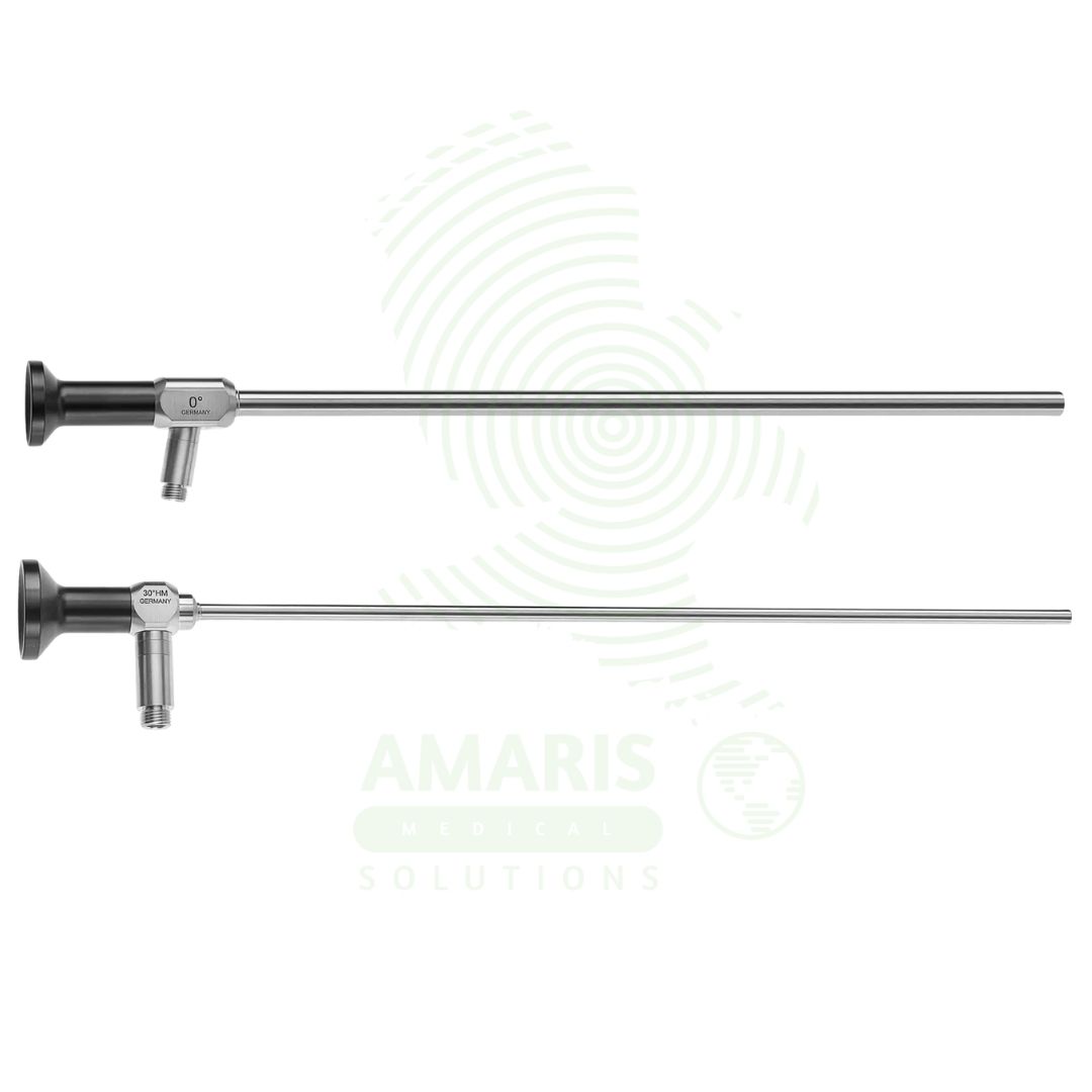
Laparoscope
$1.00
WhatsApp Order
A Laparoscope is a rigid, sterile optical telescope used in minimally invasive surgery to visualize the abdominal and pelvic cavities. Featuring a Hopkins rod-lens system for brilliant, high-resolution images, it comes in standard diameters (5mm, 10mm) and viewing angles (0° for straight-ahead or 30°/45° to look around structures). It connects to a high-intensity light source and a video camera system. Meticulous handling, mandatory pre-sterilization leak testing, and careful cleaning are required to maintain its integrity and prevent infection. As the primary visual conduit for the surgeon, its clarity and reliability are fundamental to the safety and success of any laparoscopic procedure.
Description
Laparoscope
PRIMARY CLINICAL & DIAGNOSTIC USES
1. Visualization of the Peritoneal Cavity
-
Primary Use: Provides direct optical visualization of the abdominal and pelvic organs (liver, gallbladder, stomach, intestines, spleen, uterus, ovaries, etc.) during minimally invasive laparoscopic surgery.
-
How it helps: For the surgeon, the laparoscope transforms the abdominal cavity from a dark, hidden space into a brilliantly illuminated, magnified visual field—revealing anatomy in high definition through tiny incisions. For the patient, this visualization means their surgeon can diagnose and treat conditions without the large incisions that once defined abdominal surgery, accessing the internal world through ports no larger than a fingertip.
2. Diagnostic Laparoscopy
-
Primary Use: Investigates the cause of abdominal pain, pelvic pain, infertility, trauma, and for staging cancers without a large open incision.
-
How it helps: For the surgeon facing a diagnostic dilemma—chronic pain unexplained by imaging, suspected internal injury after trauma, or the need to determine cancer spread—the laparoscope provides definitive answers through direct visualization. For the patient who has suffered months of unexplained pain, or the cancer patient whose treatment plan depends on accurate staging, diagnostic laparoscopy offers clarity that guides everything that follows, often providing answers in a single procedure.
3. Therapeutic/Surgical Laparoscopy
-
Primary Use: Enables the surgeon to perform a wide range of procedures through small keyhole incisions, including cholecystectomy, appendectomy, hernia repair, hysterectomy, and colectomy, by serving as the “eyes” of the surgical team.
-
How it helps: For the minimally invasive surgeon, the laparoscope makes possible operations that were once unthinkable through tiny incisions—removing a diseased gallbladder, repairing a hernia, or resecting a colon while watching a high-definition screen. For the patient facing surgery, laparoscopy means their procedure is performed through keyhole incisions rather than a large cut, translating to less post-operative pain, shorter hospital stays, faster return to work, and smaller scars.
SECONDARY & SUPPORTIVE USES
1. Biopsy Guidance: For the surgeon needing tissue samples from abdominal organs, the laparoscope provides precise visualization for targeting biopsies—ensuring that samples come from the most suspicious areas and that bleeding from the biopsy site is controlled. For the patient requiring tissue diagnosis, laparoscopic-guided biopsy offers a minimally invasive way to obtain definitive pathological answers without open surgery.
2. Documentation: Images and video from the laparoscope are recorded for medical records, surgical planning, and patient education—creating permanent documentation of findings and interventions. For the patient, seeing the inside of their own abdomen on a screen can demystify their condition, and recorded images can be shared with other providers for collaborative care decisions.
3. Training and Simulation: In surgical training programs, laparoscopes are essential for teaching the next generation of minimally invasive surgeons, allowing trainees to learn anatomy and technique in simulated and supervised settings. For the countless future patients who will be treated by surgeons trained on these systems, every procedure observed and every skill practiced contributes to the expertise they will bring to the operating room.
KEY PRODUCT FEATURES
1. BASIC IDENTIFICATION ATTRIBUTES
-
Device Type: A rigid optical instrument (telescope) inserted into a body cavity to transmit an image to the surgeon's eye or a video camera.
-
Designation: Defined by its application (laparoscopy) and its optical design.
-
Core Optical Designs:
-
Hopkins Rod-Lens System: The modern standard. Uses precisely aligned glass rods for superior image clarity, brightness, and wide field of view compared to older lens systems.
-
Fiber Optic Image Bundles: Used in flexible endoscopes but less common in modern rigid laparoscopes.
-
-
Key Specifications:
-
Diameter: Common sizes are 5mm and 10mm.
-
Length: Standard lengths range from 25cm to 33cm.
-
Angle of View: The direction the lens looks relative to the long axis of the scope.
-
0° (Forward Viewing): Looks straight ahead. Most common for general use.
-
30° or 45° (Angled): The viewing lens is angled at the tip. Allows the surgeon to look "around corners" and improves visualization of structures behind organs, providing more flexibility and reducing instrument clash.
-
-
2. TECHNICAL & PERFORMANCE PROPERTIES
-
Optical Quality: Determined by lens clarity, light transmission efficiency, resolution, and lack of distortion. High-quality optics are essential for safe dissection.
-
Field of View: The angular extent of the observable scene, typically 60-80 degrees.
-
Depth of Field: The range of distances within which objects appear in sharp focus.
-
Light Guide Post: Connects to a fiber-optic light cable from a high-intensity light source to illuminate the operative field.
3. PHYSICAL & OPERATIONAL PROPERTIES
-
Construction: Made of medical-grade stainless steel for durability and to withstand repeated sterilization. Contains fragile internal glass optics.
-
Compatibility: Attaches to a camera head (for video laparoscopy) or directly to an eyepiece (for direct viewing).
-
Trocar Compatibility: Designed to pass through a specific diameter surgical trocar/cannula (e.g., a 5mm scope through a 5mm port).
4. SAFETY & COMPLIANCE ATTRIBUTES
-
Regulatory Status: Classified as a Class I medical device.
-
Biocompatibility & Sterility: Must be fully sterilizable (via autoclave, chemical sterilant, or ethylene oxide) and made of biocompatible materials for internal use.
-
Optical Integrity: Lenses must be free of cracks, clouding, or delamination that could distort vision.
5. STORAGE & HANDLING ATTRIBUTES
-
Storage: Store in a protected container or on a dedicated rack to prevent damage to the lenses and tip. Often stored in sterilization trays.
-
Cleaning & Sterilization (CRITICAL):
-
Immediate Post-Use: Clean the external surface meticulously to remove blood and tissue.
-
Leak Testing: Must be performed before each sterilization cycle to check for breaches in the outer sheath, which could allow fluid ingress and cause cross-infection.
-
Sterilization: Autoclave (steam sterilization) is the gold standard for heat-tolerant, all-metal scopes. Follow the manufacturer's precise instructions for time, temperature, and drying.
-
-
Handling: Always handle with care. Do not drop or subject to impact. Hold by the sturdy body, not the eyepiece or tip.
6. LABORATORY & CLINICAL APPLICATIONS
-
Primary Application: The fundamental visual tool for all minimally invasive abdominal and pelvic (gynecological) surgeries.
-
Clinical Role: The device that makes laparoscopic surgery possible, translating the surgeon's vision into the video monitor and defining the modern standard of care for countless procedures.
SAFETY HANDLING PRECAUTIONS
1. SAFETY PRECAUTIONS
-
Thermal Injury: The distal tip of the laparoscope can become extremely hot from the light source or from proximity to electrosurgical instruments, risking internal burns. Use caution when moving it near tissues.
-
Trocar Removal: Always remove the laparoscope through its trocar under direct visualization to ensure no bowel or omentum is drawn into the port site, which could cause injury or port site hernia.
-
Image Quality: A foggy or blurry image is a major safety hazard. Use a scope warmer or anti-fog solution to prevent lens fogging upon entry into the warm peritoneal cavity.
-
Optical Damage: Never use sharp instruments or abrasive materials to clean the lens. Use only recommended soft cloths and cleaning solutions.
-
Integrity Check: Inspect the scope before each use for any damage to the tip, shaft, or lenses. A damaged scope must be removed from service immediately.
2. FIRST AID MEASURES
-
Internal Thermal Burn: If a burn is suspected from scope contact, assess the area, irrigate with cool saline if applicable, and manage the injury surgically if required.
-
Scope Breakage: If the scope tip breaks inside the patient, do not remove the trocar. Keep the fragment in view and use a grasper through another port to retrieve all pieces meticulously.
-
Loss of Visualization: If the image is lost during a critical step, the surgeon should hold position, communicate clearly, and have an assistant troubleshoot (check connections, clean lens) or replace the scope while minimizing movement.
3. FIRE FIGHTING MEASURES
-
Flammability: The scope itself is non-combustible metal and glass, but attached plastic components (cables, grips) may be.
-
Extinguishing Media: Use appropriate extinguishing media (CO2, water) for the primary fire source in the OR.
Related products
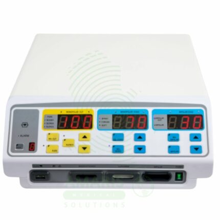
Electrosurgical Unit
An Electrosurgical Unit (ESU) is a radiofrequency generator that provides electrical energy for surgical cutting and coagulation. The ESU is a professional-grade system featuring monopolar and bipolar outputs, with safety-critical Return Electrode Monitoring (REM) for monopolar procedures. It is the universal surgical tool for precise tissue dissection with simultaneous hemostasis, used in open, laparoscopic, and endoscopic procedures across all specialties. Safe operation hinges on correct patient return pad placement, awareness of fire risks (especially with oxygen and flammable prep solutions), and adherence to protocols for patients with implanted electronic devices.
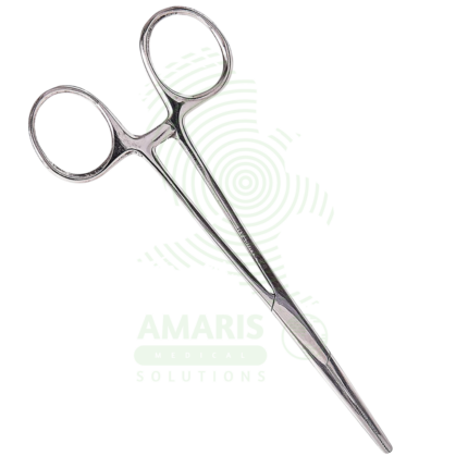
Forceps
Forceps are handheld surgical instruments used for grasping, holding, manipulating, or extracting tissues and objects during medical and surgical procedures. Available in numerous configurations including thumb forceps for delicate tissue handling, hemostatic forceps for clamping blood vessels, and specialized forceps for specific applications. Made from surgical-grade stainless steel, they are designed for repeated sterilization and use. Proper selection, handling, and maintenance are essential for safe and effective use.
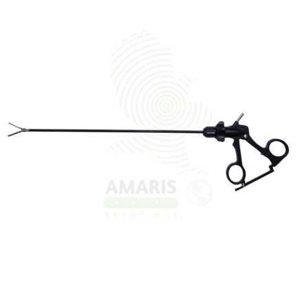
Laparoscopic Instrument
Laparoscopic Instruments are specialized surgical tools designed for use through small incisions during minimally invasive abdominal and pelvic procedures. Available in grasping, cutting, dissecting, hemostatic, and suturing configurations, they enable surgeons to perform complex operations with precision while minimizing patient trauma. With shaft diameters of 3-12 mm and lengths optimized for abdominal access, these instruments allow for the full range of surgical techniques through small incisions, resulting in reduced post-operative pain, shorter hospital stays, and faster recovery.
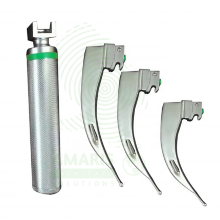
Macintosh Fiber Optic Laryngoscope
A Macintosh Fiber Optic Laryngoscope is a rigid laryngoscope with curved Macintosh blade (sizes 0-4, 70-160 mm) incorporating an integrated fiber optic light bundle that transmits bright, focused illumination (2,000-10,000+ Lux) from a handle-mounted LED or xenon bulb to the blade tip for enhanced visualization during tracheal intubation. The curved blade design allows indirect epiglottis elevation by placing the tip in the vallecula, requiring less force and neck extension than straight blades. Features stainless steel reusable blades, ergonomic handles with knurled grip, ISO standard hook-on fittings, and steam autoclave compatibility. Primary clinical applications include routine and emergency tracheal intubation during general anesthesia, difficult airway management with improved illumination, cervical spine precautions (minimal neck movement), teaching and training (enhanced visualization for instructors), rapid sequence intubation, neonatal and pediatric intubation (sizes 0-2), and oral/maxillofacial surgery. Class II medical device requiring FDA clearance. Critical safety considerations include pre-use light check (dark spots indicate broken fibers), appropriate blade size selection, proper lifting technique (not levering on teeth), battery verification, fiber optic care (avoid sharp bending), and backup device availability.
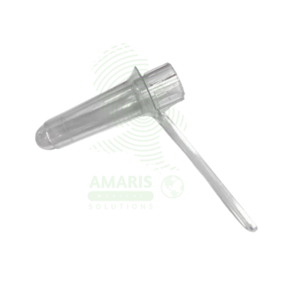
Proctoscope
A Proctoscope is a rigid, straight-tube endoscope specifically designed for the examination and treatment of the anal canal and distal rectum. It enables direct visualization to diagnose conditions like hemorrhoids, fissures, and proctitis, and serves as a conduit for therapeutic procedures such as band ligation. Available in reusable (autoclavable metal) or disposable (single-use plastic) formats, it is a fundamental tool in colorectal practice. Its effective use requires proper patient preparation, gentle insertion technique, and stringent adherence to sterilization protocols to ensure patient safety and diagnostic accuracy.
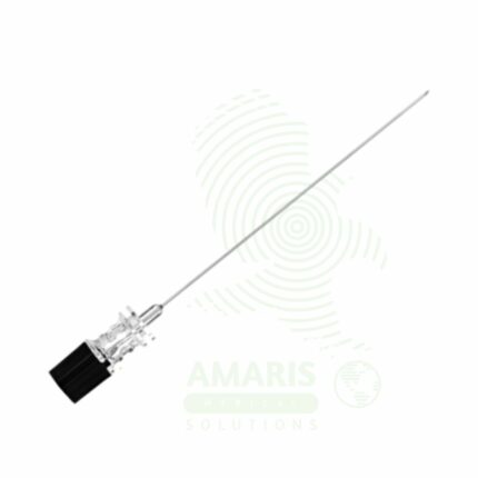
Quincke Spinal Needle
A Quincke Spinal Needle is a sterile, single-use, cutting-bevel needle designed for accessing the subarachnoid space for spinal anesthesia, diagnostic lumbar puncture, and intrathecal medication administration. Featuring a sharp cutting tip, removable stylet, and transparent hub for CSF flashback visualization, it allows precise dural penetration and reliable CSF return. Available in various gauges and lengths, it is the traditional cutting needle in neuraxial anesthesia. Strict aseptic technique, proper bevel orientation, and careful patient selection are essential to minimize complications including post-dural puncture headache and nerve injury.
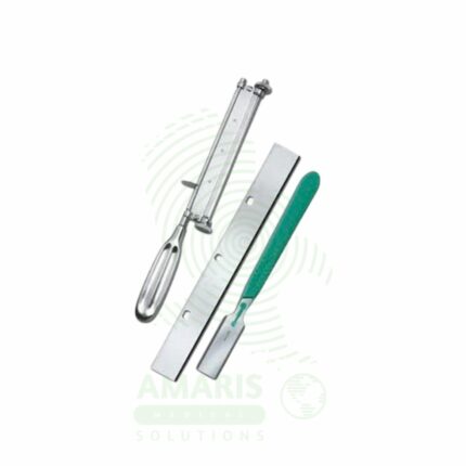
Skin Grafting Blade & Knife
A Skin Grafting Blade & Knife is a specialized, single-use surgical blade designed for use in a mechanical dermatome to harvest split-thickness skin grafts. Its exceptional sharpness is critical for obtaining a uniform sheet of skin of a predetermined thickness from a donor site. Correct use requires secure loading into the compatible dermatome, proper lubrication and preparation of the donor site, and maintenance of a consistent angle and pressure during harvest. It is a fundamental consumable in reconstructive and burn surgery, enabling the coverage of large wounds with the patient's own skin. Safety hinges on proper assembly, sharps handling, and meticulous technique to avoid donor site injury and ensure a viable graft.