Dermatoscope and Magnifiers
 Diagnostic Kits
 Vital Signs Monitors
 Stethoscopes and Accessories
 Otoscopes, Ophthalmoscopes, and Retinoscopes
 Reflex Hammers and Neurological Tools
 Scales and Measuring Devices
 Spirometers and Pulmonary Function Tests
 Electrosurgical Units and Accessories
 Cutting Instruments
 Grasping and Holding Instruments
 Hemostatic Instruments
 Specialized Surgical Sets
 Single-Use Procedure Trays and Packs
 Surgical Drapes, Gowns, and Covers
 Tissue Unifying Instruments
 Radiation Protection
 X-Ray Machines and Accessories
 Ultrasound Systems and Probes
 MRI and CT Scanners
 Radiology Consumables
 Bone Densitometers
 Fluoroscopy Equipment
 Imaging Tables and Positioning Aids
 Microscopes and Accessories
 Centrifuges and Separators
 Analyzers
 Incubators and Ovens
 Pipettes, Dispensers, and Lab Glassware
 Refrigerators, Freezers, and Storage Units
 Lab Consumables
 Sterilizers and Autoclaves for Lab Use
 Multi-Parameter Monitors
 Ventilators and Respiratory Support Devices
 Defibrillators and AEDs
 Infusion Pumps and IV Systems
 Patient Warmers and Cooling Devices
 Central Monitoring Stations
 Accessories
 Anesthesia Machines and Workstations
 Oxygen Concentrators and Delivery Systems
 Nebulizers and Inhalers
 CPAP/BiPAP Machines
 Airway Management
 Anesthesia Masks, Circuits, and Bags
 Humidifiers and Heaters
 Respiratory Therapy Accessories
 First Aid Kits and Cabinets
 Emergency Resuscitation Equipment
 Trauma Supplies
 Emergency Carts and Crash Carts
 Burn Care Products
 Bleeding Control
 Automated External Defibrillators (AEDs)
 Transport and Evacuation
 Wheelchairs and Accessories
 Walkers, Crutches, and Canes
 Prosthetics and Orthotics
 Physical Therapy Equipment
 Transfer Devices
 Bathroom Safety
 Orthopedic Traction and Tables
 Hot/Cold Therapy Packs and Units
 Beds and Mattresses
 Chairs and Stools
 Tables
 Cabinets and Storage
 Privacy Screens & Curtains
 Stands and Racks
 Linens and Textiles
 Lighting
 Autoclaves and Sterilizers
 Ultrasonic Cleaners
 Disinfectant Solutions and Wipes
 Sterilization Pouches, Wraps, and Indicators
 Instrument Trays and Containers
 UV and Ozone Disinfection Devices
 Washer Disinfectors
 Wound Care
 Gloves
 Masks and Respirators
 Catheters and Tubing
 Swabs, Applicators, and Sponges
 Incontinence Products
 Personal Protective Equipment (PPE)
 Dental Chairs and Units
 Handpieces and Burs
 Instruments
 Consumables
 Sterilization for Dental Use
 Orthodontic Supplies
 Endodontic Tools
 Slit Lamps and Tonometers
 Lensometers and Phoropters
 Ophthalmic Surgical Instruments
 Eyewear Frames and Lenses
 Contact Lens Supplies
 Vision Testing Charts and Devices
 Eye Care Consumables
 Laser Systems for Eye Care
 ENT Exam Chairs and Tables
 Endoscopes
 Audiometers and Hearing Tests
 ENT Instruments
 Nasal and Throat Packs
 Hearing Aids and Accessories
 Otology Supplies
 Fetal Dopplers and Monitors
 Delivery Beds and Tables
 Gynecological Instruments
 Neonatal Incubators and Warmers
 Breast Pumps and Accessories
 Contraceptive Devices
 Maternity Supports and Pads
 Neonatal Consumables
 Cystoscopes and Urethroscopes
 Dialysis Machines and Supplies
 Urological Catheters and Bags
 Lithotripters
 Prostate Treatment Devices
 Urinary Incontinence Products
 Kidney Stone Management Tools
 Consumables & Disposables
 EEG and EMG Machines
 Neurosurgical Instruments
 Nerve Stimulators
 Headrests and Positioning Aids
 Lumbar Puncture Kits
 Seizure Monitoring Devices
 Consumables
 Rehabilitation for Neurological Conditions
 ECG Machines and Accessories
 Holter Monitors
 Stress Test Systems
 Pacemakers and Defibrillator Accessories
 Vascular Access Devices
 Cardiac Catheters and Guidewires
 Blood Flow Meters
 Consumables
 Orthopedic Instruments
 Casts, Splints, and Padding
 Joint Replacement Supplies
 Prosthetic Limbs and Components
 Bone Grafts and Substitutes
 Traction Devices
 Orthopedic Braces and Supports
 Rehabilitation Aids for Orthopedics
 Home Oxygen Therapy
 Hospital Beds for Home Use
 Mobility Aids
 Bathroom and Daily Living Aids
 Wound Care for Home
 Monitoring Devices
 Enteral Feeding Pumps and Tubes
 Hand Sanitizers and Dispensers
 Face Shields and Goggles
 Isolation Gowns and Suits
 Biohazard Waste Containers
 Air Purifiers and HEPA Filters
 Surface Disinfectants
 Sharps Containers
 Protective Barriers
 Cardiovascular & Endurance Training
 Strength Training & Weightlifting
 Functional Training & Core Conditioning
 Physical Therapy & Rehabilitation
 Sports & Outdoor Recreation
 Gym Flooring & Facility Equipment
 Fitness Monitoring & Accessories
 Kids & Novelties
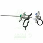
Orthopedic Arthroscope
WhatsApp Order
An Orthopedic Arthroscope is a rigid, rod-lens telescope used for the visualization and surgical treatment of joint interiors. As the cornerstone of minimally invasive joint surgery, it provides a bright, magnified view of structures like cartilage, menisci, and ligaments through tiny incisions. Available in key diameters (2.7mm, 4.0mm) and viewing angles (0°, 30°, 70°), its optical quality is paramount for surgical precision. Mandatory pre-sterilization leak testing and careful autoclaving are required to maintain sterility and integrity. Proper handling is essential to prevent costly damage to the optics and to ensure patient safety during diagnostic and therapeutic arthroscopic procedures.
Description
Orthopedic Arthroscope
PRIMARY CLINICAL & DIAGNOSTIC USES
1. Diagnostic Arthroscopy
-
Primary Use: Provides direct, magnified visualization of the interior of a joint (synovium, articular cartilage, menisci, ligaments, tendons) to diagnose the source of pain, swelling, locking, or instability when non-invasive imaging is inconclusive.
-
How it helps: For the orthopedic surgeon, the arthroscope transforms joint diagnosis from indirect imaging to direct visualization—revealing the exact condition of cartilage, the precise location of a meniscal tear, the true state of a ligament, or the source of unexplained synovitis. For the patient suffering from chronic knee pain, shoulder instability, or hip impingement, diagnostic arthroscopy often provides the definitive answer that MRI and X-ray could not, guiding treatment with certainty.
2. Therapeutic Joint Surgery (Arthroscopic Surgery)
-
Primary Use: Enables minimally invasive surgical interventions within the joint, performed through small “keyhole” portals, across multiple joints including knee, shoulder, hip, ankle, elbow, and wrist.
-
How it helps: For the orthopedic surgeon, the arthroscope is both eyes and instrument—allowing them to repair torn menisci, reconstruct cruciate ligaments, debride damaged cartilage, and stabilize shoulders through incisions measured in millimeters rather than inches. For the athlete sidelined by an ACL tear, the worker unable to lift due to rotator cuff injury, or the aging adult limited by meniscal pathology, arthroscopic surgery means their joint can be repaired through minimally invasive techniques, with less pain, faster rehabilitation, and earlier return to the activities they love.
3. Knee Arthroscopy
-
Primary Use: Performs meniscectomy, meniscal repair, ACL/PCL reconstruction, cartilage debridement, microfracture, loose body removal, and synovectomy through small portals.
-
How it helps: For the knee surgeon, the arthroscope provides access to all compartments of this complex joint—visualizing the torn meniscus catching and locking, the absent ACL causing instability, or the cartilage defect causing pain. For the patient with a locked knee from a bucket-handle meniscal tear, or the athlete whose knee gives way from ACL insufficiency, arthroscopic repair offers the chance to restore function and return to activity without the morbidity of open arthrotomy.
4. Shoulder Arthroscopy
-
Primary Use: Performs rotator cuff repair, subacromial decompression, labral repair, biceps tenodesis, and stabilization for recurrent dislocation.
-
How it helps: For the shoulder surgeon, the arthroscope navigates the complex anatomy of the glenohumeral joint and subacromial space—repairing torn rotator cuff tendons, debriding impinging bone spurs, and stabilizing recurrently dislocating shoulders. For the patient unable to lift their arm due to rotator cuff tear, or the young athlete with recurrent shoulder instability, arthroscopic repair offers the hope of restored function and stability without the large incisions and extensive soft tissue dissection of open surgery.
SECONDARY & SUPPORTIVE USES
1. Biopsy Guidance: For the orthopedic surgeon evaluating unexplained synovitis or suspected intra-articular pathology, the arthroscope allows precise, targeted biopsy of synovial tissue for histopathological diagnosis. For the patient with pigmented villonodular synovitis, inflammatory arthritis, or suspected infection, arthroscopic biopsy provides diagnostic tissue with minimal morbidity.
2. Irrigation and Debridement: In septic arthritis, the arthroscope enables thorough joint lavage to remove purulent material and debride fibrinous exudate, potentially saving the joint from rapid destruction. For the patient with an infected joint, arthroscopic washout offers a minimally invasive way to clear infection while preserving joint function.
3. Assessment and Treatment of Fractures: Arthroscopic assistance aids in the assessment and reduction of intra-articular fractures, ensuring that joint surfaces are anatomically aligned. For the patient with a tibial plateau, radial head, or other intra-articular fracture, arthroscopic visualization helps the surgeon restore the smooth joint surface essential for long-term function and prevention of post-traumatic arthritis.
4. Post-Operative Assessment and Lysis of Adhesions: Second-look arthroscopy allows assessment of healing after meniscal repair or cartilage procedures, and arthroscopic release of adhesions can treat arthrofibrosis causing joint stiffness. For the patient with persistent stiffness after surgery or injury, arthroscopic lysis of adhesions can restore motion without the trauma of open release.
5. Joint Stabilization Procedures: For patients with multidirectional instability or recurrent dislocation, arthroscopic visualization enables capsular plication or thermal capsulorrhaphy to tighten lax tissues. For the individual whose shoulder dislocates with simple daily activities, arthroscopic stabilization offers the chance for secure, stable function without the morbidity of open procedures.
6. Tendon and Cyst Evaluation: Arthroscopic techniques extend beyond joints to procedures like endoscopic carpal tunnel release and ganglion cyst excision. For the patient with carpal tunnel syndrome, endoscopic release offers faster recovery and less scar tenderness than open release, while ganglion excision through small portals minimizes cosmetic and functional impact.
KEY PRODUCT FEATURES
1. BASIC IDENTIFICATION ATTRIBUTES
-
Device Type: A rigid, rod-lens optical telescope specifically designed for insertion into joints through a small cannula.
-
Designation: Defined by its application (arthroscopy) and its optical specifications.
-
Core Specifications:
-
Diameter: Common sizes are 2.7mm (small joints: wrist, ankle, pediatric), 4.0mm (standard for knee, shoulder), and 5.0mm (larger joints or for specific optics).
-
Length: Varies by joint; typically 10-18cm.
-
Angle of View: The direction the lens looks relative to the long axis of the scope. Critical for accessing different areas of a joint.
-
0° (Forward Viewing): Provides a straightforward, panoramic view. Excellent for orientation and general visualization.
-
30° (Angled): The most commonly used. The lens is angled at 30°, allowing the surgeon to look "around corners" by rotating the scope, greatly increasing the field of view without moving the portal.
-
70° (Wide-Angle): Used for specialized visualization, such as looking into the posterior knee compartment or the acetabulum in hip arthroscopy.
-
-
2. TECHNICAL & PERFORMANCE PROPERTIES
-
Optical System: Utilizes a Hopkins rod-lens system for superior light transmission, image clarity, brightness, and wide field of view compared to traditional lens systems.
-
Field of View: The angular extent of the observable scene (e.g., 60°-90°).
-
Depth of Field: The range of distances that remain in sharp focus, allowing clear visualization from the joint capsule to the far side of the joint.
-
Light Guide Post: Connects to a high-intensity fiber-optic light cable from a xenon or LED light source to illuminate the dark joint cavity.
3. PHYSICAL & OPERATIONAL PROPERTIES
-
Construction: Made of high-grade, medical stainless steel for durability, rigidity, and to withstand repeated sterilization.
-
Integration: Attaches to a medical video camera head for display on a monitor (video arthroscopy). Older systems may have a direct eyepiece.
-
Trocar/Cannula Compatibility: Designed to pass through a specific diameter arthroscopic cannula or sheath, which maintains the portal and allows for inflow/outflow.
4. SAFETY & COMPLIANCE ATTRIBUTES
-
Regulatory Status: Classified as a Class I medical device.
-
Biocompatibility & Sterility: Must be fully sterilizable (via autoclave) and made of biocompatible materials for internal use within a sterile surgical field.
-
Optical Integrity: Lenses must be free of cracks, chips, clouding, or delamination. Even minor defects can scatter light and severely distort the image, posing a surgical risk.
5. STORAGE & HANDLING ATTRIBUTES
-
Storage: Store in a protective container or dedicated rack within a sterilization tray to prevent damage to the delicate tip and lenses. Avoid contact with other metal instruments.
-
Cleaning & Sterilization (CRITICAL):
-
Immediate Post-Use: Clean the external surface meticulously to remove biological debris.
-
Leak Testing: Must be performed before every sterilization cycle to check for breaches in the sealed outer sheath. Fluid ingress can cause cross-infection and destroy the optics.
-
Sterilization: Autoclave (steam sterilization) is the standard. Follow manufacturer's precise instructions for time, temperature, and drying cycles to prevent damage.
-
-
Handling: Always handle with extreme care. Never drop. Hold by the robust body, never by the eyepiece, camera adapter, or the distal tip. Use protective tips when not in use.
6. LABORATORY & CLINICAL APPLICATIONS
-
Primary Application: The essential visual instrument for all arthroscopic (minimally invasive joint) surgeries across orthopedic subspecialties (knee, shoulder, hip, sports medicine).
-
Clinical Role: The "eyes" of the arthroscopic surgeon, enabling precise diagnosis and complex intra-articular surgery through portals less than 1cm in size, reducing tissue trauma, pain, and recovery time compared to open surgery.
SAFETY HANDLING PRECAUTIONS
1. SAFETY PRECAUTIONS
-
Thermal Injury: The metal shaft of the scope can conduct heat from the light cable or from proximity to electrosurgical/radiofrequency probes, potentially causing cartilage or capsular burns. Awareness of scope position is key.
-
Cartilage Scuffing (Iatrogenic Damage): The sharp, metallic tip can scratch articular cartilage if inserted or moved carelessly. Always insert the scope through a cannula and move it gently within the joint.
-
Scope Breakage: Although rare, the scope can break under extreme torque or if struck. Do not use excessive force when navigating tight joints.
-
Image Artifacts: Fogging, bleeding, or debris on the lens can obscure vision. Use scope warmers, adequate irrigation pressure, and lens cleaners to maintain a clear view. A blurry image is a safety hazard.
-
Portal Placement: Incorrect portal placement can lead to poor visualization, instrument clash, and increased risk of neurovascular injury. Proper preoperative planning and anatomical knowledge are essential.
2. FIRST AID MEASURES
-
Broken Scope Tip in Joint: If the distal tip breaks, do not remove the cannula. Keep the fragment in view on the monitor. Use a grasper through a separate portal to retrieve all pieces meticulously. Confirm retrieval with fluoroscopy if necessary.
-
Significant Cartilage Damage: If iatrogenic cartilage scuffing occurs, debride the unstable edges and document the incident. The long-term impact is assessed post-operatively.
-
Loss of Visualization During Critical Step: If the image is lost (e.g., due to fogging or massive bleeding), the surgeon should announce "hold," keep instruments stationary, and have the assistant troubleshoot (clean lens, adjust inflow) before proceeding.
3. FIRE FIGHTING MEASURES
-
Flammability: The scope itself is non-combustible metal and glass. Attached light cables may have combustible components.
-
Extinguishing Media: Use appropriate extinguishing media (CO2 for electrical) for any fire source in the OR.
Related products
Arm Sling With Swathe
An Arm Sling With Swathe is a two-piece shoulder immobilization system used for complete immobilization following shoulder surgery, shoulder dislocation, proximal humerus fractures, and clavicle fractures. The sling supports the arm while the swathe wraps around the body to secure the arm against the chest, preventing abduction, external rotation, and other movements that could compromise healing. Used in orthopedic surgery, emergency departments, and post-operative care settings, it provides essential protection during the critical healing phase. Proper positioning, skin integrity monitoring, and circulation checks are essential for safe and effective use.
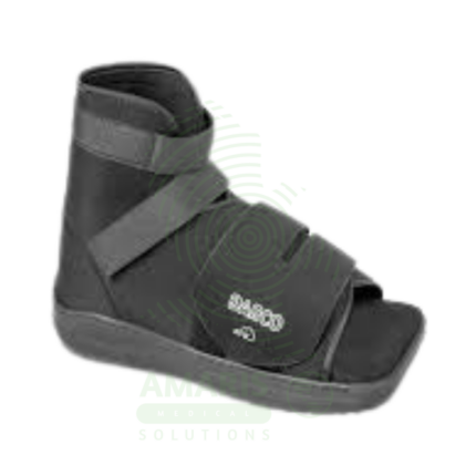
Cast Shoes & Boots
A Cast Shoes & Boots is a durable, rigid-soled overshoe designed to be worn over a below-knee cast or fracture walking boot. Its primary function is to enable safe, protected weight-bearing by providing a stable, rocker-bottom walking surface that protects the cast from wear and tear and compensates for leg length discrepancy. Featuring an adjustable strap closure to accommodate bulky dressings and swelling, it is an essential mobility aid for patients recovering from foot, ankle, or lower leg fractures, surgeries, and severe sprains. Proper fit and gait awareness are important for safe ambulation during the healing period.
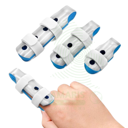
Finger Splints
Finger Splints are orthotic devices designed to immobilize, support, and protect injured or diseased fingers and thumbs. Used for treating fractures, dislocations, tendon injuries (like mallet or boutonnière deformity), joint instability, and arthritis, they promote proper healing and alignment by restricting harmful movement. Available in various forms—including rigid aluminum foam, custom thermoplastic, and soft buddy straps—they are essential tools in emergency care, orthopedics, and hand therapy. Correct application with careful attention to skin integrity and circulation is paramount for safe and effective treatment.
Orthopedic Commode Wheelchair With Flipping Armrest
The Orthopedic Commode Wheelchair with Flipping Armrest is a specialized hybrid device designed for patients with lower limb injuries or surgeries requiring non-weight-bearing status. It combines an extended-frame orthopedic wheelchair with an elevating leg rest, a commode seat opening, and a flipping armrest for lateral transfers. This allows for simultaneous fracture management (leg elevation/immobilization), pressure sore prevention (heel off-loading), and safe, hygienic toileting without dangerous transfers. Its use mandates the constant engagement of anti-tip levers due to a high risk of backwards tipping and requires careful navigation of its substantial length.

Orthopedic Padding
Orthopedic Padding is a soft, cushioning material applied directly to the skin as an essential protective layer underneath casts, splints, and braces. Its primary functions are to prevent skin breakdown by distributing pressure, absorb moisture, and provide comfort during immobilization. Available in rolls of cotton, synthetic, or foam, its correct, wrinkle-free application is a critical safety step in fracture management and orthopedic bracing, directly impacting patient tolerance and preventing complications like pressure ulcers.

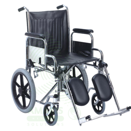
Orthopedic Wheelchair
An Orthopedic Wheelchair is a specialized manual wheelchair with an extended frame and elevating leg rests, designed to support patients who must keep one or both lower limbs in a straight, non-weight-bearing position. It is primarily used for managing lower limb fractures, post-operative recovery (e.g., hip replacements), and conditions requiring heel-off-loading to prevent pressure sores. Its key safety consideration is a heightened risk of tipping backwards due to the forward weight of the extended leg, requiring cautious use, engagement of anti-tip levers, and awareness of its increased length during navigation.

POP Bandages
POP Bandages (Plaster of Paris) are gauze rolls impregnated with calcium sulfate hemihydrate, used to fabricate custom, rigid external casts. When activated in water, they undergo a chemical reaction that generates heat and hardens into a solid structure, providing excellent immobilization for fractures, severe sprains, and post-operative protection. Valued for their superior moldability and radiolucency, they remain a fundamental tool in orthopedics. Their application requires skilled technique with strict attention to thermal safety, padding, and post-application monitoring for circulatory compromise. Proper dry storage is essential to maintain shelf life.