
 Dermatoscope and Magnifiers
 Diagnostic Kits
 Vital Signs Monitors
 Stethoscopes and Accessories
 Otoscopes, Ophthalmoscopes, and Retinoscopes
 Reflex Hammers and Neurological Tools
 Scales and Measuring Devices
 Spirometers and Pulmonary Function Tests
 Electrosurgical Units and Accessories
 Cutting Instruments
 Grasping and Holding Instruments
 Hemostatic Instruments
 Specialized Surgical Sets
 Single-Use Procedure Trays and Packs
 Surgical Drapes, Gowns, and Covers
 Tissue Unifying Instruments
 Radiation Protection
 X-Ray Machines and Accessories
 Ultrasound Systems and Probes
 MRI and CT Scanners
 Radiology Consumables
 Bone Densitometers
 Fluoroscopy Equipment
 Imaging Tables and Positioning Aids
 Microscopes and Accessories
 Centrifuges and Separators
 Analyzers
 Incubators and Ovens
 Pipettes, Dispensers, and Lab Glassware
 Refrigerators, Freezers, and Storage Units
 Lab Consumables
 Sterilizers and Autoclaves for Lab Use
 Multi-Parameter Monitors
 Ventilators and Respiratory Support Devices
 Defibrillators and AEDs
 Infusion Pumps and IV Systems
 Patient Warmers and Cooling Devices
 Central Monitoring Stations
 Accessories
 Anesthesia Machines and Workstations
 Oxygen Concentrators and Delivery Systems
 Nebulizers and Inhalers
 CPAP/BiPAP Machines
 Airway Management
 Anesthesia Masks, Circuits, and Bags
 Humidifiers and Heaters
 Respiratory Therapy Accessories
 First Aid Kits and Cabinets
 Emergency Resuscitation Equipment
 Trauma Supplies
 Emergency Carts and Crash Carts
 Burn Care Products
 Bleeding Control
 Automated External Defibrillators (AEDs)
 Transport and Evacuation
 Wheelchairs and Accessories
 Walkers, Crutches, and Canes
 Prosthetics and Orthotics
 Physical Therapy Equipment
 Transfer Devices
 Bathroom Safety
 Orthopedic Traction and Tables
 Hot/Cold Therapy Packs and Units
 Beds and Mattresses
 Chairs and Stools
 Tables
 Cabinets and Storage
 Privacy Screens & Curtains
 Stands and Racks
 Linens and Textiles
 Lighting
 Autoclaves and Sterilizers
 Ultrasonic Cleaners
 Disinfectant Solutions and Wipes
 Sterilization Pouches, Wraps, and Indicators
 Instrument Trays and Containers
 UV and Ozone Disinfection Devices
 Washer Disinfectors
 Wound Care
 Gloves
 Masks and Respirators
 Catheters and Tubing
 Swabs, Applicators, and Sponges
 Incontinence Products
 Personal Protective Equipment (PPE)
 Dental Chairs and Units
 Handpieces and Burs
 Instruments
 Consumables
 Sterilization for Dental Use
 Orthodontic Supplies
 Endodontic Tools
 Slit Lamps and Tonometers
 Lensometers and Phoropters
 Ophthalmic Surgical Instruments
 Eyewear Frames and Lenses
 Contact Lens Supplies
 Vision Testing Charts and Devices
 Eye Care Consumables
 Laser Systems for Eye Care
 ENT Exam Chairs and Tables
 Endoscopes
 Audiometers and Hearing Tests
 ENT Instruments
 Nasal and Throat Packs
 Hearing Aids and Accessories
 Otology Supplies
 Fetal Dopplers and Monitors
 Delivery Beds and Tables
 Gynecological Instruments
 Neonatal Incubators and Warmers
 Breast Pumps and Accessories
 Contraceptive Devices
 Maternity Supports and Pads
 Neonatal Consumables
 Cystoscopes and Urethroscopes
 Dialysis Machines and Supplies
 Urological Catheters and Bags
 Lithotripters
 Prostate Treatment Devices
 Urinary Incontinence Products
 Kidney Stone Management Tools
 Consumables & Disposables
 EEG and EMG Machines
 Neurosurgical Instruments
 Nerve Stimulators
 Headrests and Positioning Aids
 Lumbar Puncture Kits
 Seizure Monitoring Devices
 Consumables
 Rehabilitation for Neurological Conditions
 ECG Machines and Accessories
 Holter Monitors
 Stress Test Systems
 Pacemakers and Defibrillator Accessories
 Vascular Access Devices
 Cardiac Catheters and Guidewires
 Blood Flow Meters
 Consumables
 Orthopedic Instruments
 Casts, Splints, and Padding
 Joint Replacement Supplies
 Prosthetic Limbs and Components
 Bone Grafts and Substitutes
 Traction Devices
 Orthopedic Braces and Supports
 Rehabilitation Aids for Orthopedics
 Home Oxygen Therapy
 Hospital Beds for Home Use
 Mobility Aids
 Bathroom and Daily Living Aids
 Wound Care for Home
 Monitoring Devices
 Enteral Feeding Pumps and Tubes
 Hand Sanitizers and Dispensers
 Face Shields and Goggles
 Isolation Gowns and Suits
 Biohazard Waste Containers
 Air Purifiers and HEPA Filters
 Surface Disinfectants
 Sharps Containers
 Protective Barriers
 Cardiovascular & Endurance Training
 Strength Training & Weightlifting
 Functional Training & Core Conditioning
 Physical Therapy & Rehabilitation
 Sports & Outdoor Recreation
 Gym Flooring & Facility Equipment
 Fitness Monitoring & Accessories
 Kids & Novelties
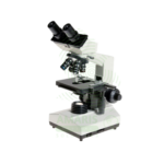
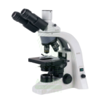
Series Multi-viewing Microscope
WhatsApp Order
A Series Multi-viewing Microscope is a specialized clinical and teaching instrument that allows multiple observers (2-5) to simultaneously view the same specimen through multiple binocular heads connected to a single main objective and stage. Available in double-headed, triple-headed, and quintuple-headed configurations with independent diopter adjustment, interpupillary distance adjustment, and illuminated LED pointers for each viewer. Features plan or plan apochromatic objectives (4×, 10×, 40×, 100× oil), 10× widefield eyepieces, high-intensity halogen or LED illumination, Abbe condenser with Köhler alignment, large mechanical stage, master focus control, and optional trinocular camera port. Magnification range 40× to 1000×. Primary clinical applications include consensus diagnosis in pathology (challenging cases, frozen sections, tumor board preparation), hematology-oncology team reviews (leukemia classification, bone marrow aspirates), cytology quality assurance (Pap smear review, FNA interpretation), microbiology infectious disease rounds (unusual organism identification, parasitology confirmation), and medical education (residency training, medical student teaching, CME workshops). Essential equipment for teaching hospitals, academic medical centers, group pathology practices, and multidisciplinary conferences requiring collaborative microscopic review.
Description
Series Multi-viewing Microscope
PRIMARY CLINICAL & DIAGNOSTIC USES
1. Pathology and Histopathology Consensus Diagnosis:
-
Primary Use: Multi-viewing microscopes enable multiple pathologists to simultaneously examine the same tissue section or cytology specimen, facilitating consensus diagnosis for challenging cases, second opinions, and quality assurance reviews in surgical pathology.
-
How it helps: Brings multiple expert eyes to complex cases, ensuring that difficult diagnoses are confirmed by consensus before treatment decisions are made, reducing the risk of misdiagnosis.
2. Hematology and Oncology Tumor Board Reviews:
-
Primary Use: Essential for tumor board conferences where oncologists, pathologists, surgeons, and radiologists jointly review bone marrow aspirates, peripheral blood smears, and tissue biopsies to develop comprehensive treatment plans for cancer patients.
-
How it helps: Allows the entire cancer care team to see exactly what the pathologist sees, ensuring that everyone understands the diagnosis and agrees on the best treatment approach for each patient.
3. Medical Education and Residency Training:
-
Primary Use: Used in teaching hospitals and medical schools for instructing residents and medical students in histopathology, hematology, microbiology, and cytology, allowing instructors to guide trainees through specimens in real-time.
-
How it helps: Transforms microscope training from a solitary experience into a shared learning opportunity, where instructors can point to exactly what students should see and ensure they understand each finding.
4. Cytology and Pap Smear Screening Quality Assurance:
-
Primary Use: Supervising pathologists can review challenging Pap smears and cytology specimens simultaneously with cytotechnologists, providing immediate feedback and educational guidance during screening.
-
How it helps: Ensures quality and consistency in cancer screening programs by allowing supervisors to review difficult cases in real-time, catching potential abnormalities before they are missed.
5. Microbiology and Infectious Disease Rounds:
-
Primary Use: Enables infectious disease specialists, microbiologists, and infection control teams to jointly review Gram stains, culture morphology, and parasite identification for complex or unusual cases.
-
How it helps: Brings the full expertise of the infectious disease team to bear on puzzling cases, ensuring that unusual organisms are correctly identified and patients receive appropriate treatment.
6. Telepathology and Remote Consultation Preparation:
-
Primary Use: Used to prepare and review specimens before digital scanning for telepathology consultation, ensuring optimal fields are selected for remote expert opinion.
-
How it helps: Ensures that when expert opinions are needed from distant specialists, the most diagnostically relevant areas are captured and shared, improving the quality of remote consultations.
7. Clinical Research and Collaborative Studies:
-
Primary Use: Facilitates collaborative research where multiple investigators need to simultaneously observe and discuss microscopic findings in translational research, drug trials, and biomarker studies.
-
How it helps: Enables research teams to work together effectively, ensuring that all investigators see exactly the same findings and can contribute their expertise to advancing scientific discovery.
SECONDARY & SUPPORTIVE USES
1. Proficiency Testing and External Quality Assurance: Used for group review of proficiency testing slides in laboratory quality assurance programs, ensuring ongoing accuracy and reliability in diagnostic testing.
2. Inter-laboratory Comparison Studies: Enables pathologists from different institutions to jointly review slides for standardization and harmonization studies, promoting consistent diagnostic criteria across laboratories.
3. Continuing Medical Education (CME) Programs: Used in workshop settings for hands-on CME courses in pathology, hematology, and microbiology, helping practicing physicians maintain and enhance their skills.
4. Veterinary Pathology Reviews: Multi-viewing microscopes facilitate case discussions in veterinary pathology and comparative medicine, extending collaborative diagnosis to animal health.
5. Forensic Pathology Case Reviews: Used in medical examiner offices for team review of forensic histology specimens, helping determine causes of death and supporting justice.
6. Pharmaceutical and Toxicology Studies: Enables team review of histopathology slides from drug safety studies and toxicology assessments, ensuring thorough evaluation of potential medication risks.
7. International Health and Global Medicine Programs: Used in training programs for pathologists and laboratory professionals in resource-limited settings, building diagnostic capacity worldwide.
KEY PRODUCT FEATURES
1. BASIC IDENTIFICATION ATTRIBUTES
-
Product Type: Specialized microscope system with multiple binocular viewing heads allowing simultaneous observation of the same specimen by multiple users.
-
Common Names: Multi-viewing Microscope, Teaching Microscope, Conference Microscope, Double-headed Microscope, Triple-headed Microscope, Multi-headed Microscope.
-
Viewing Configurations:
-
Double-headed: Two binocular viewing heads (instructor + student).
-
Triple-headed: Three binocular viewing heads (ideal for small group teaching).
-
Quintuple-headed: Five binocular viewing heads (for larger groups and conferences).
-
Multi-headed with LED pointers: Each viewer has an illuminated pointer to indicate specific features.
-
-
Optical Configuration: Multiple binocular observation tubes connected through a single main objective and specimen stage.
-
Magnification Range: 40× to 1000× (standard clinical range).
-
Objective Lenses: 4×, 10×, 40×, 100× (oil) plan or plan apochromatic objectives.
-
Eyepieces: 10× widefield with independent diopter adjustment for each viewing head.
-
Illumination: High-intensity halogen or LED transmitted light source; some models include fluorescence capability.
-
Condenser: Abbe condenser with iris diaphragm for Köhler illumination.
-
Stage: Large mechanical stage with low-position coaxial controls; ergonomic design for prolonged use.
-
Focusing: Master focus with individual diopter adjustments on each viewing head.
-
Pointer System: Illuminated LED pointers with intensity control for each viewing station.
-
Camera Port: Trinocular head option for photomicrography and digital imaging.
2. TECHNICAL & PERFORMANCE PROPERTIES
-
Optical System: Infinity-corrected plan optics ensuring consistent image quality across all viewing heads.
-
Light Distribution: Precision beam-splitting prisms ensure equal light intensity to all viewing heads.
-
Parfocality: All viewing heads maintain parfocality with master focus.
-
Image Orientation: Erect, non-reversed image at all viewing stations.
-
Interpupillary Distance: Adjustable on each binocular head (typically 48-75 mm).
-
Diopter Adjustment: Independent on each eyepiece for all viewing heads.
-
Field of View: 18-22 mm field number with 10× eyepieces.
-
Pointer System: LED pointers with adjustable brightness; centerable in field of view.
-
Focus Lock: Master focus lock to prevent accidental focus changes during multi-user observation.
-
Stage Movement: Ergonomically positioned coaxial controls for smooth X-Y movement.
-
Nosepiece: Revolving quintuple nosepiece for objective changes.
-
Köhler Illumination: Adjustable for uniform, glare-free illumination across all viewing heads.
3. PHYSICAL & OPERATIONAL PROPERTIES
-
Dimensions: Base microscope 25-30 cm W × 50-60 cm D × 45-55 cm H; additional width for multiple viewing heads.
-
Weight: 15-30 kg depending on number of viewing heads and configuration.
-
Construction: Heavy-duty cast metal base and stand for vibration-free operation.
-
Viewing Heads: Binocular tubes with adjustable inclination for comfortable viewing by multiple users.
-
Head Rotation: Some models allow viewing heads to rotate for flexible positioning around the table.
-
Stage: Large, low-position mechanical stage with ergonomic coaxial controls.
-
Focus Mechanism: Coaxial coarse and fine focus with tension adjustment; master focus control.
-
Illumination: High-intensity halogen (100W) or LED (long-life) with intensity control.
-
Condenser: Focusable Abbe condenser with centering adjustment and filter holder.
-
Pointer Controls: Individual intensity and centering controls for each pointer.
-
Camera Port: Optional trinocular tube for digital camera attachment.
-
Ergonomics: Designed for extended viewing sessions with multiple users.
-
Dust Cover: Protective cover included for storage.
4. SAFETY & COMPLIANCE ATTRIBUTES
-
Regulatory Status: Class I medical device (FDA, CE marked for IVD use).
-
Electrical Safety: Compliant with IEC 61010-1; grounded construction.
-
Optical Safety: UV-blocking optics; safe for routine clinical use.
-
Heat Dissipation: High-intensity illumination systems include heat filters and cooling fans.
-
Chemical Resistance: Stage and frame resistant to common laboratory disinfectants.
-
Stability: Heavy base prevents tipping; designed for multiple users leaning in.
-
Cleaning: Surfaces designed for easy cleaning with mild detergents and disinfectants.
-
Quality Management: Manufactured under ISO 13485 or ISO 9001 certified processes.
-
Warranty: Typically 2-5 years depending on manufacturer and configuration.
5. STORAGE & HANDLING ATTRIBUTES
-
Storage: Store in a clean, dry environment when not in use; always use dust cover.
-
Installation: Professional installation recommended; place on rigid, vibration-free table or custom multi-viewing station; ensure adequate space for all users.
-
Cleaning: Clean lenses with lens paper and approved optical cleaner. Clean stage and frame with mild detergent and soft cloth.
-
Objective Care: Keep objectives clean; use immersion oil only with 100× objective; clean immediately after use.
-
Condenser Care: Keep condenser and filters clean; align for Köhler illumination.
-
Bulb Replacement (Halogen): Allow to cool; use specified bulb type; avoid touching glass with fingers; record hours.
-
LED Maintenance: LED modules typically have a long-life (50,000+ hours).
-
Annual Maintenance: Professional cleaning, alignment, and calibration recommended.
-
Inspection: Before each use, check all viewing heads, objectives, eyepieces, and illumination; verify pointer function.
6. LABORATORY & CLINICAL APPLICATIONS
-
Primary Application: Simultaneous viewing of microscopic specimens by multiple observers for consensus diagnosis, teaching, and collaborative review.
-
Pathology and Histopathology Applications:
-
Consensus Diagnosis: Multiple pathologists reviewing challenging cases (tumor margins, unusual morphology, rare entities).
-
Frozen Section Consultation: Real-time intraoperative consultation with surgical team.
-
Quality Assurance: Second reviews and random case audits with supervisor.
-
Tumor Board Preparation: Case review before multidisciplinary conferences.
-
-
Hematology Applications:
-
Leukemia/Lymphoma Classification: Team review of bone marrow aspirates and biopsies.
-
Difficult Differential Diagnoses: Atypical cells, rare hematologic conditions.
-
Peripheral Blood Smear Review: Consensus on malaria species, parasite identification.
-
-
Cytology Applications:
-
Pap Smear Review: Supervisor review of abnormal and challenging cases.
-
Fine Needle Aspirate Interpretation: Team review of adequacy and diagnosis.
-
Body Fluid Cytology: Consensus on malignant cells vs. reactive changes.
-
-
Microbiology Applications:
-
Unusual Organism Identification: Team review of Gram stains, AFB stains, fungal morphology.
-
Parasitology Confirmation: Species identification for malaria, intestinal parasites, tissue parasites.
-
Mycobacteriology: AFB smear review for treatment decisions.
-
-
Medical Education Applications:
-
Resident Training: Guided instruction in histopathology, hematology, microbiology.
-
Medical Student Teaching: Small group instruction in normal and abnormal histology.
-
Continuing Education: Workshop-based CME courses with hands-on microscopy.
-
-
Research Applications:
-
Collaborative Studies: Multiple investigators reviewing study specimens.
-
Drug Safety Assessment: Team review of toxicology histopathology.
-
Biomarker Validation: Consensus scoring of immunohistochemistry results.
-
SAFETY HANDLING PRECAUTIONS
1. SAFETY PRECAUTIONS
-
Lens Care: Never touch lenses with fingers; use only lens paper and approved cleaners.
-
Oil Immersion: Use only with 100× objective; clean immediately after use to prevent hardening.
-
Light Source: High-intensity halogen bulbs become very hot; allow to cool before handling; ensure proper ventilation.
-
Electrical Safety: Keep cords away from water; unplug before cleaning; use only specified voltage.
-
Multiple Users: Ensure all users are aware of focus and stage controls to prevent collisions or damage.
-
Pointer Safety: LED pointers are low intensity but avoid shining directly into eyes.
-
Ergonomics: Adjust viewing heads and chairs for comfortable posture; take regular breaks.
-
Vibration: Ensure stable table; avoid heavy foot traffic during critical observations.
-
Cleaning: Never use household glass cleaners, acetone, or xylene on lenses.
-
Training: All users should be trained on proper microscope use and care.
2. FIRST AID MEASURES
-
Eye Contact with Cleaning Solution: Flush eyes with copious water for 15 minutes; seek medical attention.
-
Broken Slide or Cover Glass: Carefully remove fragments with forceps; dispose in sharps container; clean stage and objectives carefully.
-
Specimen Spill on Microscope: Disconnect power; carefully clean with appropriate disinfectant; dry thoroughly before reuse.
-
Bulb Breakage (Halogen): Disconnect power; allow to cool; carefully remove fragments; dispose properly.
-
Electrical Malfunction: Disconnect power; do not use until serviced by qualified personnel.
3. FIRE FIGHTING MEASURES
-
Flammability: Plastic components and immersion oil are combustible; metal parts non-combustible.
-
Extinguishing Media: For electrical fire, use CO₂ or dry chemical (Class C) extinguisher.
-
Power Off: Disconnect power if safe to do so.
-
High-Intensity Light Source: May generate significant heat; keep away from flammable materials.


