Dermatoscope and Magnifiers
 Diagnostic Kits
 Vital Signs Monitors
 Stethoscopes and Accessories
 Otoscopes, Ophthalmoscopes, and Retinoscopes
 Reflex Hammers and Neurological Tools
 Scales and Measuring Devices
 Spirometers and Pulmonary Function Tests
 Electrosurgical Units and Accessories
 Cutting Instruments
 Grasping and Holding Instruments
 Hemostatic Instruments
 Specialized Surgical Sets
 Single-Use Procedure Trays and Packs
 Surgical Drapes, Gowns, and Covers
 Tissue Unifying Instruments
 Radiation Protection
 X-Ray Machines and Accessories
 Ultrasound Systems and Probes
 MRI and CT Scanners
 Radiology Consumables
 Bone Densitometers
 Fluoroscopy Equipment
 Imaging Tables and Positioning Aids
 Microscopes and Accessories
 Centrifuges and Separators
 Analyzers
 Incubators and Ovens
 Pipettes, Dispensers, and Lab Glassware
 Refrigerators, Freezers, and Storage Units
 Lab Consumables
 Sterilizers and Autoclaves for Lab Use
 Multi-Parameter Monitors
 Ventilators and Respiratory Support Devices
 Defibrillators and AEDs
 Infusion Pumps and IV Systems
 Patient Warmers and Cooling Devices
 Central Monitoring Stations
 Accessories
 Anesthesia Machines and Workstations
 Oxygen Concentrators and Delivery Systems
 Nebulizers and Inhalers
 CPAP/BiPAP Machines
 Airway Management
 Anesthesia Masks, Circuits, and Bags
 Humidifiers and Heaters
 Respiratory Therapy Accessories
 First Aid Kits and Cabinets
 Emergency Resuscitation Equipment
 Trauma Supplies
 Emergency Carts and Crash Carts
 Burn Care Products
 Bleeding Control
 Automated External Defibrillators (AEDs)
 Transport and Evacuation
 Wheelchairs and Accessories
 Walkers, Crutches, and Canes
 Prosthetics and Orthotics
 Physical Therapy Equipment
 Transfer Devices
 Bathroom Safety
 Orthopedic Traction and Tables
 Hot/Cold Therapy Packs and Units
 Beds and Mattresses
 Chairs and Stools
 Tables
 Cabinets and Storage
 Privacy Screens & Curtains
 Stands and Racks
 Linens and Textiles
 Lighting
 Autoclaves and Sterilizers
 Ultrasonic Cleaners
 Disinfectant Solutions and Wipes
 Sterilization Pouches, Wraps, and Indicators
 Instrument Trays and Containers
 UV and Ozone Disinfection Devices
 Washer Disinfectors
 Wound Care
 Gloves
 Masks and Respirators
 Catheters and Tubing
 Swabs, Applicators, and Sponges
 Incontinence Products
 Personal Protective Equipment (PPE)
 Dental Chairs and Units
 Handpieces and Burs
 Instruments
 Consumables
 Sterilization for Dental Use
 Orthodontic Supplies
 Endodontic Tools
 Slit Lamps and Tonometers
 Lensometers and Phoropters
 Ophthalmic Surgical Instruments
 Eyewear Frames and Lenses
 Contact Lens Supplies
 Vision Testing Charts and Devices
 Eye Care Consumables
 Laser Systems for Eye Care
 ENT Exam Chairs and Tables
 Endoscopes
 Audiometers and Hearing Tests
 ENT Instruments
 Nasal and Throat Packs
 Hearing Aids and Accessories
 Otology Supplies
 Fetal Dopplers and Monitors
 Delivery Beds and Tables
 Gynecological Instruments
 Neonatal Incubators and Warmers
 Breast Pumps and Accessories
 Contraceptive Devices
 Maternity Supports and Pads
 Neonatal Consumables
 Cystoscopes and Urethroscopes
 Dialysis Machines and Supplies
 Urological Catheters and Bags
 Lithotripters
 Prostate Treatment Devices
 Urinary Incontinence Products
 Kidney Stone Management Tools
 Consumables & Disposables
 EEG and EMG Machines
 Neurosurgical Instruments
 Nerve Stimulators
 Headrests and Positioning Aids
 Lumbar Puncture Kits
 Seizure Monitoring Devices
 Consumables
 Rehabilitation for Neurological Conditions
 ECG Machines and Accessories
 Holter Monitors
 Stress Test Systems
 Pacemakers and Defibrillator Accessories
 Vascular Access Devices
 Cardiac Catheters and Guidewires
 Blood Flow Meters
 Consumables
 Orthopedic Instruments
 Casts, Splints, and Padding
 Joint Replacement Supplies
 Prosthetic Limbs and Components
 Bone Grafts and Substitutes
 Traction Devices
 Orthopedic Braces and Supports
 Rehabilitation Aids for Orthopedics
 Home Oxygen Therapy
 Hospital Beds for Home Use
 Mobility Aids
 Bathroom and Daily Living Aids
 Wound Care for Home
 Monitoring Devices
 Enteral Feeding Pumps and Tubes
 Hand Sanitizers and Dispensers
 Face Shields and Goggles
 Isolation Gowns and Suits
 Biohazard Waste Containers
 Air Purifiers and HEPA Filters
 Surface Disinfectants
 Sharps Containers
 Protective Barriers
 Cardiovascular & Endurance Training
 Strength Training & Weightlifting
 Functional Training & Core Conditioning
 Physical Therapy & Rehabilitation
 Sports & Outdoor Recreation
 Gym Flooring & Facility Equipment
 Fitness Monitoring & Accessories
 Kids & Novelties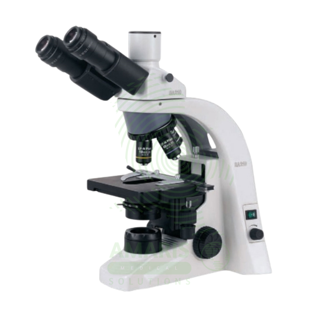
Biological Microscope
$1.00
WhatsApp Order
A Biological Microscope is a Class I medical device (optical instrument) designed for examining stained and unstained biological specimens in clinical, research, and educational laboratories. Available in monocular, binocular, or trinocular configurations with magnification from 40× to 1000× (oil immersion) using 4×, 10×, 40×, and 100× achromatic or plan achromatic objectives, 10× widefield eyepieces, Abbe condenser, mechanical stage, and built-in halogen or LED illumination. Features include coaxial coarse/fine focusing, revolving nosepiece, Köhler illumination alignment, and ergonomic design for comfortable use. Primary clinical applications include microbiological examination (Gram stains, acid-fast stains, wet mounts), hematology (differential counts, RBC morphology, platelet estimation), histopathology (tissue sections, H&E, special stains), cytology (Pap smears, FNAs, body fluids), urinalysis (sediment examination), parasitology (malaria, ova, parasites), and fertility (semen analysis). Essential equipment in every clinical laboratory for infectious disease diagnosis, cancer detection, hematological disorder evaluation, and countless other diagnostic applications requiring direct visualization of specimens. Biological microscopes are the foundation of laboratory medicine, enabling pathologists, microbiologists, hematologists, and laboratory scientists to visualize and interpret the cellular and microbial world for patient diagnosis and care.
Description
Biological Microscope
PRIMARY CLINICAL & DIAGNOSTIC USES
1. Microbiological Examination and Pathogen Identification:
-
Primary Use: Biological microscopes are essential for identifying bacteria, fungi, and parasites in clinical specimens including sputum, urine, blood cultures, cerebrospinal fluid, and tissue samples, enabling diagnosis of infectious diseases and guiding antimicrobial therapy decisions.
-
How it helps: Reveals the invisible organisms causing infections, allowing doctors to choose antibiotics that target the specific bacteria or treat the exact parasite making a patient sick.
2. Hematology and Blood Cell Analysis:
-
Primary Use: Used to perform manual differential white blood cell counts, evaluate red blood cell morphology, identify abnormal cells, and assess platelet morphology in patients with hematological disorders including leukemias, anemias, and thrombocytopenias.
-
How it helps: Gives hematologists a direct view of blood cells, revealing the telltale changes that signal leukemia, the characteristic shapes of sickle cell disease, and the subtle abnormalities that guide diagnosis and treatment.
3. Histopathology and Tissue Examination:
-
Primary Use: Essential for examining stained tissue sections to diagnose cancers, inflammatory conditions, infections, and other pathological processes in surgical pathology and biopsy evaluation.
-
How it helps: Enables pathologists to see cancer cells in tissue biopsies, identify the extent of disease, and determine whether all abnormal tissue has been removed during surgery.
4. Cytology and Cancer Screening:
-
Primary Use: Employed for examining Pap smears, fine needle aspiration biopsies, and body fluid cytology to detect cancerous and precancerous conditions at early, treatable stages.
-
How it helps: Catches cancer at its earliest, most treatable stages, often before patients have any symptoms, and monitors for recurrence after treatment.
5. Urinalysis and Sediment Examination:
-
Primary Use: Used to identify cells, casts, crystals, bacteria, and parasites in urine sediment for diagnosis of urinary tract infections, renal diseases, and metabolic disorders.
-
How it helps: Reveals the hidden story in a urine sample, showing doctors whether kidney damage, urinary tract infections, or metabolic disorders are affecting a patient’s health.
6. Parasitology and Tropical Medicine:
-
Primary Use: Critical for identifying malaria parasites in blood films, intestinal parasites in stool specimens, and tissue parasites in patients with suspected parasitic infections, particularly in endemic regions.
-
How it helps: Spots the parasites that cause devastating tropical diseases, from malaria in blood films to worms in stool samples, ensuring patients receive the right antiparasitic treatment.
7. Fertility and Reproductive Medicine:
-
Primary Use: Used for semen analysis including sperm count, motility assessment, and morphology evaluation in infertility workups and assisted reproduction procedures.
-
How it helps: Helps couples understand the factors affecting their fertility, providing essential information that guides treatment decisions and brings them closer to achieving their dream of starting a family.
SECONDARY & SUPPORTIVE USES
1. Research and Clinical Studies: Used in translational research, drug development studies, and clinical trials requiring microscopic examination of biological specimens, advancing medical knowledge and treatment.
2. Medical Education and Training: Fundamental tool for teaching histology, pathology, microbiology, and hematology to medical students, residents, and laboratory professionals, training the next generation of healthcare providers.
3. Veterinary Diagnostic Medicine: Employed in veterinary laboratories for examining animal blood, tissues, and body fluids, helping veterinarians diagnose and treat illnesses in animals.
4. Food and Beverage Industry: Used in quality control laboratories for microbiological testing of food products, helping ensure the safety of the food supply.
5. Environmental and Occupational Health: Employed to analyze water samples, air samples, and occupational exposure specimens for microorganisms and particulates, protecting public health.
6. Pharmaceutical Quality Control: Used for particulate analysis, crystal identification, and microbiological testing in pharmaceutical manufacturing, ensuring medications are safe and pure.
7. Forensic Medicine: Employed in forensic laboratories for analysis of trace evidence, bloodstains, and tissue samples, helping solve crimes and bring justice to victims.
KEY PRODUCT FEATURES
1. BASIC IDENTIFICATION ATTRIBUTES
-
Product Type: Compound optical microscope designed for examining stained and unstained biological specimens in clinical and research laboratories.
-
Common Names: Biological Microscope, Compound Microscope, Laboratory Microscope, Clinical Microscope, Research Microscope.
-
Optical Configuration: Monocular, binocular, or trinocular observation tubes.
-
Magnification Range: 40× to 1000× (standard); up to 1500× with oil immersion.
-
Objective Lenses: 4× (scanning), 10× (low power), 40× (high dry), 100× (oil immersion) - typically achromatic or plan achromatic.
-
Eyepieces: 10× or 15× widefield with diopter adjustment.
-
Illumination: Built-in halogen or LED transmitted light source with intensity control.
-
Condenser: Abbe condenser NA 1.25 with iris diaphragm and filter holder.
-
Stage: Mechanical stage with X-Y controls for precise specimen positioning.
-
Focusing: Coaxial coarse and fine focus knobs with tension adjustment.
-
Nosepiece: Revolving quadruple or quintuple nosepiece for objective changes.
-
Power Supply: 100-240 VAC, 50/60 Hz with built-in transformer.
2. TECHNICAL & PERFORMANCE PROPERTIES
-
Optical System: Finite or infinity-corrected optics; plan objectives provide flat-field images edge-to-edge.
-
Numerical Aperture (NA): 10×: 0.25, 40×: 0.65, 100×: 1.25 (oil).
-
Resolution: Capable of resolving details to 0.2-0.3 microns with oil immersion.
-
Working Distance: 10×: 6-8 mm, 40×: 0.5-1 mm, 100×: 0.1-0.2 mm (oil).
-
Field of View: 10× eyepieces provide 18-22 mm field number.
-
Parfocality: Objectives remain in focus when rotating nosepiece (minimal adjustment needed).
-
Köhler Illumination: Adjustable for uniform, glare-free illumination across the field.
-
Light Source: Halogen (6V 20W or 12V 100W) or LED (long-life, 50,000+ hours) with intensity control.
-
Condenser: Focusable Abbe condenser with centering adjustment.
-
Interpupillary Distance (Binocular): Adjustable 48-75 mm.
-
Diopter Adjustment: Compensates for vision differences between eyes.
3. PHYSICAL & OPERATIONAL PROPERTIES
-
Dimensions: 20-30 cm W × 40-50 cm D × 40-50 cm H.
-
Weight: 5-15 kg depending on configuration and materials.
-
Construction: Cast metal base and arm for stability; some models with high-strength polymers.
-
Stand: Robust, ergonomic design with inclined eyepieces for comfortable viewing.
-
Focus Mechanism: Coaxial coarse and fine focus knobs with tension adjustment; fine focus graduation typically 2 microns.
-
Nosepiece: Revolving quadruple or quintuple ball-bearing mounted.
-
Stage: Mechanical stage 140×140 mm with 75×30 mm movement range; low-position coaxial controls.
-
Condenser: Rack and pinion focusing with centering screws; filter holder for daylight or contrast filters.
-
Light Source: Built-in with intensity control; LED (long-life) or halogen (replaceable bulb).
-
Polarization: Some models include polarizer/analyzer for birefringent specimen examination.
-
Dust Cover: Protective cover included for storage.
-
Certifications: RoHS compliant; CE marked; ISO 9001 manufacturing.
4. SAFETY & COMPLIANCE ATTRIBUTES
-
Regulatory Status: Class I medical device (FDA, CE marked for IVD use when used with IVD applications).
-
Electrical Safety: Compliant with IEC 61010-1 for laboratory equipment.
-
Optical Safety: UV-blocking optics; safe for routine clinical use.
-
Chemical Resistance: Stage and frame resistant to common laboratory disinfectants.
-
Cleaning: Surfaces designed for easy cleaning with mild detergents and disinfectants.
-
Heat Dissipation: LED models produce minimal heat; halogen models have heat shields.
-
Ergonomics: Designed to reduce operator fatigue during prolonged use.
-
Quality Management: Manufactured under ISO 13485 or ISO 9001 certified processes.
-
Warranty: Typically 2-5 years depending on manufacturer.
5. STORAGE & HANDLING ATTRIBUTES
-
Storage: Store in a clean, dry environment when not in use; always use dust cover.
-
Installation: Place on rigid, vibration-free surface; avoid direct sunlight, drafts, and temperature extremes.
-
Cleaning: Clean lenses with lens paper and approved optical cleaner; never use regular tissues or paper. Clean stage and frame with mild detergent and soft cloth.
-
Objective Care: Keep objectives clean; use immersion oil only with 100× objective; clean immediately after use.
-
Condenser Care: Keep condenser and filters clean; align per manufacturer instructions for Köhler illumination.
-
Bulb Replacement (Halogen): Allow to cool; use specified bulb type; avoid touching glass with fingers.
-
LED Maintenance: LED modules typically non-replaceable; entire unit replacement if failure.
-
Annual Maintenance: Professional cleaning, alignment, and calibration recommended.
-
Inspection: Before each use, check objectives, eyepieces, and illumination; clean as needed.
6. LABORATORY & CLINICAL APPLICATIONS
-
Primary Application: Visual examination of stained and unstained biological specimens for clinical diagnosis across all laboratory disciplines.
-
Microbiology Applications:
-
Gram Stain: Bacterial morphology and Gram reaction at 100× oil.
-
Acid-Fast Stain: Mycobacteria identification at 100× oil.
-
Wet Mounts: Motility, fungi, parasites at 10× and 40×.
-
KOH Preparations: Fungal elements from skin, hair, nails at 10× and 40×.
-
India Ink: Cryptococcus from CSF at 40×.
-
Culture Examination: Colony morphology, Gram stain from colonies.
-
-
Hematology Applications:
-
Differential Count: 100-cell or 200-cell leukocyte differential at 100× oil.
-
RBC Morphology: Size, shape, color, inclusions at 100× oil.
-
Platelet Estimation: Adequacy and morphology at 100× oil.
-
Reticulocyte Count: New methylene blue stain at 100× oil.
-
Bone Marrow Aspirates: Cell lineage and maturation at 100× oil.
-
-
Histopathology Applications:
-
Routine H&E Staining: Tissue architecture at 4×, 10×; cellular detail at 40×.
-
Special Stains: Connective tissue (Trichrome), microorganisms (GMS, PAS) at 40×, 100× oil.
-
Immunohistochemistry: Chromogen localization at 10×, 40×.
-
-
Cytology Applications:
-
Pap Smears: Cervical cytology screening at 10×, 40×.
-
Fine Needle Aspirates: Cell block and smear examination at 40×, 100× oil.
-
Body Fluids: Cell identification at 40×, 100× oil.
-
-
Urinalysis Applications:
-
Sediment Examination: Cells, casts, crystals, bacteria at 10×, 40×, 100× oil.
-
-
Parasitology Applications:
-
Malaria Smears: Thick and thin blood films at 100× oil.
-
Stool Ova and Parasites: Direct mounts and concentrates at 10×, 40×.
-
Blood Parasites: Trypanosomes, filaria at 40×, 100× oil.
-
-
Fertility Applications:
-
Semen Analysis: Sperm count, motility, morphology at 10×, 40×, 100× oil.
-
SAFETY HANDLING PRECAUTIONS
1. SAFETY PRECAUTIONS
-
Lens Care: Never touch lenses with fingers; use only lens paper and approved cleaners. Avoid excessive solvent that may damage lens coatings.
-
Oil Immersion: Use only with 100× objective; clean immediately after use to prevent hardening. Use only immersion oil specified for microscopy.
-
Light Source: Halogen bulbs become hot; allow to cool before handling. LED sources remain cool.
-
Electrical Safety: Keep cords away from water; unplug before cleaning; use only specified voltage.
-
Chemical Safety: Specimens may contain infectious agents; follow universal precautions; clean spills immediately.
-
Ergonomics: Maintain good posture; adjust eyepieces and stage height for comfort; take regular breaks during prolonged use.
-
Köhler Illumination: Proper alignment essential for optimal image quality; realign after bulb changes or if disturbed.
-
Vibration: Place on vibration-free surface; avoid traffic areas; use anti-vibration tables if needed.
-
Cleaning: Never use household glass cleaners, acetone, or xylene on lenses; use only approved optical cleaners.
-
Training: Operators should be trained on proper microscope use, care, and cleaning procedures.
2. FIRST AID MEASURES
-
Eye Contact with Cleaning Solution: Flush eyes with copious water for 15 minutes; seek medical attention.
-
Broken Slide or Cover Glass: Carefully remove fragments with forceps; dispose in sharps container; clean stage and objectives carefully.
-
Specimen Spill on Microscope: Disconnect power; carefully clean with appropriate disinfectant; dry thoroughly before reuse.
-
Chemical Spill (Stains, Reagents): Follow chemical spill protocol; use appropriate PPE; clean affected areas immediately.
-
Electrical Malfunction: Disconnect power; do not use until serviced by qualified personnel.
3. FIRE FIGHTING MEASURES
-
Flammability: Plastic components and immersion oil are combustible; metal parts non-combustible.
-
Extinguishing Media: For electrical fire, use CO₂ or dry chemical (Class C) extinguisher.
-
Power Off: Disconnect power if safe to do so.
-
Immersion Oil: May be flammable; store away from ignition sources.
Related products
ECG Machine
An ECG Machine is a Class II medical device that records and displays the electrical activity of the heart through surface electrodes, producing an electrocardiogram for diagnosis of cardiac conditions. Standard diagnostic machines record 12 simultaneous leads (3 limb, 6 precordial, 1 ground) with frequency response 0.05-150 Hz, sampling rate 500-1,000 Hz, and high-resolution (5-10 µV) signal acquisition. Features include color touchscreen display, thermal array printer, computerized interpretation algorithms, internal memory (50-500+ ECGs), and network connectivity for EMR integration. Lead wires (AHA or IEC color coding) connect to disposable adhesive electrodes. Portable models (5-15 kg) with rechargeable batteries enable bedside and mobile use; cart-mounted units provide full diagnostic capability. Primary clinical applications include diagnosis of arrhythmias (AF, VT, bradycardia), detection of myocardial ischemia/infarction (STEMI, NSTEMI), evaluation of chest pain, preoperative cardiac risk assessment, monitoring electrolyte imbalances, assessment of chamber enlargement, and drug effect/toxicity monitoring. Essential diagnostic equipment in emergency departments, cardiology clinics, ICUs, operating rooms, and primary care settings worldwide.
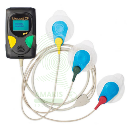
Holter ECG Monitor
A Holter ECG Monitor is a portable, wearable medical device used for continuous, long-term recording of a patient's heart's electrical activity over 24 to 48 hours or more. It is the cornerstone diagnostic tool for detecting intermittent cardiac arrhythmias, evaluating unexplained symptoms like syncope or palpitations, and monitoring the efficacy of antiarrhythmic treatments or implanted cardiac device function. Consisting of a small digital recorder connected to chest electrodes (or an all-in-one adhesive patch), it allows patients to maintain their normal daily routine while capturing a comprehensive electrocardiographic record.
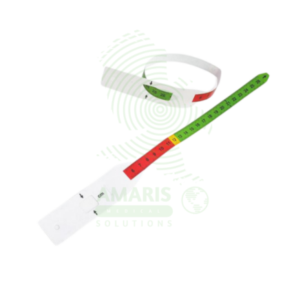
MUAC Tape
A MUAC Tape (Mid-Upper Arm Circumference) is a specialized, color-coded measuring tape used globally as the primary field tool for screening children aged 6-59 months for acute malnutrition. Its simple design—featuring red, yellow, and green zones corresponding to Severe Acute Malnutrition, Moderate Acute Malnutrition, and normal nutritional status—allows for instant, non-invasive assessment without the need for scales, calculation, or literacy. Durable, portable, and inexpensive, it is the cornerstone of community-based nutrition programs, emergency relief efforts, and public health surveillance, enabling early detection and life-saving intervention for malnourished children in even the most resource-constrained settings.
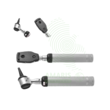
Ophthalmoscope
An Ophthalmoscope is a handheld diagnostic instrument used to examine the interior of the eye, specifically the fundus (retina, optic disc, blood vessels, and macula). By providing illuminated magnification, it serves as a vital screening tool for detecting ocular diseases like glaucoma and retinal disorders, and more importantly, for identifying signs of systemic conditions such as diabetes, hypertension, and increased intracranial pressure. Its operation involves selecting appropriate apertures and compensating lenses to obtain a clear view. While offering a detailed but narrow field of vision, mastery of the direct ophthalmoscope remains a cornerstone skill in general medicine, neurology, and ophthalmology for assessing vascular and neurological health.
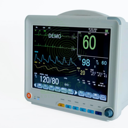
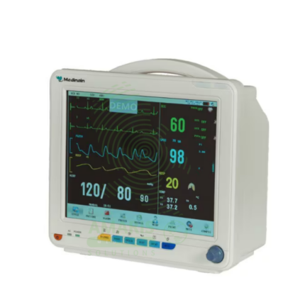
Patient Monitor
The Patient Monitor (6 Parameters BCCMS8000) is a versatile multi-parameter monitor designed for continuous surveillance of core vital signs in various clinical settings. It tracks six essential parameters—ECG, SpO2, Non-Invasive Blood Pressure (NIBP), Respiration, Temperature, and Pulse Rate—providing clinicians with real-time waveforms and numerical data on a clear color display. With its robust alarm system, battery backup for transport, and reliable performance, it is a fundamental tool for ensuring patient safety on general hospital wards, during procedures, and in emergency departments. Its design balances comprehensive monitoring capability with user-friendly operation.
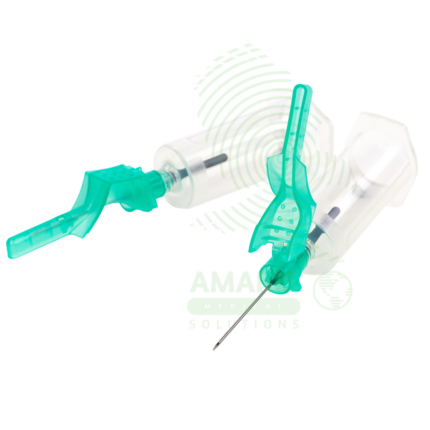
SAFE SERIES Blood Collection Needles
The SAFE SERIES (AM-SAFE) by Beijing Precil (Mindray subsidiary) is a comprehensive line of safety-engineered blood collection devices designed to prevent accidental needlestick injuries in healthcare workers. The series includes venous blood collection needles (AM-SAFE-V), standard safety needles (AM-SAFE1), winged infusion sets/butterfly needles (AM-SAFE2), high-gauge thin-wall needles for fragile veins (AM-SAFE3), premium coated needles for enhanced patient comfort (AM-GOLD), and nurse-friendly one-handed safety variants (AM-NURSE). All devices incorporate integral safety mechanisms that shield the contaminated needle immediately after withdrawal, complying with OSHA Needlestick Safety and Prevention Act requirements. Available in various gauges (18G-25G) and configurations for routine venipuncture, difficult vein access, pediatric/geriatric patients, and frequent blood draws. Sterile, single-use, latex-free, and color-coded for easy gauge identification. Essential safety devices for blood collection in hospitals, clinics, laboratories, and home healthcare settings.
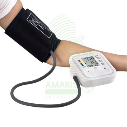
Upper Arm Blood Pressure Monitor
The Upper Arm Blood Pressure Monitor is a clinically validated, automatic digital device designed for accurate and comfortable blood pressure and pulse measurement at home or in professional settings. Known for its user-friendly features like Intellisense inflation technology, a large clear display, and comprehensive functions including irregular heartbeat detection, multi-user memory, and a WHO-based hypertension indicator, it provides reliable data for effective hypertension management. Its robust construction, adherence to international safety standards (CE, FDA), and focus on patient comfort make it a trusted choice for individuals and healthcare providers seeking a dependable tool for cardiovascular health monitoring.