Dermatoscope and Magnifiers
 Diagnostic Kits
 Vital Signs Monitors
 Stethoscopes and Accessories
 Otoscopes, Ophthalmoscopes, and Retinoscopes
 Reflex Hammers and Neurological Tools
 Scales and Measuring Devices
 Spirometers and Pulmonary Function Tests
 Electrosurgical Units and Accessories
 Cutting Instruments
 Grasping and Holding Instruments
 Hemostatic Instruments
 Specialized Surgical Sets
 Single-Use Procedure Trays and Packs
 Surgical Drapes, Gowns, and Covers
 Tissue Unifying Instruments
 Radiation Protection
 X-Ray Machines and Accessories
 Ultrasound Systems and Probes
 MRI and CT Scanners
 Radiology Consumables
 Bone Densitometers
 Fluoroscopy Equipment
 Imaging Tables and Positioning Aids
 Microscopes and Accessories
 Centrifuges and Separators
 Analyzers
 Incubators and Ovens
 Pipettes, Dispensers, and Lab Glassware
 Refrigerators, Freezers, and Storage Units
 Lab Consumables
 Sterilizers and Autoclaves for Lab Use
 Multi-Parameter Monitors
 Ventilators and Respiratory Support Devices
 Defibrillators and AEDs
 Infusion Pumps and IV Systems
 Patient Warmers and Cooling Devices
 Central Monitoring Stations
 Accessories
 Anesthesia Machines and Workstations
 Oxygen Concentrators and Delivery Systems
 Nebulizers and Inhalers
 CPAP/BiPAP Machines
 Airway Management
 Anesthesia Masks, Circuits, and Bags
 Humidifiers and Heaters
 Respiratory Therapy Accessories
 First Aid Kits and Cabinets
 Emergency Resuscitation Equipment
 Trauma Supplies
 Emergency Carts and Crash Carts
 Burn Care Products
 Bleeding Control
 Automated External Defibrillators (AEDs)
 Transport and Evacuation
 Wheelchairs and Accessories
 Walkers, Crutches, and Canes
 Prosthetics and Orthotics
 Physical Therapy Equipment
 Transfer Devices
 Bathroom Safety
 Orthopedic Traction and Tables
 Hot/Cold Therapy Packs and Units
 Beds and Mattresses
 Chairs and Stools
 Tables
 Cabinets and Storage
 Privacy Screens & Curtains
 Stands and Racks
 Linens and Textiles
 Lighting
 Autoclaves and Sterilizers
 Ultrasonic Cleaners
 Disinfectant Solutions and Wipes
 Sterilization Pouches, Wraps, and Indicators
 Instrument Trays and Containers
 UV and Ozone Disinfection Devices
 Washer Disinfectors
 Wound Care
 Gloves
 Masks and Respirators
 Catheters and Tubing
 Swabs, Applicators, and Sponges
 Incontinence Products
 Personal Protective Equipment (PPE)
 Dental Chairs and Units
 Handpieces and Burs
 Instruments
 Consumables
 Sterilization for Dental Use
 Orthodontic Supplies
 Endodontic Tools
 Slit Lamps and Tonometers
 Lensometers and Phoropters
 Ophthalmic Surgical Instruments
 Eyewear Frames and Lenses
 Contact Lens Supplies
 Vision Testing Charts and Devices
 Eye Care Consumables
 Laser Systems for Eye Care
 ENT Exam Chairs and Tables
 Endoscopes
 Audiometers and Hearing Tests
 ENT Instruments
 Nasal and Throat Packs
 Hearing Aids and Accessories
 Otology Supplies
 Fetal Dopplers and Monitors
 Delivery Beds and Tables
 Gynecological Instruments
 Neonatal Incubators and Warmers
 Breast Pumps and Accessories
 Contraceptive Devices
 Maternity Supports and Pads
 Neonatal Consumables
 Cystoscopes and Urethroscopes
 Dialysis Machines and Supplies
 Urological Catheters and Bags
 Lithotripters
 Prostate Treatment Devices
 Urinary Incontinence Products
 Kidney Stone Management Tools
 Consumables & Disposables
 EEG and EMG Machines
 Neurosurgical Instruments
 Nerve Stimulators
 Headrests and Positioning Aids
 Lumbar Puncture Kits
 Seizure Monitoring Devices
 Consumables
 Rehabilitation for Neurological Conditions
 ECG Machines and Accessories
 Holter Monitors
 Stress Test Systems
 Pacemakers and Defibrillator Accessories
 Vascular Access Devices
 Cardiac Catheters and Guidewires
 Blood Flow Meters
 Consumables
 Orthopedic Instruments
 Casts, Splints, and Padding
 Joint Replacement Supplies
 Prosthetic Limbs and Components
 Bone Grafts and Substitutes
 Traction Devices
 Orthopedic Braces and Supports
 Rehabilitation Aids for Orthopedics
 Home Oxygen Therapy
 Hospital Beds for Home Use
 Mobility Aids
 Bathroom and Daily Living Aids
 Wound Care for Home
 Monitoring Devices
 Enteral Feeding Pumps and Tubes
 Hand Sanitizers and Dispensers
 Face Shields and Goggles
 Isolation Gowns and Suits
 Biohazard Waste Containers
 Air Purifiers and HEPA Filters
 Surface Disinfectants
 Sharps Containers
 Protective Barriers
 Cardiovascular & Endurance Training
 Strength Training & Weightlifting
 Functional Training & Core Conditioning
 Physical Therapy & Rehabilitation
 Sports & Outdoor Recreation
 Gym Flooring & Facility Equipment
 Fitness Monitoring & Accessories
 Kids & Novelties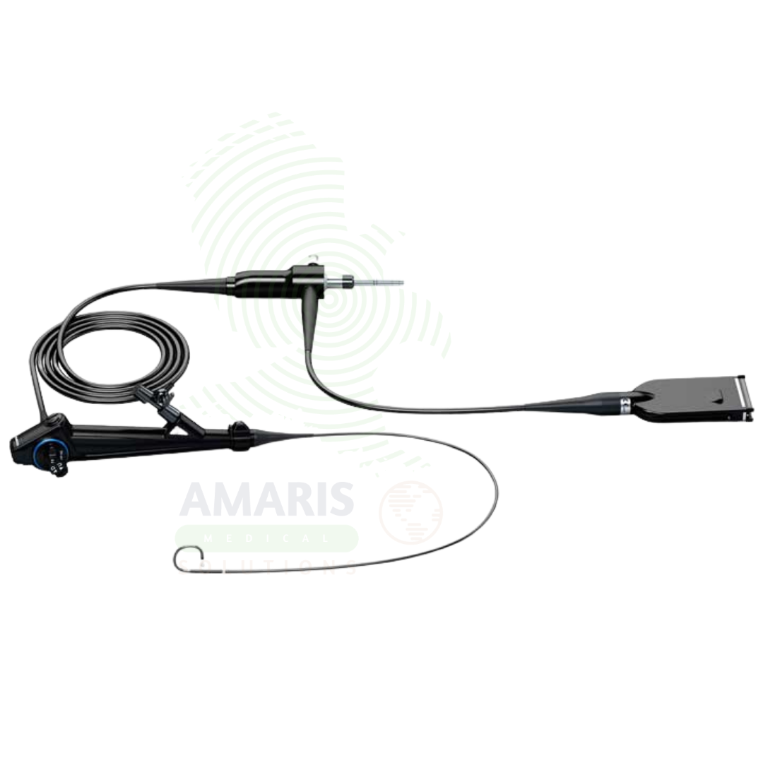
Ureteroscope
$1.00
WhatsApp Order
A Ureteroscope is a specialized endoscope used for visualization and treatment of the ureter and renal collecting system. Available in semi-rigid and flexible configurations, it enables diagnostic evaluation of ureteral pathology, laser lithotripsy for stone fragmentation, biopsy of upper tract tumors, and treatment of ureteral strictures. Essential in endourology, it offers minimally invasive diagnosis and treatment with preservation of renal function and rapid recovery.
Description
Ureteroscope
PRIMARY CLINICAL & DIAGNOSTIC USES
1. Diagnostic Visualization of the Ureter and Renal Pelvis
-
Primary Use: Provides direct visualization of the ureter, ureteropelvic junction, and renal collecting system for diagnostic evaluation of ureteral stones, strictures, tumors, and congenital anomalies. The ureteroscope is inserted through the urethra and bladder into the ureter.
-
How it helps: For the urologist and endourologist, the ureteroscope transforms the ureter from a narrow, inaccessible tube into a clearly visualized structure—revealing stones that obstruct, strictures that narrow, and tumors that may be missed on imaging. For the patient with flank pain, hematuria, or suspected ureteral pathology, ureteroscopy provides definitive diagnosis and guides treatment.
2. Ureteroscopic Stone Fragmentation and Removal
-
Primary Use: Enables endoscopic fragmentation and removal of ureteral and renal stones using laser, pneumatic, or ultrasonic lithotripsy. The ureteroscope provides direct visualization for precise targeting of stones and retrieval of fragments.
-
How it helps: For the urologist, ureteroscopic stone treatment offers a highly effective, minimally invasive approach to stone removal—achieving high stone-free rates with a single procedure. For the patient with ureteral or kidney stones, ureteroscopy offers rapid relief of obstruction, preservation of renal function, and quick return to normal activities.
3. Laser Lithotripsy for Ureteral and Renal Stones
-
Primary Use: Used in conjunction with holmium laser fibers for intracorporeal laser lithotripsy. The flexible ureteroscope allows access to the entire upper urinary tract, enabling laser fragmentation of stones in the ureter and kidney.
-
How it helps: For the urologist, laser lithotripsy through a flexible ureteroscope provides the ability to fragment stones of all compositions throughout the upper urinary tract—turning stones into dust or retrievable fragments with minimal tissue injury. For the patient, laser ureteroscopy offers a minimally invasive, highly effective treatment with high stone-free rates.
4. Ureteroscopic Biopsy of Urothelial Lesions
-
Primary Use: Allows directed biopsy of suspicious ureteral or renal pelvic lesions under direct visualization. Biopsy forceps and brushes passed through the ureteroscope obtain tissue samples for histopathological diagnosis.
-
How it helps: For the urologic oncologist, ureteroscopic biopsy provides definitive diagnosis of upper tract urothelial carcinoma—enabling accurate staging and treatment planning without open surgery. For the patient with suspected upper tract cancer, ureteroscopic biopsy offers minimally invasive diagnosis and may allow kidney-sparing treatment.
5. Ureteral Stricture Evaluation and Treatment
-
Primary Use: Used to evaluate the location, length, and severity of ureteral strictures, and to perform endoscopic treatment including balloon dilation, laser incision, and stent placement.
-
How it helps: For the urologist, ureteroscopic stricture management offers a minimally invasive alternative to open ureteral reconstruction—relieving obstruction and preserving renal function. For the patient with a ureteral stricture, endoscopic treatment offers rapid relief with minimal morbidity.
SECONDARY & SUPPORTIVE USES
1. Ureteral Stent Placement and Removal: Placement and removal of ureteral stents under direct visualization.
2. Ureteroscopic Retrieval of Foreign Bodies: Removal of migrated stents, stone fragments, and other foreign bodies.
3. Pyeloscopy: Visualization of the renal pelvis and calyces through the ureteroscope.
4. Antegrade Ureteroscopy: Combined with percutaneous access for complex stone treatment.
5. Diagnostic Ureteroscopy: Evaluation of unexplained hematuria, positive cytology, or filling defects on imaging.
6. Pediatric Ureteroscopy: Smaller caliber ureteroscopes for pediatric patients.
KEY PRODUCT FEATURES
1. BASIC IDENTIFICATION ATTRIBUTES
-
Device Type: A specialized endoscope designed for visualization and treatment of the ureter and renal collecting system.
-
Designation: Ureteroscope, Flexible Ureteroscope, Semi-Rigid Ureteroscope, Digital Ureteroscope.
-
Types:
-
Semi-Rigid Ureteroscope: Rigid shaft with flexible tip; ideal for distal and mid-ureter.
-
Flexible Ureteroscope: Fully flexible shaft with active tip deflection; allows access to the entire upper urinary tract.
-
Digital Ureteroscope: Integrated camera at the distal tip for high-definition visualization.
-
-
Key Components:
-
Telescope: Rod lens system or digital camera.
-
Working Channel: 1-3 mm for laser fibers, baskets, and instruments.
-
Light Source: Fiberoptic or LED illumination.
-
Tip Deflection: Active tip deflection (180-270°) for flexible models.
-
Irrigation: Continuous flow channels for clear visualization.
-
2. TECHNICAL & PERFORMANCE PROPERTIES
-
Diameter: 6-9 French for flexible; 6-12 French for semi-rigid.
-
Length: 30-70 cm.
-
Working Channel: 1-3 mm for instrument passage.
-
Tip Deflection: 180-270° for flexible ureteroscopes.
-
Field of View: Wide angle for comprehensive visualization.
-
Resolution: HD for digital scopes.
3. PHYSICAL & OPERATIONAL PROPERTIES
-
Construction: Precision-machined stainless steel (semi-rigid); polymer with fiber optics (flexible).
-
Ergonomics: Designed for single-handed operation.
-
Sterilization: Steam autoclave for semi-rigid; low-temperature sterilization for flexible.
-
Portability: Fixed in endoscopy suites or mobile for bedside use.
4. SAFETY & COMPLIANCE ATTRIBUTES
-
Regulatory Status: Class II medical device regulated by FDA.
-
Biocompatibility: Materials safe for intraureteral and intrarenal use.
-
Laser Compatibility: Compatible with holmium and other medical lasers.
-
Electrical Safety: Compliant for use with light sources and cameras.
5. STORAGE & HANDLING ATTRIBUTES
-
Storage: Store in protective cases; hang flexible ureteroscopes in storage cabinets.
-
Cleaning: Thorough cleaning after each use; ultrasonic cleaning recommended.
-
Sterilization: Steam autoclave for semi-rigid; high-level disinfection for flexibility.
-
Inspection: Inspect for lens damage, light transmission, and tip deflection.
6. LABORATORY & CLINICAL APPLICATIONS
-
Primary Application: Diagnosis and treatment of ureteral and renal pathology.
-
Clinical Role: Essential in endourology for stone treatment, stricture management, and upper tract tumor diagnosis.
SAFETY HANDLING PRECAUTIONS
1. SAFETY PRECAUTIONS
-
Guidewire Safety: Always use guidewire for ureteral access to prevent injury.
-
Laser Safety: Use appropriate laser safety precautions; protect eyes.
-
Irrigation Pressure: Monitor irrigation pressure to prevent fluid overload and pyelovenous reflux.
-
Ureteral Access Sheath: Consider use for prolonged procedures to protect ureter.
-
Temperature Monitoring: Monitor irrigation temperature during laser use.
2. FIRST AID MEASURES
-
Ureteral Perforation: If perforation occurs, stop procedure; place ureteral stent; manage expectantly.
-
Ureteral Avulsion: Rare but serious; if suspected, abort procedure; immediate surgical consultation.
-
Fluid Overload: If signs of fluid overload, stop procedure; administer diuretics; monitor.
3. FIRE FIGHTING MEASURES
-
Flammability: Metal components are non-flammable; plastic parts may burn.
-
Extinguishing Media: Use water, foam, or CO₂ as appropriate for surrounding materials.
Related products
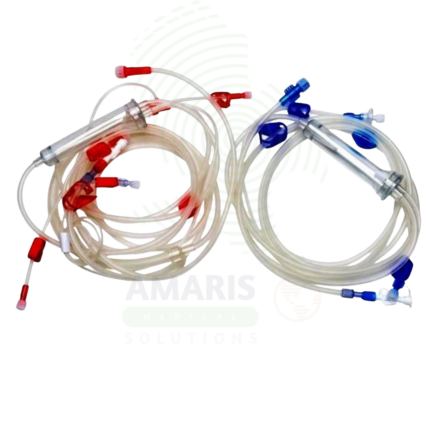
Bloodlines for Dialysis
Bloodlines for Dialysis are sterile, single-use tubing sets that form the extracorporeal circuit for hemodialysis and other blood purification therapies. Transporting blood from the patient to the dialyzer and back, they include pressure monitoring ports, air trap chambers, and medication administration sites. Essential for dialysis centers, hospitals, and home hemodialysis.
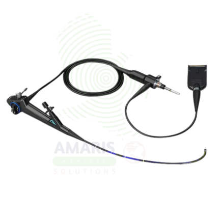
Cystoscope
A Cystoscope is a specialized endoscope used for visualization of the urethra and bladder for diagnostic and therapeutic urologic procedures. Rigid cystoscopes provide superior optics for operative procedures, while flexible cystoscopes offer enhanced patient comfort for diagnostic examinations. Essential for evaluation of hematuria, diagnosis and surveillance of bladder cancer, stent management, and treatment of bladder stones and urethral strictures, cystoscopy is the cornerstone of urologic practice.
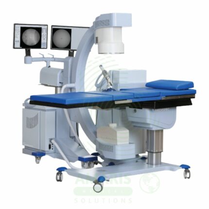
ESWL Lithotripter Systems
A ESWL Lithotripter Systems is a non-invasive surgical device that uses focused, high-energy acoustic shock waves generated outside the body to fragment kidney and upper ureteral stones. The patient lies on a table while integrated imaging (fluoroscopy/ultrasound) is used to target the stone precisely at the shock wave focal point. Treatment success depends on stone size, location, and composition. While avoiding surgical incisions, the procedure requires meticulous safety protocols to manage risks such as renal injury, bleeding, and radiation exposure. It is a capital-intensive, fixed equipment system that defines the modern, non-invasive approach to urolithiasis.
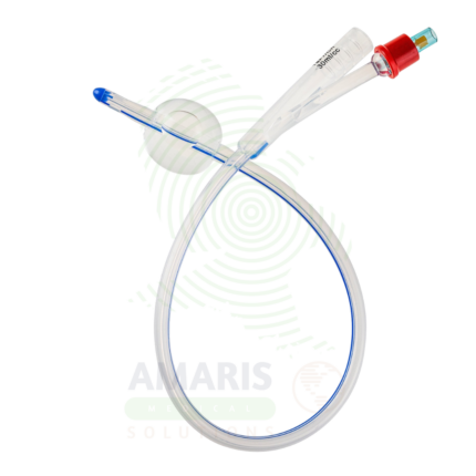
Silicone Urinary Catheters
Silicone Urinary Catheters are sterile, single-use, latex-free indwelling (Foley) catheters manufactured from 100% medical-grade silicone. They are indicated for patients requiring short- or long-term bladder drainage, particularly those with latex allergy, anticipated extended indwelling time (>7 days), or history of rapid catheter encrustation and blockage. Compared to latex and silicone-coated latex catheters, 100% silicone offers superior biocompatibility, enhanced resistance to biofilm formation and mineral encrustation, a larger internal lumen for improved flow, and complete absence of latex proteins. Available in French sizes 6 Fr-30 Fr with various tip configurations (straight, Coude, three-way irrigation) and balloon volumes (5-50 mL). Strict aseptic insertion technique, daily site care, maintenance of closed drainage, and prompt removal when no longer indicated are essential to prevent catheter-associated urinary tract infection (CAUTI) and urethral trauma.
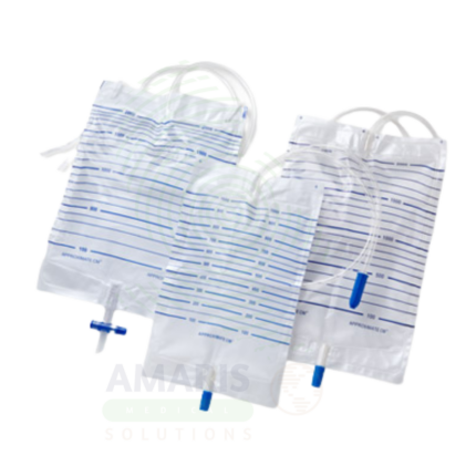
Urine Bags
Urine Bags are sterile, single-use, closed-system medical devices designed for gravity drainage, collection, containment, and measurement of urine from indwelling urinary catheters. Available in two primary configurations: large-capacity drainage bags (2000-4000 mL) for bedridden patients and overnight use, and smaller leg bags (350-750 mL) for ambulatory, community-dwelling patients. Core components include a transparent graduated collection bag, flexible drainage tubing, anti-reflux valve, needleless sampling port, and secure outlet valve. Manufactured from medical-grade PVC (DEHP-free alternatives available) or non-PVC materials, all components are latex-free and terminally sterilized. Critical safety requirements include maintaining the bag below bladder level at all times to prevent retrograde flow, preserving closed system integrity, and adhering to strict single-patient-use protocols. An indispensable component of CAUTI prevention bundles and urinary drainage management across acute care, long-term care, and home healthcare settings.
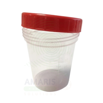
Urine Containers
Urine Containers are sterile or non-sterile, single-use medical devices manufactured from medical-grade polypropylene or polyethylene for the collection, containment, preservation, and transport of urine specimens for diagnostic testing. Available in capacities ranging from 30 mL to 3 liters, they include standard universal containers (plain), preserved containers with boric acid for urine culture, large-volume containers for 24-hour collections, pediatric adhesive collection bags, and DOT-specification drug testing kits with integrated temperature strips and tamper-evident features. Critical performance attributes include leak-proof sealing, chemical inertness, clear volume graduations, and preservative efficacy at specified fill volumes. Strict single-use protocol, adherence to fill line requirements, and handling under universal precautions are essential for specimen integrity and healthcare worker safety. An indispensable consumable in clinical diagnostics, occupational health, and forensic toxicology.
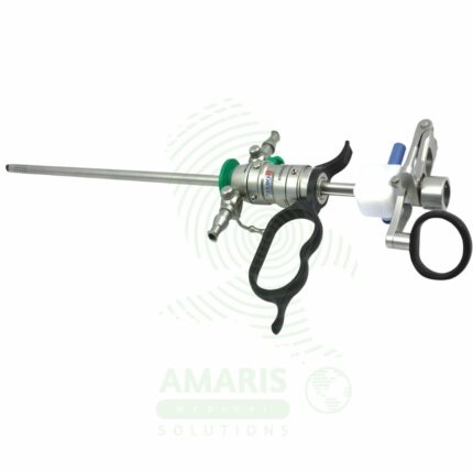
Urology Endoscopes
Urology Endoscopes are specialized instruments for visualizing and operating within the urinary tract. They include rigid and flexible cystoscopes for the bladder, semi-rigid and flexible ureteroscopes for the ureters and kidneys, and nephroscopes for percutaneous kidney surgery. These devices enable critical procedures like TURP, TURBT, and laser lithotripsy for stones. Flexible scopes, essential for complex cases, require meticulous high-level disinfection and careful handling to prevent damage. Safe use hinges on managing irrigation fluid absorption, preventing perforation, and protecting the delicate optics during energy-based treatments, making them fundamental yet sophisticated tools in urological practice.