Dermatoscope and Magnifiers
 Diagnostic Kits
 Vital Signs Monitors
 Stethoscopes and Accessories
 Otoscopes, Ophthalmoscopes, and Retinoscopes
 Reflex Hammers and Neurological Tools
 Scales and Measuring Devices
 Spirometers and Pulmonary Function Tests
 Electrosurgical Units and Accessories
 Cutting Instruments
 Grasping and Holding Instruments
 Hemostatic Instruments
 Specialized Surgical Sets
 Single-Use Procedure Trays and Packs
 Surgical Drapes, Gowns, and Covers
 Tissue Unifying Instruments
 Radiation Protection
 X-Ray Machines and Accessories
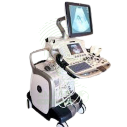
 Ultrasound Systems and Probes
 MRI and CT Scanners
 Radiology Consumables
 Bone Densitometers
 Fluoroscopy Equipment
 Imaging Tables and Positioning Aids
 Microscopes and Accessories
 Centrifuges and Separators
 Analyzers
 Incubators and Ovens
 Pipettes, Dispensers, and Lab Glassware
 Refrigerators, Freezers, and Storage Units
 Lab Consumables
 Sterilizers and Autoclaves for Lab Use
 Multi-Parameter Monitors
 Ventilators and Respiratory Support Devices
 Defibrillators and AEDs
 Infusion Pumps and IV Systems
 Patient Warmers and Cooling Devices
 Central Monitoring Stations
 Accessories
 Anesthesia Machines and Workstations
 Oxygen Concentrators and Delivery Systems
 Nebulizers and Inhalers
 CPAP/BiPAP Machines
 Airway Management
 Anesthesia Masks, Circuits, and Bags
 Humidifiers and Heaters
 Respiratory Therapy Accessories
 First Aid Kits and Cabinets
 Emergency Resuscitation Equipment
 Trauma Supplies
 Emergency Carts and Crash Carts
 Burn Care Products
 Bleeding Control
 Automated External Defibrillators (AEDs)
 Transport and Evacuation
 Wheelchairs and Accessories
 Walkers, Crutches, and Canes
 Prosthetics and Orthotics
 Physical Therapy Equipment
 Transfer Devices
 Bathroom Safety
 Orthopedic Traction and Tables
 Hot/Cold Therapy Packs and Units
 Beds and Mattresses
 Chairs and Stools
 Tables
 Cabinets and Storage
 Privacy Screens & Curtains
 Stands and Racks
 Linens and Textiles
 Lighting
 Autoclaves and Sterilizers
 Ultrasonic Cleaners
 Disinfectant Solutions and Wipes
 Sterilization Pouches, Wraps, and Indicators
 Instrument Trays and Containers
 UV and Ozone Disinfection Devices
 Washer Disinfectors
 Wound Care
 Gloves
 Masks and Respirators
 Catheters and Tubing
 Swabs, Applicators, and Sponges
 Incontinence Products
 Personal Protective Equipment (PPE)
 Dental Chairs and Units
 Handpieces and Burs
 Instruments
 Consumables
 Sterilization for Dental Use
 Orthodontic Supplies
 Endodontic Tools
 Slit Lamps and Tonometers
 Lensometers and Phoropters
 Ophthalmic Surgical Instruments
 Eyewear Frames and Lenses
 Contact Lens Supplies
 Vision Testing Charts and Devices
 Eye Care Consumables
 Laser Systems for Eye Care
 ENT Exam Chairs and Tables
 Endoscopes
 Audiometers and Hearing Tests
 ENT Instruments
 Nasal and Throat Packs
 Hearing Aids and Accessories
 Otology Supplies
 Fetal Dopplers and Monitors
 Delivery Beds and Tables
 Gynecological Instruments
 Neonatal Incubators and Warmers
 Breast Pumps and Accessories
 Contraceptive Devices
 Maternity Supports and Pads
 Neonatal Consumables
 Cystoscopes and Urethroscopes
 Dialysis Machines and Supplies
 Urological Catheters and Bags
 Lithotripters
 Prostate Treatment Devices
 Urinary Incontinence Products
 Kidney Stone Management Tools
 Consumables & Disposables
 EEG and EMG Machines
 Neurosurgical Instruments
 Nerve Stimulators
 Headrests and Positioning Aids
 Lumbar Puncture Kits
 Seizure Monitoring Devices
 Consumables
 Rehabilitation for Neurological Conditions
 ECG Machines and Accessories
 Holter Monitors
 Stress Test Systems
 Pacemakers and Defibrillator Accessories
 Vascular Access Devices
 Cardiac Catheters and Guidewires
 Blood Flow Meters
 Consumables
 Orthopedic Instruments
 Casts, Splints, and Padding
 Joint Replacement Supplies
 Prosthetic Limbs and Components
 Bone Grafts and Substitutes
 Traction Devices
 Orthopedic Braces and Supports
 Rehabilitation Aids for Orthopedics
 Home Oxygen Therapy
 Hospital Beds for Home Use
 Mobility Aids
 Bathroom and Daily Living Aids
 Wound Care for Home
 Monitoring Devices
 Enteral Feeding Pumps and Tubes
 Hand Sanitizers and Dispensers
 Face Shields and Goggles
 Isolation Gowns and Suits
 Biohazard Waste Containers
 Air Purifiers and HEPA Filters
 Surface Disinfectants
 Sharps Containers
 Protective Barriers
 Cardiovascular & Endurance Training
 Strength Training & Weightlifting
 Functional Training & Core Conditioning
 Physical Therapy & Rehabilitation
 Sports & Outdoor Recreation
 Gym Flooring & Facility Equipment
 Fitness Monitoring & Accessories
 Kids & Novelties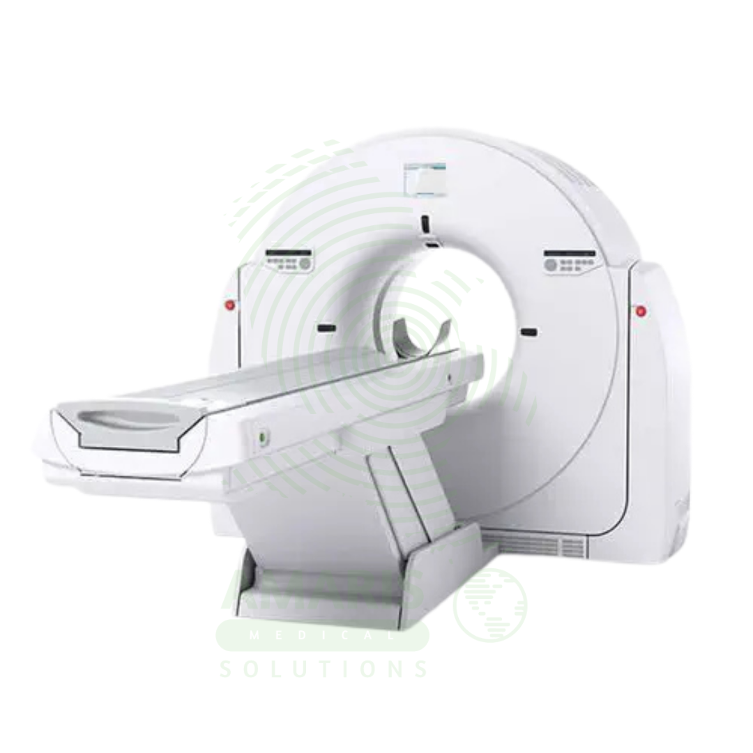
CT Scanner
$1.00
WhatsApp Order
A CT Scanner is an advanced diagnostic imaging device that uses a rotating X-ray source and detector array to create detailed cross-sectional images (“slices”) of the body. By combining these slices, it generates comprehensive 2D and 3D views of bones, organs, and blood vessels, making it indispensable for trauma evaluation, cancer staging, vascular assessment, and guiding complex procedures. While offering unparalleled diagnostic clarity, its operation requires strict adherence to radiation safety principles (ALARA) and protocols for the safe use of contrast agents to maximize patient benefit and minimize risk.
Description
CT Scanner
PRIMARY CLINICAL & DIAGNOSTIC USES
1. Comprehensive Cross-Sectional Anatomical Imaging
-
Primary Use: Produces detailed, high-resolution, two-dimensional and three-dimensional images of internal body structures, including bones, blood vessels, soft tissues, and organs, providing critical information that is often not visible on standard X-rays.
-
How it helps: For the radiologist and referring physician, the CT scanner transforms the internal body from a series of overlapping shadows into clearly defined, slice-by-slice images—revealing the exact size, shape, and location of tumors, the precise extent of fractures, and the detailed anatomy of organs and blood vessels. For the patient, a CT scan means their condition can be diagnosed with certainty, their treatment planned with precision, and their prognosis determined with confidence, often without the need for exploratory surgery.
2. Trauma and Emergency Diagnosis
-
Primary Use: The rapid imaging capability is essential in emergency medicine for quickly assessing patients with traumatic injuries to identify internal bleeding, organ damage, spinal injuries, and skull or complex bone fractures.
-
How it helps: For the emergency physician and trauma surgeon, the CT scanner is a life-saving tool that provides immediate answers in the golden hour—revealing hidden internal bleeding, identifying spinal fractures that threaten the cord, and detecting brain injuries that require urgent intervention. For the trauma patient arriving unconscious or with multiple injuries, a rapid CT scan means their life-threatening conditions are identified and treated immediately, without the delay of exploratory surgery or the risk of missed injuries.
3. Cancer Detection, Staging, and Treatment Planning
-
Primary Use: Used to detect tumors, determine their size and precise location, assess the extent of disease spread, guide biopsies, and plan for radiation therapy or surgical intervention.
-
How it helps: For the oncologist, radiation therapist, and surgical oncologist, the CT scanner provides the detailed anatomical roadmap essential for cancer care—identifying primary tumors, revealing metastatic spread, guiding biopsy needles to suspicious lesions, and mapping radiation fields to target cancer while sparing healthy tissue. For the patient facing a cancer diagnosis, CT imaging provides the answers that guide everything that follows: what type of cancer, how advanced, where has it spread, and what treatments offer the best chance.
4. Vascular and Cardiac Imaging
-
Primary Use: Enables detailed visualization of blood vessels to diagnose conditions like aneurysms, blockages, dissections, and pulmonary embolisms, while cardiac CT can assess coronary arteries and heart structure.
-
How it helps: For the vascular surgeon, cardiologist, and interventional radiologist, CT angiography provides a non-invasive window into the vascular system—revealing the exact location and extent of blockages, the size and shape of aneurysms, and the presence of life-threatening pulmonary emboli. For the patient with chest pain, suspected aneurysm, or stroke symptoms, a CT angiogram can provide definitive answers without the risks of invasive catheter angiography.
5. Guiding Interventional Procedures and Surgery
-
Primary Use: Provides real-time imaging guidance for precise needle placement in biopsies, abscess drainages, and minimally invasive surgeries, reducing the need for open exploratory procedures.
-
How it helps: For the interventional radiologist and surgeon performing minimally invasive procedures, CT guidance transforms a blind needle pass into a precisely planned intervention—confirming that the biopsy needle is within the tumor, that the drain is positioned correctly in an abscess, and that critical structures are avoided. For the patient undergoing a biopsy or drainage, CT guidance means the procedure is more likely to succeed on the first attempt, with fewer complications and less discomfort.
SECONDARY & SUPPORTIVE USES
1. Monitoring Disease Progression and Treatment Response: Used to track the size of tumors during chemotherapy or the healing of infections and injuries over time. For the patient undergoing cancer treatment, serial CT scans provide objective evidence of whether the therapy is working, guiding decisions about continuing, changing, or stopping treatment.
2. Pre-surgical Planning: Provides a detailed roadmap of patient anatomy for surgeons, especially in complex cases involving the brain, spine, face, or major joints. For the patient facing complex surgery, a preoperative CT scan means their surgeon can plan the approach in advance, anticipating anatomical variations and reducing operative time and risk.
3. Detection of Infectious Processes: Can identify the location and extent of infections such as pneumonia, abdominal abscesses, or osteomyelitis. For the patient with fever of unknown origin or suspected deep infection, CT imaging can locate the source and guide treatment.
4. Screening: Used in specific high-risk screening programs, such as low-dose CT for lung cancer screening in heavy smokers or CT colonography. For the high-risk individual, screening CT can detect cancer at an early, curable stage, long before symptoms develop.
5. Neurological Assessment: Critical for diagnosing strokes, brain tumors, and other neurological conditions by imaging the brain and cerebral vessels. For the patient with sudden neurological symptoms, a rapid CT scan distinguishes between bleeding and clot, determining whether they need clot-busting drugs or emergency surgery.
KEY PRODUCT FEATURES
1. BASIC IDENTIFICATION ATTRIBUTES
-
Type: A diagnostic imaging device that uses X-rays and computer processing.
-
Designation: Computed Tomography Scanner, also historically called a CAT (Computed Axial Tomography) Scanner.
-
Common Variants/Specifications:
-
Slice Count: Defined by the number of image slices acquired per rotation (e.g., 16-slice, 64-slice, 128-slice, 256-slice, Dual-Source). Higher slice counts allow for faster scanning and higher resolution.
-
Gantry Aperture: The diameter of the opening (typically 70-85 cm), determining the size of patients or objects that can be scanned.
-
Specialized CTs: Cardiac CT, Cone-Beam CT (CBCT - used in dentistry and interventional radiology), Portable CT (for ICU or operating room).
-
2. TECHNICAL & PERFORMANCE PROPERTIES
-
Imaging Principle: Rotates an X-ray tube and a ring of detectors around the patient. The computer uses mathematical algorithms (tomographic reconstruction) to combine the data from multiple X-ray projections into cross-sectional images.
-
Key Performance Metrics:
-
Temporal Resolution: How fast an image can be acquired, critical for freezing cardiac motion.
-
Spatial Resolution: The ability to distinguish between two small, closely spaced objects.
-
Contrast Resolution: The ability to distinguish differences in tissue density (e.g., tumor from normal tissue).
-
-
Radiation Dose: CT scans involve a higher dose of ionizing radiation than standard X-rays. Modern systems incorporate dose-reduction technologies (iterative reconstruction, automatic exposure control).
3. PHYSICAL & OPERATIONAL PROPERTIES
-
Major Components: A gantry (ring containing X-ray tube and detectors), a patient table that moves through the gantry, and a separate computer workstation for image reconstruction and viewing.
-
Scan Speed: Modern multi-slice scanners can acquire images of large body areas (e.g., chest/abdomen/pelvis) in seconds.
-
Image Reconstruction: Creates images in axial, coronal, sagittal, and oblique planes, as well as 3D volume-rendered images.
4. SAFETY & COMPLIANCE ATTRIBUTES
-
Regulatory Status: Class II medical device (varies by region), highly regulated.
-
Radiation Safety: Must comply with stringent national and international regulations for radiation-producing equipment. Requires licensed operators and a radiation safety program.
-
Contrast Agent Safety: Many CT exams use iodinated intravenous contrast agents to enhance vascular and organ visualization. Protocols must exist for screening and managing patients at risk for allergic reactions or contrast-induced nephropathy.
5. STORAGE & HANDLING ATTRIBUTES
-
Environment: Requires a controlled environment (specific temperature, humidity) and stable, dedicated electrical power.
-
Service and Calibration: Requires scheduled preventive maintenance and calibration by specialized service engineers to ensure image quality and dose accuracy.
-
Software Updates: Regular software updates are provided by the manufacturer for performance improvements and new applications.
6. LABORATORY & CLINICAL APPLICATIONS
-
Primary Application: A cornerstone of modern diagnostic radiology, deployed in hospital radiology departments, outpatient imaging centers, and emergency rooms.
-
Workflow Integration: Images are stored and transmitted via Picture Archiving and Communication System (PACS) and interpreted by radiologists on dedicated diagnostic workstations.
SAFETY HANDLING PRECAUTIONS
1. SAFETY PRECAUTIONS
-
Radiation Dose Justification (ALARA Principle): Every scan must be medically justified. The protocol should use the lowest radiation dose necessary to achieve diagnostic image quality (As Low As Reasonably Achievable).
-
Pregnancy Screening: All females of childbearing age must be screened for possible pregnancy before a CT scan of the abdomen/pelvis or any high-dose scan.
-
Contrast Agent Precautions: Patients must be screened for renal impairment, prior contrast reactions, and allergies. Pre-medication protocols may be required for high-risk patients.
-
Patient Positioning and Immobilization: Proper positioning and instructions to hold breath are critical to prevent motion artifacts that degrade image quality.
2. FIRST AID MEASURES
-
Acute Contrast Reaction: Stop injection immediately. Call for emergency medical support. Treat according to reaction severity (e.g., antihistamines for mild, epinephrine for anaphylaxis).
-
Extravasation of Contrast: Stop injection. Elevate the extremity. Apply warm or cold compresses as per protocol. Monitor for compartment syndrome.
3. FIRE FIGHTING MEASURES
-
Electrical Fire Hazard: Contains high-voltage components and electronics.
-
Extinguishing Media: Use CO₂ or dry chemical extinguishers. Do not use water on electrical fires. Evacuate area and alert fire department.
Related products
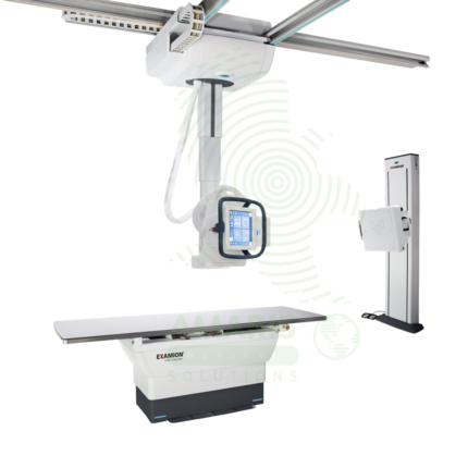
Digital Ceiling X-ray
A Digital Ceiling X-ray is a ceiling-mounted digital radiography system for general diagnostic imaging of the skeletal, chest, abdominal, and extremity anatomy. The ceiling-mounted tube assembly provides full room coverage for flexible patient positioning, while digital flat panel detectors produce immediate high-resolution images for rapid diagnosis. Integrated with PACS and RIS, it supports efficient digital workflow from image acquisition to interpretation. Used in radiology departments, emergency rooms, and outpatient imaging centers.
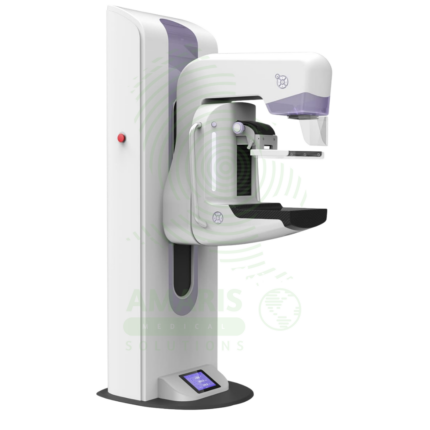
Digital Mammography System
A Digital Mammography System is a specialized X-ray imaging system designed for breast cancer screening and diagnosis. Using high-resolution digital detectors, it provides superior image quality with lower radiation dose compared to film mammography. Advanced systems offer tomosynthesis (3D) imaging, which improves cancer detection rates and reduces false positives. Used in breast imaging centers, radiology departments, and women's health facilities, it is the gold standard for early detection of breast cancer.

Lead Glass
Lead Glass is a transparent radiation shielding material used in X-ray rooms, CT suites, fluoroscopy suites, and radiation therapy control areas. Impregnated with lead oxide, it provides radiation attenuation equivalent to lead sheet while allowing direct visual observation of patients during procedures. Used for observation windows in control booths and procedure rooms, lead glass maintains the integrity of the radiation shielding envelope while enabling staff to monitor patient positioning, movement, and comfort. Proper installation with lead-lined frames and seals is essential for continuous radiation protection.
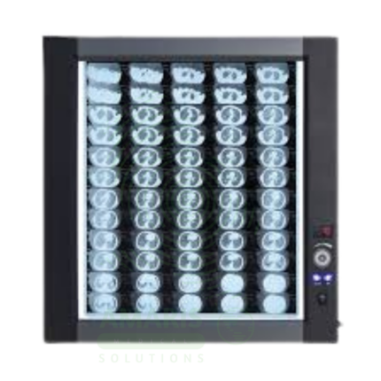
LED Medical Film Viewer
An LED Medical Film Viewer is a light box designed for viewing and interpreting analog X-ray films. Using LED backlight technology, it provides uniform, high-luminance illumination with instant on capability and long life. Available in single, dual, and multi-panel configurations, it supports side-by-side comparison of current and prior studies, pre-operative planning, and group teaching. Essential for radiology departments, orthopedic clinics, emergency departments, and operating rooms where film-based imaging is still used.
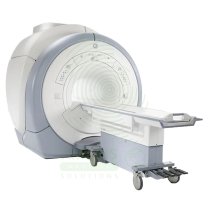
Magnetic Resonance Imaging
Magnetic Resonance Imaging (MRI) is a non-invasive diagnostic imaging modality that uses powerful magnetic fields and radiofrequency waves to produce detailed images of soft tissues, organs, and internal structures without ionizing radiation. It is the gold standard for imaging the brain, spinal cord, joints, muscles, and ligaments, and is essential for neurological, musculoskeletal, oncologic, and cardiovascular diagnosis. MRI provides exceptional soft tissue contrast, enabling precise anatomical characterization, tumor staging, and treatment planning. Strict safety protocols for ferromagnetic screening and contrast administration are essential for patient safety.
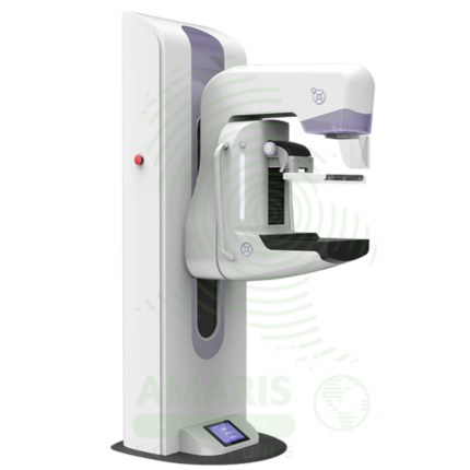
Mammography Machine
A Mammography Machine is a specialized, low-dose X-ray system designed exclusively for imaging the breast. It is the gold-standard tool for breast cancer screening and diagnostic evaluation, utilizing firm breast compression and high-resolution digital detectors to produce detailed images of breast tissue. Modern systems often incorporate Digital Breast Tomosynthesis (DBT or "3D mammography") to reduce tissue overlap and improve cancer detection. Its operation is highly regulated, requiring certified technologists, qualified interpreting physicians, and a rigorous quality assurance program to ensure patient safety, optimal image quality, and accurate early detection of breast cancer, which is vital for reducing mortality.
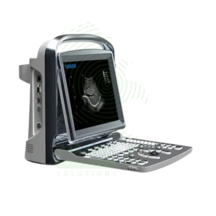
Portable Ultrasound Machine
A Portable Ultrasound Machine is a compact, battery-powered imaging device designed for point-of-care use, bringing diagnostic capability directly to the patient's bedside. It is essential for rapid triage in emergency and critical care settings, procedural guidance, and basic examinations in clinics and remote locations. While offering core imaging modes like B-mode and Color Doppler in a lightweight, durable package, its effective use requires clinician training to recognize both its diagnostic value and its limitations compared to comprehensive departmental systems. Proper cleaning, battery management, and data security are paramount for its safe and effective deployment.