Dermatoscope and Magnifiers
 Diagnostic Kits
 Vital Signs Monitors
 Stethoscopes and Accessories
 Otoscopes, Ophthalmoscopes, and Retinoscopes
 Reflex Hammers and Neurological Tools
 Scales and Measuring Devices
 Spirometers and Pulmonary Function Tests
 Electrosurgical Units and Accessories
 Cutting Instruments
 Grasping and Holding Instruments
 Hemostatic Instruments
 Specialized Surgical Sets
 Single-Use Procedure Trays and Packs
 Surgical Drapes, Gowns, and Covers
 Tissue Unifying Instruments
 Radiation Protection
 X-Ray Machines and Accessories
 Ultrasound Systems and Probes
 MRI and CT Scanners
 Radiology Consumables
 Bone Densitometers
 Fluoroscopy Equipment
 Imaging Tables and Positioning Aids
 Microscopes and Accessories
 Centrifuges and Separators
 Analyzers
 Incubators and Ovens
 Pipettes, Dispensers, and Lab Glassware
 Refrigerators, Freezers, and Storage Units
 Lab Consumables
 Sterilizers and Autoclaves for Lab Use
 Multi-Parameter Monitors
 Ventilators and Respiratory Support Devices
 Defibrillators and AEDs
 Infusion Pumps and IV Systems
 Patient Warmers and Cooling Devices
 Central Monitoring Stations
 Accessories
 Anesthesia Machines and Workstations
 Oxygen Concentrators and Delivery Systems
 Nebulizers and Inhalers
 CPAP/BiPAP Machines
 Airway Management
 Anesthesia Masks, Circuits, and Bags
 Humidifiers and Heaters
 Respiratory Therapy Accessories
 First Aid Kits and Cabinets
 Emergency Resuscitation Equipment
 Trauma Supplies
 Emergency Carts and Crash Carts
 Burn Care Products
 Bleeding Control
 Automated External Defibrillators (AEDs)
 Transport and Evacuation
 Wheelchairs and Accessories
 Walkers, Crutches, and Canes
 Prosthetics and Orthotics
 Physical Therapy Equipment
 Transfer Devices
 Bathroom Safety
 Orthopedic Traction and Tables
 Hot/Cold Therapy Packs and Units
 Beds and Mattresses
 Chairs and Stools
 Tables
 Cabinets and Storage
 Privacy Screens & Curtains
 Stands and Racks
 Linens and Textiles
 Lighting
 Autoclaves and Sterilizers
 Ultrasonic Cleaners
 Disinfectant Solutions and Wipes
 Sterilization Pouches, Wraps, and Indicators
 Instrument Trays and Containers
 UV and Ozone Disinfection Devices
 Washer Disinfectors
 Wound Care
 Gloves
 Masks and Respirators
 Catheters and Tubing
 Swabs, Applicators, and Sponges
 Incontinence Products
 Personal Protective Equipment (PPE)
 Dental Chairs and Units
 Handpieces and Burs
 Instruments
 Consumables
 Sterilization for Dental Use
 Orthodontic Supplies
 Endodontic Tools
 Slit Lamps and Tonometers
 Lensometers and Phoropters
 Ophthalmic Surgical Instruments
 Eyewear Frames and Lenses
 Contact Lens Supplies
 Vision Testing Charts and Devices
 Eye Care Consumables
 Laser Systems for Eye Care
 ENT Exam Chairs and Tables
 Endoscopes
 Audiometers and Hearing Tests
 ENT Instruments
 Nasal and Throat Packs
 Hearing Aids and Accessories
 Otology Supplies
 Fetal Dopplers and Monitors
 Delivery Beds and Tables
 Gynecological Instruments
 Neonatal Incubators and Warmers
 Breast Pumps and Accessories
 Contraceptive Devices
 Maternity Supports and Pads
 Neonatal Consumables
 Cystoscopes and Urethroscopes
 Dialysis Machines and Supplies
 Urological Catheters and Bags
 Lithotripters
 Prostate Treatment Devices
 Urinary Incontinence Products
 Kidney Stone Management Tools
 Consumables & Disposables
 EEG and EMG Machines
 Neurosurgical Instruments
 Nerve Stimulators
 Headrests and Positioning Aids
 Lumbar Puncture Kits
 Seizure Monitoring Devices
 Consumables
 Rehabilitation for Neurological Conditions
 ECG Machines and Accessories
 Holter Monitors
 Stress Test Systems
 Pacemakers and Defibrillator Accessories
 Vascular Access Devices
 Cardiac Catheters and Guidewires
 Blood Flow Meters
 Consumables
 Orthopedic Instruments
 Casts, Splints, and Padding
 Joint Replacement Supplies
 Prosthetic Limbs and Components
 Bone Grafts and Substitutes
 Traction Devices
 Orthopedic Braces and Supports
 Rehabilitation Aids for Orthopedics
 Home Oxygen Therapy
 Hospital Beds for Home Use
 Mobility Aids
 Bathroom and Daily Living Aids
 Wound Care for Home
 Monitoring Devices
 Enteral Feeding Pumps and Tubes
 Hand Sanitizers and Dispensers
 Face Shields and Goggles
 Isolation Gowns and Suits
 Biohazard Waste Containers
 Air Purifiers and HEPA Filters
 Surface Disinfectants
 Sharps Containers
 Protective Barriers
 Cardiovascular & Endurance Training
 Strength Training & Weightlifting
 Functional Training & Core Conditioning
 Physical Therapy & Rehabilitation
 Sports & Outdoor Recreation
 Gym Flooring & Facility Equipment
 Fitness Monitoring & Accessories
 Kids & Novelties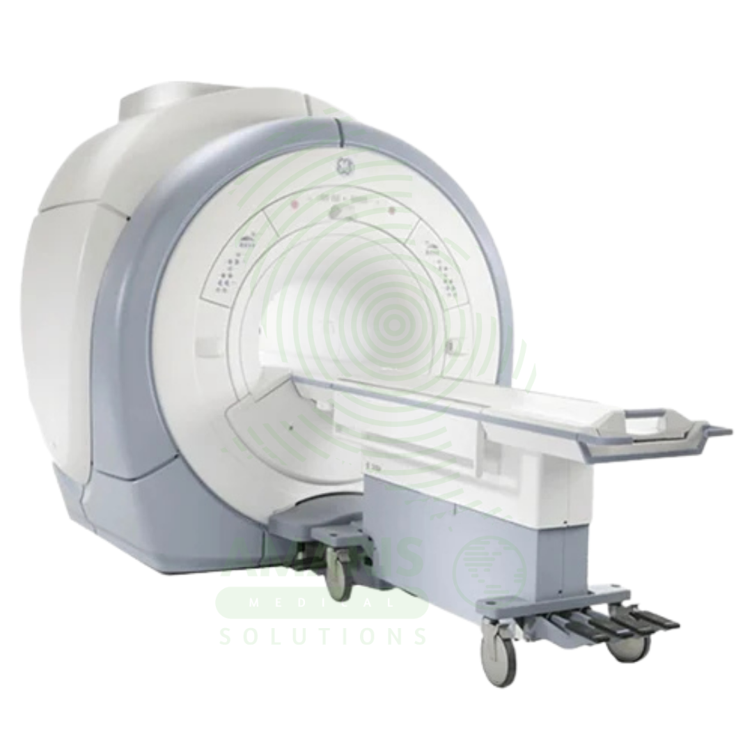
Magnetic Resonance Imaging
$1.00
WhatsApp Order
Magnetic Resonance Imaging (MRI) is a non-invasive diagnostic imaging modality that uses powerful magnetic fields and radiofrequency waves to produce detailed images of soft tissues, organs, and internal structures without ionizing radiation. It is the gold standard for imaging the brain, spinal cord, joints, muscles, and ligaments, and is essential for neurological, musculoskeletal, oncologic, and cardiovascular diagnosis. MRI provides exceptional soft tissue contrast, enabling precise anatomical characterization, tumor staging, and treatment planning. Strict safety protocols for ferromagnetic screening and contrast administration are essential for patient safety.
Description
Magnetic Resonance Imaging (MRI)
PRIMARY CLINICAL & DIAGNOSTIC USES
1. High-Contrast Soft Tissue Imaging
-
Primary Use: Produces exceptionally detailed images of soft tissues using powerful magnetic fields and radiofrequency waves without ionizing radiation. MRI is the gold standard for imaging the brain, spinal cord, nerves, muscles, ligaments, tendons, cartilage, and internal organs including the liver, prostate, uterus, and heart.
-
How it helps: For the radiologist and referring physician, MRI reveals the internal architecture of the human body with a clarity that no other imaging modality can match—distinguishing grey matter from white matter in the brain, revealing the internal structure of tumors, and showing the fine detail of ligaments, tendons, and cartilage. For the patient, an MRI means their condition can be diagnosed with confidence, often without exposure to radiation, and with sufficient detail to guide surgical planning, treatment decisions, and monitoring of disease progression.
2. Neurological and Neurosurgical Diagnosis
-
Primary Use: Critical for evaluating conditions including strokes (in subacute and chronic phases), multiple sclerosis, brain tumors, aneurysms, pituitary gland disorders, and degenerative diseases of the brain and spine such as Alzheimer’s disease, disc herniations, and spinal stenosis.
-
How it helps: For the neurologist and neurosurgeon, MRI provides an unprecedented window into the central nervous system—revealing the demyelinating plaques of multiple sclerosis, mapping the precise location and extent of brain tumors before surgery, and showing the compression of neural elements by herniated discs or spinal stenosis. For the patient with neurological symptoms, an MRI can provide definitive answers, distinguishing between treatable conditions and those requiring monitoring, and guiding decisions about surgery, medication, or watchful waiting.
3. Musculoskeletal System Evaluation
-
Primary Use: Provides unmatched detail for assessing joint injuries including knee meniscus and ligament tears, shoulder labral and rotator cuff tears, cartilage defects, bone marrow disorders, osteomyelitis, and soft tissue tumors.
-
How it helps: For the orthopedic surgeon and sports medicine specialist, MRI is indispensable for evaluating injuries that cannot be seen on X-ray—revealing torn menisci and cruciate ligaments in the knee, detecting labral tears in the shoulder, and identifying occult fractures and bone bruises. For the athlete with a suspected ligament tear or the patient with unexplained joint pain, an MRI provides the detailed anatomical information needed to determine whether surgery is necessary and to plan the procedure if it is.
4. Oncologic Imaging and Staging
-
Primary Use: Used for detecting, characterizing, and staging cancers, especially in the brain, spine, breast (with dedicated breast MRI), prostate (with multiparametric MRI), liver, and rectum. MRI is essential for surgical and radiation therapy planning, providing precise anatomical localization of tumors.
-
How it helps: For the oncologist, radiation therapist, and surgical oncologist, MRI provides the detailed anatomical roadmap essential for cancer care—defining the exact boundaries of tumors, revealing invasion into adjacent structures, detecting metastatic disease, and guiding biopsy and treatment planning. For the patient facing a cancer diagnosis, MRI provides the information needed to stage the disease accurately, plan treatment with precision, and monitor response to therapy over time.
5. Cardiovascular Imaging (Cardiac MRI)
-
Primary Use: Evaluates heart structure and function, detects myocardial damage from heart attacks, identifies cardiomyopathies, tumors of the heart, pericardial disease, and diseases of the large blood vessels including aortic dissection and aneurysms.
-
How it helps: For the cardiologist and cardiothoracic surgeon, cardiac MRI provides a comprehensive assessment of heart structure and function in a single examination—measuring ejection fraction, identifying areas of scar from prior heart attacks, detecting infiltrative diseases of the heart muscle, and evaluating complex congenital heart disease. For the patient with heart failure, suspected cardiomyopathy, or complex congenital heart disease, cardiac MRI provides the detailed information needed to guide treatment and predict outcomes.
SECONDARY & SUPPORTIVE USES
1. Functional MRI (fMRI): Maps brain activity by detecting blood flow changes during cognitive tasks, used in neurosurgery planning to identify eloquent cortex (speech, motor, sensory areas) before tumor resection, and in cognitive research.
2. Diffusion Weighted Imaging (DWI): Sensitive to the movement of water molecules, crucial for early detection of acute stroke within minutes of onset, and for characterizing tumors by assessing cellular density.
3. Magnetic Resonance Spectroscopy (MRS): Analyzes the biochemical composition of tissues, providing metabolic information that complements anatomical imaging for tumor characterization and metabolic disorder diagnosis.
4. MR Angiography (MRA): Visualizes blood vessels without contrast injection, used to detect aneurysms, arterial stenosis, dissections, and vascular malformations.
5. Diffusion Tensor Imaging (DTI): Maps white matter tracts in the brain, used for surgical planning and assessment of traumatic brain injury.
6. Interventional MRI: Provides real-time imaging guidance for biopsy needle placement and minimally invasive thermal ablation therapies.
7. Breast MRI: Used for screening high-risk patients, evaluating extent of breast cancer, and assessing response to neoadjuvant chemotherapy.
8. MR Cholangiopancreatography (MRCP): Non-invasive imaging of the biliary and pancreatic ducts for evaluation of stones, strictures, and tumors.
KEY PRODUCT FEATURES
1. BASIC IDENTIFICATION ATTRIBUTES
-
Device Type: A medical imaging system that uses powerful magnetic fields and radiofrequency waves to generate detailed images of internal body structures.
-
Designation: MRI, Magnetic Resonance Imaging, MRI Scanner, MRI System.
-
Magnet Types:
-
Closed MRI: High-field cylindrical system providing superior image quality.
-
Open MRI: Lower-field system with open sides for claustrophobic or larger patients.
-
Wide-Bore MRI: Larger diameter bore for increased patient comfort.
-
Standing/Upright MRI: Allows imaging in weight-bearing positions.
-
-
Field Strength: Ranges from 0.2T to 3.0T for clinical use; 1.5T and 3.0T most common; higher field strengths (7T) used for research.
2. TECHNICAL & PERFORMANCE PROPERTIES
-
Magnetic Field Strength: Measured in Tesla (T); higher field strength provides better signal-to-noise ratio and spatial resolution.
-
Magnet Type: Superconducting (most common), permanent, or resistive.
-
Bore Size: Standard bore 60-70 cm; wide bore 70-80 cm.
-
Image Acquisition: Multiple sequences (T1-weighted, T2-weighted, FLAIR, DWI, etc.) for different tissue characterization.
-
Contrast Agents: Gadolinium-based contrast used for enhancement of vascular structures, tumors, and inflammatory processes.
3. PHYSICAL & OPERATIONAL PROPERTIES
-
Construction: Large cylindrical magnet with patient table; housed in shielded room to contain magnetic field.
-
Patient Positioning: Patient lies supine on table that moves into magnet bore.
-
Noise Level: Significant acoustic noise during scanning; hearing protection required.
-
Scan Time: 20-60 minutes depending on protocol.
4. SAFETY & COMPLIANCE ATTRIBUTES
-
Regulatory Status: Class II medical device regulated by FDA.
-
Magnetic Field Safety: Strict screening for ferromagnetic implants, devices, and foreign bodies.
-
Contraindications: Cardiac pacemakers, implantable defibrillators, certain aneurysm clips, cochlear implants, ferromagnetic foreign bodies.
-
Acoustic Safety: Hearing protection required due to high noise levels.
-
Claustrophobia: Some patients require sedation or open MRI systems.
5. STORAGE & HANDLING ATTRIBUTES
-
Storage: Permanent installation in shielded MRI suite.
-
Quench Venting: Superconducting magnets require a venting system for emergency magnet quench.
-
Cryogen Management: Liquid helium required for superconducting magnet cooling.
-
Maintenance: Regular calibration and cryogen refills required.
6. LABORATORY & CLINICAL APPLICATIONS
-
Primary Application: Soft tissue imaging for neurological, musculoskeletal, oncologic, and cardiovascular diagnosis.
-
Clinical Role: Essential imaging modality in radiology, neurology, orthopedics, oncology, and cardiology.
SAFETY HANDLING PRECAUTIONS
1. SAFETY PRECAUTIONS
-
Ferromagnetic Screening: Strict screening of all patients and personnel for ferromagnetic implants and objects.
-
Zone Management: Designated MRI safety zones (I-IV) with controlled access.
-
Quench Protocol: Emergency procedures for magnet quench with oxygen monitoring.
-
Contrast Safety: Screen for renal function before gadolinium administration to prevent nephrogenic systemic fibrosis.
-
Patient Monitoring: Monitor patients during scan; provide communication system for patient anxiety.
2. FIRST AID MEASURES
-
Quench: In event of magnet quench, evacuate area; monitor for oxygen displacement.
-
Patient Distress: If a patient experiences claustrophobia or panic, remove from the scanner immediately.
-
Contrast Reaction: Treat gadolinium contrast reactions per protocol; monitor for allergic response.
3. FIRE FIGHTING MEASURES
-
Flammability: Equipment is non-flammable; fire risk from electrical components.
-
Extinguishing Media: Use CO₂ or dry chemical extinguisher for electrical fires.
-
Magnetic Field Hazard: Use non-ferromagnetic extinguishers in MRI suite.
Related products
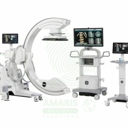
C-Arm Surgical System
A C-Arm Surgical System is a mobile fluoroscopic X-ray imaging device with a distinctive C-shaped arm connecting the X-ray tube and detector. It is an indispensable tool in modern operating rooms and interventional suites, providing real-time live imaging to guide complex procedures in orthopedics, spine surgery, pain management, and vascular interventions. Its mobility allows precise positioning around the patient, while features like pulsed fluoroscopy and dose monitoring are critical for radiation safety. Modern flat-panel systems offer high-resolution imaging and advanced capabilities like 3D Cone-Beam CT. Safe operation demands rigorous adherence to radiation protection protocols (ALARA) for both patients and the surgical team.
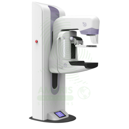
Digital Mammography System
A Digital Mammography System is a specialized X-ray imaging system designed for breast cancer screening and diagnosis. Using high-resolution digital detectors, it provides superior image quality with lower radiation dose compared to film mammography. Advanced systems offer tomosynthesis (3D) imaging, which improves cancer detection rates and reduces false positives. Used in breast imaging centers, radiology departments, and women's health facilities, it is the gold standard for early detection of breast cancer.
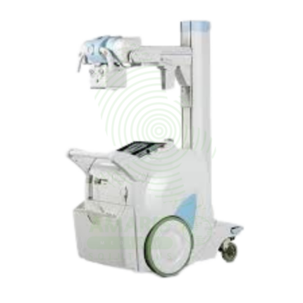
Digital Mobile X-ray
A Digital Mobile X-ray is a battery-powered, portable digital radiography system designed for bedside imaging in intensive care units, neonatal intensive care units, emergency departments, and operating rooms. The mobile unit enables high-quality chest, abdominal, and extremity imaging at the patient's bedside, eliminating the risks associated with transporting critically ill patients. Wireless digital detectors provide immediate image capture and transmission to PACS, supporting rapid diagnosis and treatment decisions.
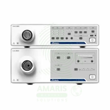
Endoscopy System
The Endoscopy System is a complete video processor and light source stack that forms the core of a modern digital endoscopy suite. It provides high-definition imaging for compatible Fujinon flexible video endoscopes, enabling diagnostic and therapeutic procedures in gastroenterology, pulmonology, and urology. Key features include advanced image processing and enhancement technologies like Narrow Band Imaging (NBI) for improved diagnostic accuracy. As a critical piece of capital equipment, it requires careful handling, the use of compatible accessories, and regular professional maintenance to ensure optimal performance and safety.

Lead Glass
Lead Glass is a transparent radiation shielding material used in X-ray rooms, CT suites, fluoroscopy suites, and radiation therapy control areas. Impregnated with lead oxide, it provides radiation attenuation equivalent to lead sheet while allowing direct visual observation of patients during procedures. Used for observation windows in control booths and procedure rooms, lead glass maintains the integrity of the radiation shielding envelope while enabling staff to monitor patient positioning, movement, and comfort. Proper installation with lead-lined frames and seals is essential for continuous radiation protection.
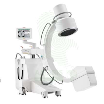
Mobile C-arm Surgical System
A Mobile C-arm Surgical System is a portable fluoroscopic imaging device used for real-time intraoperative guidance during orthopedic, spinal, vascular, and pain management procedures. The C-shaped arm allows flexible positioning around the patient, providing AP, lateral, and oblique views to verify instrument placement, fracture reduction, and device deployment. Essential for minimally invasive surgery, it enables surgeons to achieve precision and accuracy while reducing operative time and improving patient outcomes.
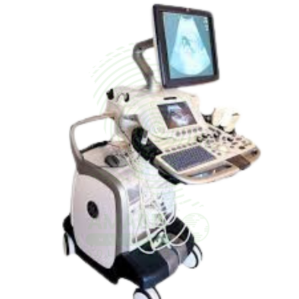
Ultrasound Machine
The Ultrasound Machine is a diagnostic imaging system that uses high-frequency sound waves to produce real-time, dynamic images of internal body structures, including soft tissues, organs, blood vessels, and developing fetuses. It provides non-invasive, radiation-free visualization using multiple transducer probes for abdominal, cardiac, obstetric, gynecological, vascular, and musculoskeletal examinations. With core features like B-mode grayscale imaging, M-mode for motion assessment, and comprehensive Doppler capabilities (Color, Power, Spectral), it offers essential diagnostic functionality for evaluating anatomy, physiology, and blood flow. Its portability, digital image storage, and user-friendly interface make it a practical tool for hospitals, clinics, emergency departments, and point-of-care settings across virtually all medical specialties, requiring proper operator training and adherence to probe disinfection protocols for safe and effective use.