Dermatoscope and Magnifiers
 Diagnostic Kits
 Vital Signs Monitors
 Stethoscopes and Accessories
 Otoscopes, Ophthalmoscopes, and Retinoscopes
 Reflex Hammers and Neurological Tools
 Scales and Measuring Devices
 Spirometers and Pulmonary Function Tests
 Electrosurgical Units and Accessories
 Cutting Instruments
 Grasping and Holding Instruments
 Hemostatic Instruments
 Specialized Surgical Sets
 Single-Use Procedure Trays and Packs
 Surgical Drapes, Gowns, and Covers
 Tissue Unifying Instruments
 Radiation Protection
 X-Ray Machines and Accessories
 Ultrasound Systems and Probes
 MRI and CT Scanners
 Radiology Consumables
 Bone Densitometers
 Fluoroscopy Equipment
 Imaging Tables and Positioning Aids
 Microscopes and Accessories
 Centrifuges and Separators
 Analyzers
 Incubators and Ovens
 Pipettes, Dispensers, and Lab Glassware
 Refrigerators, Freezers, and Storage Units
 Lab Consumables
 Sterilizers and Autoclaves for Lab Use
 Multi-Parameter Monitors
 Ventilators and Respiratory Support Devices
 Defibrillators and AEDs
 Infusion Pumps and IV Systems
 Patient Warmers and Cooling Devices
 Central Monitoring Stations
 Accessories
 Anesthesia Machines and Workstations
 Oxygen Concentrators and Delivery Systems
 Nebulizers and Inhalers
 CPAP/BiPAP Machines
 Airway Management
 Anesthesia Masks, Circuits, and Bags
 Humidifiers and Heaters
 Respiratory Therapy Accessories
 First Aid Kits and Cabinets
 Emergency Resuscitation Equipment
 Trauma Supplies
 Emergency Carts and Crash Carts
 Burn Care Products
 Bleeding Control
 Automated External Defibrillators (AEDs)
 Transport and Evacuation
 Wheelchairs and Accessories
 Walkers, Crutches, and Canes
 Prosthetics and Orthotics
 Physical Therapy Equipment
 Transfer Devices
 Bathroom Safety
 Orthopedic Traction and Tables
 Hot/Cold Therapy Packs and Units
 Beds and Mattresses
 Chairs and Stools
 Tables
 Cabinets and Storage
 Privacy Screens & Curtains
 Stands and Racks
 Linens and Textiles
 Lighting
 Autoclaves and Sterilizers
 Ultrasonic Cleaners
 Disinfectant Solutions and Wipes
 Sterilization Pouches, Wraps, and Indicators
 Instrument Trays and Containers
 UV and Ozone Disinfection Devices
 Washer Disinfectors
 Wound Care
 Gloves
 Masks and Respirators
 Catheters and Tubing
 Swabs, Applicators, and Sponges
 Incontinence Products
 Personal Protective Equipment (PPE)
 Dental Chairs and Units
 Handpieces and Burs
 Instruments
 Consumables
 Sterilization for Dental Use
 Orthodontic Supplies
 Endodontic Tools
 Slit Lamps and Tonometers
 Lensometers and Phoropters
 Ophthalmic Surgical Instruments
 Eyewear Frames and Lenses
 Contact Lens Supplies
 Vision Testing Charts and Devices
 Eye Care Consumables
 Laser Systems for Eye Care
 ENT Exam Chairs and Tables
 Endoscopes
 Audiometers and Hearing Tests
 ENT Instruments
 Nasal and Throat Packs
 Hearing Aids and Accessories
 Otology Supplies
 Fetal Dopplers and Monitors
 Delivery Beds and Tables
 Gynecological Instruments
 Neonatal Incubators and Warmers
 Breast Pumps and Accessories
 Contraceptive Devices
 Maternity Supports and Pads
 Neonatal Consumables
 Cystoscopes and Urethroscopes
 Dialysis Machines and Supplies
 Urological Catheters and Bags
 Lithotripters
 Prostate Treatment Devices
 Urinary Incontinence Products
 Kidney Stone Management Tools
 Consumables & Disposables
 EEG and EMG Machines
 Neurosurgical Instruments
 Nerve Stimulators
 Headrests and Positioning Aids
 Lumbar Puncture Kits
 Seizure Monitoring Devices
 Consumables
 Rehabilitation for Neurological Conditions
 ECG Machines and Accessories
 Holter Monitors
 Stress Test Systems
 Pacemakers and Defibrillator Accessories
 Vascular Access Devices
 Cardiac Catheters and Guidewires
 Blood Flow Meters
 Consumables
 Orthopedic Instruments
 Casts, Splints, and Padding
 Joint Replacement Supplies
 Prosthetic Limbs and Components
 Bone Grafts and Substitutes
 Traction Devices
 Orthopedic Braces and Supports
 Rehabilitation Aids for Orthopedics
 Home Oxygen Therapy
 Hospital Beds for Home Use
 Mobility Aids
 Bathroom and Daily Living Aids
 Wound Care for Home
 Monitoring Devices
 Enteral Feeding Pumps and Tubes
 Hand Sanitizers and Dispensers
 Face Shields and Goggles
 Isolation Gowns and Suits
 Biohazard Waste Containers
 Air Purifiers and HEPA Filters
 Surface Disinfectants
 Sharps Containers
 Protective Barriers
 Cardiovascular & Endurance Training
 Strength Training & Weightlifting
 Functional Training & Core Conditioning
 Physical Therapy & Rehabilitation
 Sports & Outdoor Recreation
 Gym Flooring & Facility Equipment
 Fitness Monitoring & Accessories
 Kids & Novelties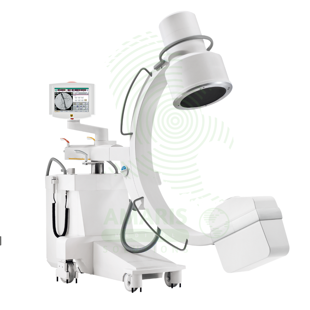

Mobile C-arm Surgical System
$1.00
WhatsApp Order
A Mobile C-arm Surgical System is a portable fluoroscopic imaging device used for real-time intraoperative guidance during orthopedic, spinal, vascular, and pain management procedures. The C-shaped arm allows flexible positioning around the patient, providing AP, lateral, and oblique views to verify instrument placement, fracture reduction, and device deployment. Essential for minimally invasive surgery, it enables surgeons to achieve precision and accuracy while reducing operative time and improving patient outcomes.
Description
Mobile C-arm Surgical System
PRIMARY CLINICAL & DIAGNOSTIC USES
1. Intraoperative Fluoroscopic Imaging
-
Primary Use: Provides real-time, mobile X-ray imaging during surgical procedures, allowing surgeons to visualize anatomy, instrument placement, and device deployment without closing the incision. The mobile C-arm can be positioned around the patient in the operating room, enabling immediate feedback during surgery.
-
How it helps: For the surgeon and operating room team, the mobile C-arm transforms surgery from a partially blind procedure into a visually guided precision operation—revealing anatomy hidden beneath tissue and bone, confirming hardware position before the patient leaves the OR, and allowing for immediate adjustments without reopening the incision. For the patient, this real-time imaging means their surgeon can place implants, reduce fractures, and deploy devices with confidence, reducing the need for repeat surgeries and ensuring the best possible outcome.
2. Orthopedic and Trauma Surgery
-
Primary Use: Essential for fracture reduction and fixation of long bones, pelvis, and extremities; placement of intramedullary nails, plates, and screws; spinal instrumentation including pedicle screw placement; and joint replacement procedures. The mobile C-arm allows for multi-planar imaging to verify alignment and hardware position.
-
How it helps: For the orthopedic and trauma surgeon, the mobile C-arm is indispensable for confirming that a reduced fracture is perfectly aligned, that pedicle screws are safely within bone and clear of nerves, and that joint replacement components are positioned correctly for optimal function. For the patient with a broken leg, a fractured spine, or a replaced joint, intraoperative imaging means their surgeon can achieve perfect alignment and hardware placement without guesswork, maximizing the chance of full recovery.
3. Spinal Surgery
-
Primary Use: Critical for pedicle screw placement in spinal fusion, vertebroplasty, kyphoplasty, discectomy, and deformity correction. The C-arm provides AP and lateral views to confirm trajectory and depth of instrumentation, minimizing risk of neurological injury.
-
How it helps: For the spine surgeon, the mobile C-arm provides the critical visualization needed to place screws millimeters from the spinal cord and nerve roots—confirming trajectory, depth, and position in real time. For the patient undergoing spinal fusion or deformity correction, this precision means their surgeon can stabilize their spine with confidence, minimizing the risk of catastrophic neurological injury that could leave them with paralysis or chronic pain.
4. Vascular and Endovascular Surgery
-
Primary Use: Used for guiding catheter placement, stent deployment, embolization procedures, and thrombectomy in vascular and endovascular surgery. The mobile C-arm enables visualization of guidewires, catheters, and devices within blood vessels.
-
How it helps: For the vascular surgeon and interventional radiologist, the mobile C-arm’s real-time imaging allows them to navigate catheters through blood vessels, deploy stents precisely at blockages, and confirm complete occlusion of aneurysms or bleeding vessels. For the patient with a life-threatening aneurysm, a critical artery blockage, or active bleeding, this image guidance means their condition can be treated through a tiny puncture rather than open surgery, with less pain, faster recovery, and fewer complications.
5. Pain Management and Interventional Procedures
-
Primary Use: Guides needle placement for epidural steroid injections, nerve blocks, facet joint injections, radiofrequency ablations, and spinal cord stimulator lead placements. The mobile C-arm allows for precise targeting of therapeutic injections.
-
How it helps: For the pain management specialist and interventional radiologist, the mobile C-arm ensures that therapeutic injections and ablation probes reach exactly the intended target—epidural space, nerve root, or facet joint—rather than missing and providing no relief. For the patient suffering from chronic back pain, radiculopathy, or nerve-related pain, precise image guidance means their procedure has the best chance of providing meaningful relief, potentially avoiding more invasive surgery.
SECONDARY & SUPPORTIVE USES
1. Urological Procedures: Guides percutaneous nephrolithotomy for kidney stones, ureteroscopy with stone fragmentation, and stent placements.
2. Gastroenterological and ERCP Procedures: Assists in complex endoscopic retrograde cholangiopancreatography for stone removal and stent placement in the biliary tree.
3. Cardiac Electrophysiology Studies: Used in EP labs for catheter ablation procedures to treat arrhythmias.
4. Foreign Body Localization and Removal: Helps locate and remove radiopaque foreign objects in soft tissue or body cavities.
5. Dental and Maxillofacial Surgery: For complex reconstructive surgeries and implant placements.
6. Orthopedic Oncology: Guides tumor resection and limb salvage procedures.
KEY PRODUCT FEATURES
1. BASIC IDENTIFICATION ATTRIBUTES
-
Device Type: A mobile fluoroscopic imaging system with C-arm configuration for intraoperative use.
-
Designation: Mobile C-arm, Surgical C-arm, Intraoperative Fluoroscopy, Mini C-arm, O-arm.
-
Key Components:
-
C-Arm: C-shaped structure with X-ray tube and image intensifier or flat panel detector at opposite ends.
-
Mobile Cart: Battery-powered or wheeled base for maneuverability.
-
X-ray Generator: High-frequency generator for continuous fluoroscopy.
-
Image Detector: Image intensifier or flat panel detector.
-
Monitor: High-resolution display for real-time imaging.
-
Controls: Foot pedal, hand switches, and touchscreen interface.
-
2. TECHNICAL & PERFORMANCE PROPERTIES
-
Detector Type: Image intensifier (traditional) or flat panel detector (digital).
-
Field of View: Typically 6-12 inches; some models offer variable fields.
-
C-Arm Rotation: Multi-axis rotation for AP, lateral, and oblique views.
-
Generator Power: 5-20 kW for mobile systems.
-
Fluoroscopy Modes: Pulsed and continuous fluoroscopy; digital subtraction angiography available on some models.
-
3D Imaging: Some advanced models offer 3D imaging capability.
3. PHYSICAL & OPERATIONAL PROPERTIES
-
Mobility: Battery-powered or manual movement; lockable wheels for stability.
-
C-Arm Dimensions: Compact for maneuverability in operating rooms.
-
Controls: Intuitive interface; foot pedal for exposure control.
-
Image Storage: Digital archiving; DICOM connectivity.
4. SAFETY & COMPLIANCE ATTRIBUTES
-
Regulatory Status: Class II medical device regulated by FDA.
-
Radiation Safety: Pulsed fluoroscopy, last image hold, and dose tracking features.
-
Sterile Field Compatibility: Drapes available for sterile use in the operating room.
-
Collision Avoidance: Sensors to prevent equipment collision.
5. STORAGE & HANDLING ATTRIBUTES
-
Storage: Stored in the operating room or designated storage area; battery charged overnight.
-
Cleaning: Wipe with hospital-grade disinfectants; follow manufacturer guidelines.
-
Maintenance: Regular calibration and preventive maintenance required.
6. LABORATORY & CLINICAL APPLICATIONS
-
Primary Application: Intraoperative imaging for orthopedic, spinal, vascular, and pain management procedures.
-
Clinical Role: Essential equipment in operating rooms, interventional suites, and pain management clinics.
SAFETY HANDLING PRECAUTIONS
1. SAFETY PRECAUTIONS
-
Radiation Protection: Use lead aprons, thyroid shields, and maintain distance during exposure.
-
C-Arm Positioning: Ensure C-arm is properly positioned before use; avoid collisions with patient and equipment.
-
Sterile Technique: Maintain sterile field; use sterile drapes for C-arm.
-
Battery Management: Keep unit charged; monitor battery status during procedures.
2. FIRST AID MEASURES
-
Collision: If C-arm collides with patient or equipment, stop movement; assess for injury; inspect unit for damage.
-
Equipment Malfunction: If equipment fails, complete procedure with alternative imaging if available; contact service provider.
3. FIRE FIGHTING MEASURES
-
Flammability: Equipment is non-flammable; fire risk from electrical components.
-
Extinguishing Media: For electrical fire, use CO₂ or dry chemical extinguisher.
Related products
Analogue Fixed X-ray Machine
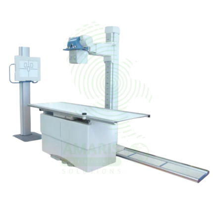
An Analogue Fixed X-ray Machine is a permanent installation X-ray system using traditional film cassettes for general radiography in radiology departments and imaging centers. Featuring ceiling-mounted tube assemblies, tilting tables, and wall stands, it provides essential diagnostic imaging for skeletal, chest, abdominal, and extremity examinations using film technology. Film cassettes are processed in darkroom facilities, producing permanent physical images for patient records and consultation. Used in facilities without digital radiography, as backup for digital systems, and in resource-limited settings.
Analogue Mobile X-ray Machine
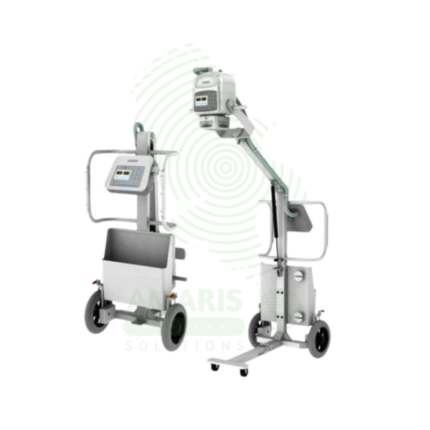
An Analogue Mobile X-ray Machine is a battery-powered, portable X-ray system using traditional film cassettes for bedside imaging in intensive care units, neonatal intensive care units, emergency departments, and operating rooms. The mobile unit enables chest, abdominal, and extremity imaging at the patient's bedside, eliminating the risks associated with transporting critically ill patients. Film cassettes are processed in darkroom facilities for image development. Used in hospitals without digital radiography, as backup for digital systems, and in resource-limited settings where digital infrastructure is not available.
Computed Tomography (CT)
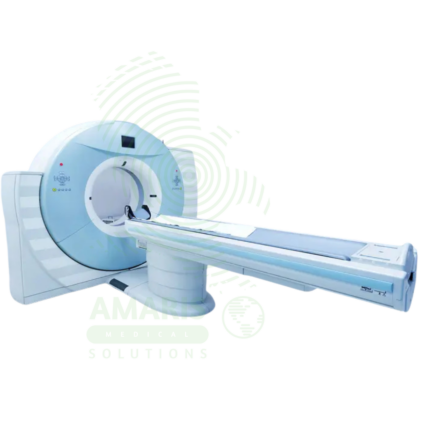
Computed Tomography (CT) is a diagnostic imaging modality that uses X-rays and computer processing to create detailed cross-sectional images of the body. Essential for trauma evaluation, cancer diagnosis, vascular imaging, and surgical planning, CT provides rapid, high-resolution images that guide life-saving decisions in emergency medicine, oncology, and surgery. Advanced multi-slice systems enable whole-body scanning in seconds with sub-millimeter resolution. Radiation dose optimization and contrast safety protocols are essential for patient safety.
Dental X-ray Machine
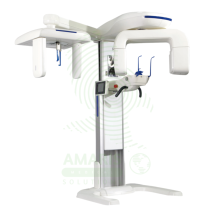
A Dental X-ray Machine is a specialized radiographic system designed for imaging teeth, jaws, and facial structures. It encompasses intraoral units for detailed tooth-specific views, panoramic machines for wide screening shots, and advanced Cone Beam CT (CBCT) scanners for 3D surgical planning. Utilizing low-dose radiation and digital imaging technology, it is indispensable for diagnosing cavities, gum disease, infections, and planning treatments like implants, orthodontics, and oral surgery. Its safe operation requires strict adherence to radiation protection protocols, including the use of lead aprons, proper collimation, and operator training to ensure patient and staff safety while obtaining critical diagnostic information.
Digital U-arm X-ray
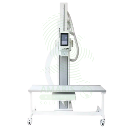
A Digital U-arm X-ray is a versatile digital radiography system designed for emergency departments, urgent care centers, and outpatient clinics. The U-arm configuration provides flexible positioning for chest, abdominal, skeletal, and extremity imaging with easy patient access for stretcher and wheelchair patients. Digital detectors produce immediate high-resolution images for rapid diagnosis, while the compact footprint allows installation in space-constrained settings. Essential for rapid, high-quality imaging in emergency and ambulatory care environments.
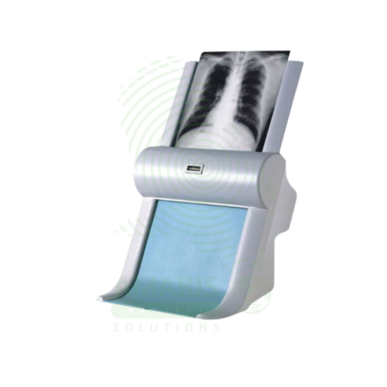
Film Digitizer
A Film Digitizer is a specialized scanner that converts analog radiographic films into high-fidelity digital images (DICOM format). It is the essential tool for migrating historical film archives into a modern digital PACS, enabling filmless workflow, remote access, and long-term preservation of diagnostic records. Its critical performance characteristics are a wide optical density range (to capture all film details) and high spatial resolution. By creating secure, accessible digital copies, it protects against the loss of physical films and integrates past patient history with current digital imaging, supporting comprehensive care and efficient radiology practice.
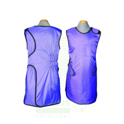
Lead Apron
A Lead Apron is a protective garment worn by healthcare workers to shield against scatter radiation during fluoroscopic procedures, X-ray examinations, and interventional radiology. Made of lead-impregnated material, it attenuates scatter radiation to the thyroid, chest, and reproductive organs, ensuring occupational radiation exposure remains within safe limits. Available in frontal, wrap-around, and two-piece designs with lead equivalence ranging from 0.25 mm to 0.5 mm, proper storage, annual inspection, and use of thyroid shields are essential for effective radiation protection.