Dermatoscope and Magnifiers
 Diagnostic Kits
 Vital Signs Monitors
 Stethoscopes and Accessories
 Otoscopes, Ophthalmoscopes, and Retinoscopes
 Reflex Hammers and Neurological Tools
 Scales and Measuring Devices
 Spirometers and Pulmonary Function Tests
 Electrosurgical Units and Accessories
 Cutting Instruments
 Grasping and Holding Instruments
 Hemostatic Instruments
 Specialized Surgical Sets
 Single-Use Procedure Trays and Packs
 Surgical Drapes, Gowns, and Covers
 Tissue Unifying Instruments
 Radiation Protection
 X-Ray Machines and Accessories
 Ultrasound Systems and Probes
 MRI and CT Scanners
 Radiology Consumables
 Bone Densitometers
 Fluoroscopy Equipment
 Imaging Tables and Positioning Aids
 Microscopes and Accessories
 Centrifuges and Separators
 Analyzers
 Incubators and Ovens
 Pipettes, Dispensers, and Lab Glassware
 Refrigerators, Freezers, and Storage Units
 Lab Consumables
 Sterilizers and Autoclaves for Lab Use
 Multi-Parameter Monitors
 Ventilators and Respiratory Support Devices
 Defibrillators and AEDs
 Infusion Pumps and IV Systems
 Patient Warmers and Cooling Devices
 Central Monitoring Stations
 Accessories
 Anesthesia Machines and Workstations
 Oxygen Concentrators and Delivery Systems
 Nebulizers and Inhalers
 CPAP/BiPAP Machines
 Airway Management
 Anesthesia Masks, Circuits, and Bags
 Humidifiers and Heaters
 Respiratory Therapy Accessories
 First Aid Kits and Cabinets
 Emergency Resuscitation Equipment
 Trauma Supplies
 Emergency Carts and Crash Carts
 Burn Care Products
 Bleeding Control
 Automated External Defibrillators (AEDs)
 Transport and Evacuation
 Wheelchairs and Accessories
 Walkers, Crutches, and Canes
 Prosthetics and Orthotics
 Physical Therapy Equipment
 Transfer Devices
 Bathroom Safety
 Orthopedic Traction and Tables
 Hot/Cold Therapy Packs and Units
 Beds and Mattresses
 Chairs and Stools
 Tables
 Cabinets and Storage
 Privacy Screens & Curtains
 Stands and Racks
 Linens and Textiles
 Lighting
 Autoclaves and Sterilizers
 Ultrasonic Cleaners
 Disinfectant Solutions and Wipes
 Sterilization Pouches, Wraps, and Indicators
 Instrument Trays and Containers
 UV and Ozone Disinfection Devices
 Washer Disinfectors
 Wound Care
 Gloves
 Masks and Respirators
 Catheters and Tubing
 Swabs, Applicators, and Sponges
 Incontinence Products
 Personal Protective Equipment (PPE)
 Dental Chairs and Units
 Handpieces and Burs
 Instruments
 Consumables
 Sterilization for Dental Use
 Orthodontic Supplies
 Endodontic Tools
 Slit Lamps and Tonometers
 Lensometers and Phoropters
 Ophthalmic Surgical Instruments
 Eyewear Frames and Lenses
 Contact Lens Supplies
 Vision Testing Charts and Devices
 Eye Care Consumables
 Laser Systems for Eye Care
 ENT Exam Chairs and Tables
 Endoscopes
 Audiometers and Hearing Tests
 ENT Instruments
 Nasal and Throat Packs
 Hearing Aids and Accessories
 Otology Supplies
 Fetal Dopplers and Monitors
 Delivery Beds and Tables
 Gynecological Instruments
 Neonatal Incubators and Warmers
 Breast Pumps and Accessories
 Contraceptive Devices
 Maternity Supports and Pads
 Neonatal Consumables
 Cystoscopes and Urethroscopes
 Dialysis Machines and Supplies
 Urological Catheters and Bags
 Lithotripters
 Prostate Treatment Devices
 Urinary Incontinence Products
 Kidney Stone Management Tools
 Consumables & Disposables
 EEG and EMG Machines
 Neurosurgical Instruments
 Nerve Stimulators
 Headrests and Positioning Aids
 Lumbar Puncture Kits
 Seizure Monitoring Devices
 Consumables
 Rehabilitation for Neurological Conditions
 ECG Machines and Accessories
 Holter Monitors
 Stress Test Systems
 Pacemakers and Defibrillator Accessories
 Vascular Access Devices
 Cardiac Catheters and Guidewires
 Blood Flow Meters
 Consumables
 Orthopedic Instruments
 Casts, Splints, and Padding
 Joint Replacement Supplies
 Prosthetic Limbs and Components
 Bone Grafts and Substitutes
 Traction Devices
 Orthopedic Braces and Supports
 Rehabilitation Aids for Orthopedics
 Home Oxygen Therapy
 Hospital Beds for Home Use
 Mobility Aids
 Bathroom and Daily Living Aids
 Wound Care for Home
 Monitoring Devices
 Enteral Feeding Pumps and Tubes
 Hand Sanitizers and Dispensers
 Face Shields and Goggles
 Isolation Gowns and Suits
 Biohazard Waste Containers
 Air Purifiers and HEPA Filters
 Surface Disinfectants
 Sharps Containers
 Protective Barriers
 Cardiovascular & Endurance Training
 Strength Training & Weightlifting
 Functional Training & Core Conditioning
 Physical Therapy & Rehabilitation
 Sports & Outdoor Recreation
 Gym Flooring & Facility Equipment
 Fitness Monitoring & Accessories
 Kids & Novelties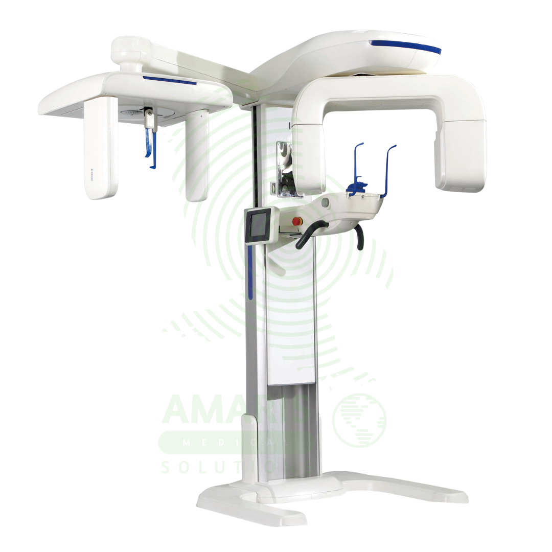

Dental X-ray Machine
$1.00
WhatsApp Order
A Dental X-ray Machine is a specialized radiographic system designed for imaging teeth, jaws, and facial structures. It encompasses intraoral units for detailed tooth-specific views, panoramic machines for wide screening shots, and advanced Cone Beam CT (CBCT) scanners for 3D surgical planning. Utilizing low-dose radiation and digital imaging technology, it is indispensable for diagnosing cavities, gum disease, infections, and planning treatments like implants, orthodontics, and oral surgery. Its safe operation requires strict adherence to radiation protection protocols, including the use of lead aprons, proper collimation, and operator training to ensure patient and staff safety while obtaining critical diagnostic information.
Description
Dental X-ray Machine
PRIMARY CLINICAL & DIAGNOSTIC USES
1. Intraoral Radiography
-
Primary Use: Captures detailed images of individual teeth, their roots, and the surrounding bone structure, including bitewing X-rays to detect cavities between teeth and assess bone levels, periapical X-rays to view the entire tooth from crown to root tip and diagnose abscesses, cysts, and root fractures, and occlusal X-rays to show larger areas of the upper or lower jaw.
-
How it helps: For the dentist and dental specialist, intraoral radiography transforms the hidden structures of the tooth and jaw into visible diagnostic information—revealing cavities hidden between teeth, infections at the root tips, bone loss from periodontal disease, and fractures invisible to the naked eye. For the patient, these images mean that dental problems are detected early, before they cause pain or require extensive treatment, and that procedures like root canals and extractions are performed with complete knowledge of the underlying anatomy.
2. Extraoral Radiography
-
Primary Use: Used for broader imaging of the skull and jaws, including panoramic X-rays to view all teeth, upper and lower jaws, sinuses, and temporomandibular joints on a single film for screening, orthodontic assessment, and wisdom tooth evaluation, and cephalometric X-rays for orthodontic treatment planning to analyze the relationship between teeth, jaws, and facial skeleton.
-
How it helps: For the orthodontist and oral surgeon, panoramic and cephalometric imaging provides a comprehensive view of the entire facial structure—revealing impacted wisdom teeth, assessing jaw relationships for orthodontic treatment, evaluating the temporomandibular joints, and detecting pathology that might be missed on smaller intraoral films. For the patient facing orthodontic treatment or wisdom tooth extraction, these images ensure that treatment is planned with complete information, avoiding surprises and improving outcomes.
3. Cone Beam Computed Tomography
-
Primary Use: A specific type of advanced dental X-ray machine that provides 3D volumetric images, used for precise planning of dental implant placement, evaluation of complex root canals, assessment of jaw tumors and pathologies, and orthodontic and surgical planning.
-
How it helps: For the implantologist, endodontist, and oral surgeon, CBCT technology transforms dental imaging from 2D to 3D—revealing the exact dimensions of bone available for implants, showing the precise relationship of roots to vital structures like the inferior alveolar nerve, and providing a virtual roadmap for complex surgical procedures. For the patient receiving dental implants or undergoing complex root canal treatment, 3D imaging means their procedure can be planned with millimeter precision, reducing the risk of nerve injury, implant failure, and surgical complications.
4. Diagnosis of Dental and Maxillofacial Pathology
-
Primary Use: Essential for detecting and diagnosing dental caries, periodontal disease, periapical infections, impacted teeth, jaw fractures, cysts, tumors, and sinus issues related to dental health.
-
How it helps: For the dentist and oral pathologist, X-ray imaging is the primary tool for seeing beyond what the eye can see—detecting decay before it destroys teeth, identifying bone loss before teeth are lost, and discovering cysts and tumors before they cause irreversible damage. For the patient, regular dental X-rays mean that problems are caught early, when they are smaller, easier to treat, and less expensive to manage.
5. Guiding Dental Procedures
-
Primary Use: Provides real-time imaging or precise pre-operative maps to guide complex procedures such as root canal therapy, surgical extractions, and dental implant surgery.
-
How it helps: For the dentist performing root canal therapy, X-ray guidance ensures that the entire root canal system is cleaned and sealed—confirming that instruments reach the tip of the root and that filling material completely obturates the space. For the patient undergoing a root canal, extraction, or implant placement, image guidance means the procedure is performed with precision, reducing the risk of failure and the need for retreatment.
SECONDARY & SUPPORTIVE USES
1. Forensic Dentistry: Used for dental identification in forensic investigations, where the unique characteristics of teeth and dental restorations can provide positive identification of individuals when other methods are unavailable. For families seeking answers about missing loved ones, dental X-rays can provide closure when it is most needed.
2. Monitoring Growth and Development: In pediatric dentistry and orthodontics, serial X-rays monitor jaw growth and tooth eruption, guiding decisions about the timing and type of orthodontic intervention. For the growing child, these images ensure that treatment is timed optimally for the best and most stable results.
3. Assessment of Trauma: To evaluate teeth and jaws following facial injury, revealing fractures, displacements, and injuries that may not be visible on clinical examination. For the patient who has sustained facial trauma, prompt X-ray evaluation ensures that all injuries are identified and treated appropriately.
4. Quality Control and Verification: Used post-operatively to verify the success of root canal fillings, the placement of implants, or the fit of dental restorations. For the patient who has undergone dental treatment, post-operative X-rays provide reassurance that the procedure was successful and that their investment in dental health was worthwhile.
KEY PRODUCT FEATURES
1. BASIC IDENTIFICATION ATTRIBUTES
-
Type: An X-ray generator and imaging system optimized for the dental and maxillofacial region.
-
Designation: Categorized by imaging technique: Intraoral X-ray Unit, Panoramic X-ray Machine, Cephalometric Unit, CBCT Scanner.
-
Common Variants:
-
Intraoral X-ray Unit: Consists of a wall-mounted or handheld X-ray tubehead, an exposure control, and a digital sensor or film holder. Used for bitewing and periapical views.
-
Panoramic Machine (OPG): The patient stands or sits while a rotating arm (with an X-ray tube and a detector) moves around the head, capturing a single wide image of the entire dentition.
-
Cephalometric Machine: A specialized lateral skull X-ray unit with a consistent distance and alignment for orthodontic analysis.
-
Cone Beam CT (CBCT) Scanner: A rotating gantry that captures a cone-shaped X-ray beam, producing a 3D dataset. May be a standalone unit or combined with a panoramic machine.
-
2. TECHNICAL & PERFORMANCE PROPERTIES
-
Imaging Principle: Uses a focused, low-dose X-ray beam. Digital systems use a solid-state sensor (CCD/CMOS) or a phosphor plate (PSP) instead of film.
-
Key Components:
-
X-ray Tubehead: Contains the high-voltage generator and tube, often with a very small focal spot for high-resolution images.
-
Position Indicating Device (PID): A lead-lined cylinder or rectangular collimator that shapes and directs the beam.
-
Image Receptor: Digital sensor, phosphor plate, or traditional film.
-
Cephalostat: A head-positioning device used in cephalometric radiography.
-
-
Radiation Dose: Dental X-rays use very low doses of radiation. Digital sensors require significantly less exposure than film. CBCT doses are higher than 2D imaging but lower than medical CT scans.
3. PHYSICAL & OPERATIONAL PROPERTIES
-
Mounting: Intraoral units are wall-mounted or on a flexible arm. Panoramic/CBCT units are large floor-standing devices.
-
Digital Workflow: Digital images appear instantly on a computer monitor, can be enhanced, stored electronically, and easily shared.
-
Ease of Use: Designed for frequent use by dental staff, with features like preset exposure settings for different teeth and patient sizes.
4. SAFETY & COMPLIANCE ATTRIBUTES
-
Regulatory Status: Class II medical device (radiation-emitting).
-
Radiation Safety: Must comply with national regulations for dental radiology. Requires proper shielding (lead-lined walls), collimation to restrict the beam size, use of thyroid collars and lead aprons, and adherence to the ALARA (As Low As Reasonably Achievable) principle.
-
Quality Assurance: Requires regular testing and calibration to ensure consistent output and image quality.
5. STORAGE & HANDLING ATTRIBUTES
-
Storage: N/A for fixed units. Handheld intraoral units have a docking station.
-
Cleaning & Disinfection: All patient contact surfaces (PID, chin rests, handles) must be cleaned and disinfected between patients. Digital sensors require high-level disinfection or barrier protection.
-
Sensor Care: Digital sensors are fragile and must be handled carefully to avoid damage.
6. LABORATORY & CLINICAL APPLICATIONS
-
Primary Application: Found in every general dental practice, orthodontic office, oral surgery clinic, and endodontic (root canal) specialist office.
-
Operators: Used by dentists and dental assistants trained in dental radiography safety and techniques.
SAFETY HANDLING PRECAUTIONS
1. SAFETY PRECAUTIONS
-
Pregnancy Screening: Always ask female patients of childbearing age about possible pregnancy. Use abdominal/thyroid shielding and postpone elective X-rays if pregnant.
-
Proper Technique and Positioning: Accurate positioning minimizes retakes and unnecessary exposure. Use the fastest image receptor (digital sensor) to reduce dose.
-
Personal Protective Equipment (PPE): The operator must leave the room or stand behind a suitable radiation barrier during exposure. A radiation badge should be worn to monitor occupational exposure.
-
Equipment Safety: Never hold the tubehead or sensor in place during an exposure. Use device-holders.
2. FIRST AID MEASURES
-
Radiation Overexposure (Extremely Rare): In case of malfunction leading to unintended exposure, report immediately to the Radiation Safety Officer.
-
Patient Injury from Equipment (e.g., pinch): Stop the procedure. Provide first aid. Report the incident.
3. FIRE FIGHTING MEASURES
-
Electrical Fire Hazard: Contains high-voltage components.
-
Extinguishing Media: Use CO₂ or dry chemical extinguishers. Evacuate and alert the fire department.
Related products
Analogue Mobile X-ray Machine
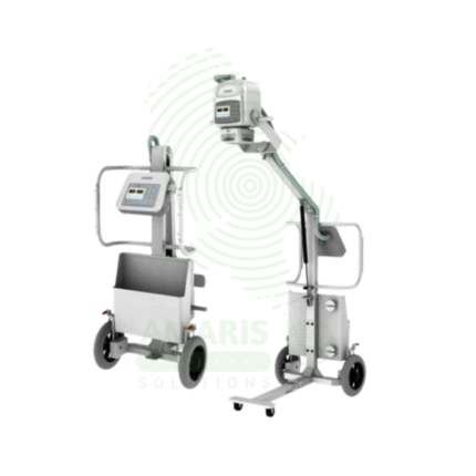
An Analogue Mobile X-ray Machine is a battery-powered, portable X-ray system using traditional film cassettes for bedside imaging in intensive care units, neonatal intensive care units, emergency departments, and operating rooms. The mobile unit enables chest, abdominal, and extremity imaging at the patient's bedside, eliminating the risks associated with transporting critically ill patients. Film cassettes are processed in darkroom facilities for image development. Used in hospitals without digital radiography, as backup for digital systems, and in resource-limited settings where digital infrastructure is not available.
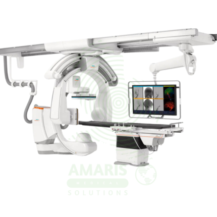
Angiographic Intervention System
An Angiographic Intervention System is a high-end fluoroscopic imaging system designed for guiding minimally invasive vascular and interventional procedures. Essential for cardiac catheterization laboratories, interventional radiology suites, and hybrid operating rooms, it provides real-time, high-resolution visualization of blood vessels, catheters, and devices during coronary interventions, peripheral vascular procedures, neurovascular interventions, and structural heart procedures. Advanced features include rotational angiography for 3D reconstruction, digital subtraction angiography, and dose reduction technologies, enabling precise treatment with minimal radiation exposure.
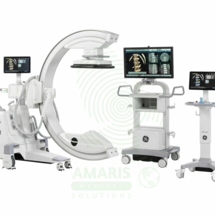
C-Arm Surgical System
A C-Arm Surgical System is a mobile fluoroscopic X-ray imaging device with a distinctive C-shaped arm connecting the X-ray tube and detector. It is an indispensable tool in modern operating rooms and interventional suites, providing real-time live imaging to guide complex procedures in orthopedics, spine surgery, pain management, and vascular interventions. Its mobility allows precise positioning around the patient, while features like pulsed fluoroscopy and dose monitoring are critical for radiation safety. Modern flat-panel systems offer high-resolution imaging and advanced capabilities like 3D Cone-Beam CT. Safe operation demands rigorous adherence to radiation protection protocols (ALARA) for both patients and the surgical team.
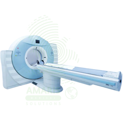
Computed Tomography (CT)
Computed Tomography (CT) is a diagnostic imaging modality that uses X-rays and computer processing to create detailed cross-sectional images of the body. Essential for trauma evaluation, cancer diagnosis, vascular imaging, and surgical planning, CT provides rapid, high-resolution images that guide life-saving decisions in emergency medicine, oncology, and surgery. Advanced multi-slice systems enable whole-body scanning in seconds with sub-millimeter resolution. Radiation dose optimization and contrast safety protocols are essential for patient safety.
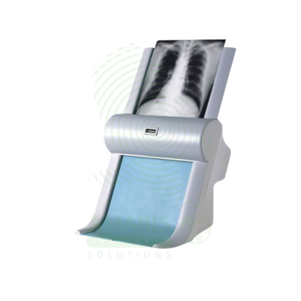
Film Digitizer
A Film Digitizer is a specialized scanner that converts analog radiographic films into high-fidelity digital images (DICOM format). It is the essential tool for migrating historical film archives into a modern digital PACS, enabling filmless workflow, remote access, and long-term preservation of diagnostic records. Its critical performance characteristics are a wide optical density range (to capture all film details) and high spatial resolution. By creating secure, accessible digital copies, it protects against the loss of physical films and integrates past patient history with current digital imaging, supporting comprehensive care and efficient radiology practice.
LED Medical Film Viewer
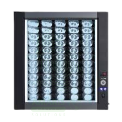
An LED Medical Film Viewer is a light box designed for viewing and interpreting analog X-ray films. Using LED backlight technology, it provides uniform, high-luminance illumination with instant on capability and long life. Available in single, dual, and multi-panel configurations, it supports side-by-side comparison of current and prior studies, pre-operative planning, and group teaching. Essential for radiology departments, orthopedic clinics, emergency departments, and operating rooms where film-based imaging is still used.
Panoramic X-ray Dental Machine
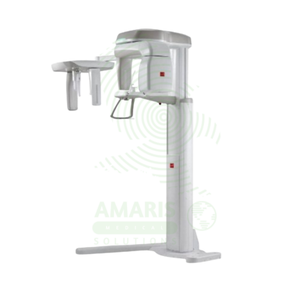
A Panoramic X-ray Dental Machine is a rotating extraoral radiographic system that produces a single, broad 2D image of the entire jaws, teeth, TMJs, and sinuses. By focusing on a curved "focal trough," it provides an efficient screening tool for wisdom teeth evaluation, orthodontic planning, and detecting large jaw pathologies. While offering a valuable overview with a relatively low radiation dose, its diagnostic utility is entirely dependent on precise patient positioning to avoid blurring and distortion. It is a cornerstone of modern dental diagnostic imaging, serving as a crucial first step in comprehensive oral assessment and treatment planning.