Dermatoscope and Magnifiers
 Diagnostic Kits
 Vital Signs Monitors
 Stethoscopes and Accessories
 Otoscopes, Ophthalmoscopes, and Retinoscopes
 Reflex Hammers and Neurological Tools
 Scales and Measuring Devices
 Spirometers and Pulmonary Function Tests
 Electrosurgical Units and Accessories
 Cutting Instruments
 Grasping and Holding Instruments
 Hemostatic Instruments
 Specialized Surgical Sets
 Single-Use Procedure Trays and Packs
 Surgical Drapes, Gowns, and Covers
 Tissue Unifying Instruments
 Radiation Protection
 X-Ray Machines and Accessories
 Ultrasound Systems and Probes
 MRI and CT Scanners
 Radiology Consumables
 Bone Densitometers
 Fluoroscopy Equipment
 Imaging Tables and Positioning Aids
 Microscopes and Accessories
 Centrifuges and Separators
 Analyzers
 Incubators and Ovens
 Pipettes, Dispensers, and Lab Glassware
 Refrigerators, Freezers, and Storage Units
 Lab Consumables
 Sterilizers and Autoclaves for Lab Use
 Multi-Parameter Monitors
 Ventilators and Respiratory Support Devices
 Defibrillators and AEDs
 Infusion Pumps and IV Systems
 Patient Warmers and Cooling Devices
 Central Monitoring Stations
 Accessories
 Anesthesia Machines and Workstations
 Oxygen Concentrators and Delivery Systems
 Nebulizers and Inhalers
 CPAP/BiPAP Machines
 Airway Management
 Anesthesia Masks, Circuits, and Bags
 Humidifiers and Heaters
 Respiratory Therapy Accessories
 First Aid Kits and Cabinets
 Emergency Resuscitation Equipment
 Trauma Supplies
 Emergency Carts and Crash Carts
 Burn Care Products
 Bleeding Control
 Automated External Defibrillators (AEDs)
 Transport and Evacuation
 Wheelchairs and Accessories
 Walkers, Crutches, and Canes
 Prosthetics and Orthotics
 Physical Therapy Equipment
 Transfer Devices
 Bathroom Safety
 Orthopedic Traction and Tables
 Hot/Cold Therapy Packs and Units
 Beds and Mattresses
 Chairs and Stools
 Tables
 Cabinets and Storage
 Privacy Screens & Curtains
 Stands and Racks
 Linens and Textiles
 Lighting
 Autoclaves and Sterilizers
 Ultrasonic Cleaners
 Disinfectant Solutions and Wipes
 Sterilization Pouches, Wraps, and Indicators
 Instrument Trays and Containers
 UV and Ozone Disinfection Devices
 Washer Disinfectors
 Wound Care
 Gloves
 Masks and Respirators
 Catheters and Tubing
 Swabs, Applicators, and Sponges
 Incontinence Products
 Personal Protective Equipment (PPE)
 Dental Chairs and Units
 Handpieces and Burs
 Instruments
 Consumables
 Sterilization for Dental Use
 Orthodontic Supplies
 Endodontic Tools
 Slit Lamps and Tonometers
 Lensometers and Phoropters
 Ophthalmic Surgical Instruments
 Eyewear Frames and Lenses
 Contact Lens Supplies
 Vision Testing Charts and Devices
 Eye Care Consumables
 Laser Systems for Eye Care
 ENT Exam Chairs and Tables
 Endoscopes
 Audiometers and Hearing Tests
 ENT Instruments
 Nasal and Throat Packs
 Hearing Aids and Accessories
 Otology Supplies
 Fetal Dopplers and Monitors
 Delivery Beds and Tables
 Gynecological Instruments
 Neonatal Incubators and Warmers
 Breast Pumps and Accessories
 Contraceptive Devices
 Maternity Supports and Pads
 Neonatal Consumables
 Cystoscopes and Urethroscopes
 Dialysis Machines and Supplies
 Urological Catheters and Bags
 Lithotripters
 Prostate Treatment Devices
 Urinary Incontinence Products
 Kidney Stone Management Tools
 Consumables & Disposables
 EEG and EMG Machines
 Neurosurgical Instruments
 Nerve Stimulators
 Headrests and Positioning Aids
 Lumbar Puncture Kits
 Seizure Monitoring Devices
 Consumables
 Rehabilitation for Neurological Conditions
 ECG Machines and Accessories
 Holter Monitors
 Stress Test Systems
 Pacemakers and Defibrillator Accessories
 Vascular Access Devices
 Cardiac Catheters and Guidewires
 Blood Flow Meters
 Consumables
 Orthopedic Instruments
 Casts, Splints, and Padding
 Joint Replacement Supplies
 Prosthetic Limbs and Components
 Bone Grafts and Substitutes
 Traction Devices
 Orthopedic Braces and Supports
 Rehabilitation Aids for Orthopedics
 Home Oxygen Therapy
 Hospital Beds for Home Use
 Mobility Aids
 Bathroom and Daily Living Aids
 Wound Care for Home
 Monitoring Devices
 Enteral Feeding Pumps and Tubes
 Hand Sanitizers and Dispensers
 Face Shields and Goggles
 Isolation Gowns and Suits
 Biohazard Waste Containers
 Air Purifiers and HEPA Filters
 Surface Disinfectants
 Sharps Containers
 Protective Barriers
 Cardiovascular & Endurance Training
 Strength Training & Weightlifting
 Functional Training & Core Conditioning
 Physical Therapy & Rehabilitation
 Sports & Outdoor Recreation
 Gym Flooring & Facility Equipment
 Fitness Monitoring & Accessories
 Kids & Novelties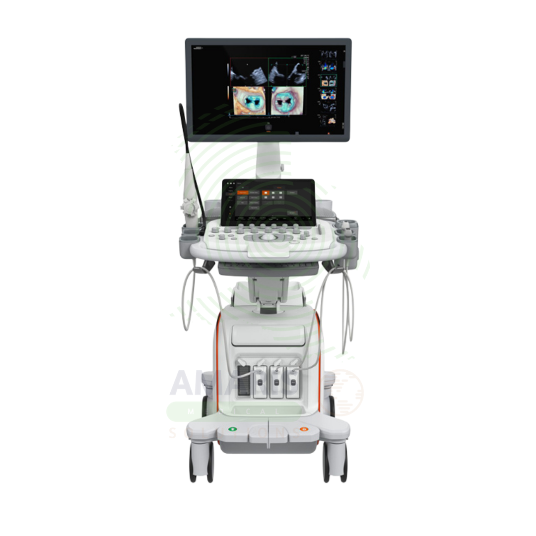
Cardiovascular Ultrasound
$1.00
WhatsApp Order
A Cardiovascular Ultrasound System is a specialized echocardiography device designed for non-invasive assessment of cardiac structure, function, and hemodynamics. Using dedicated phased array transducers and advanced Doppler techniques, it evaluates ventricular function, valvular disease, congenital heart defects, and myocardial ischemia. Essential for cardiology, cardiac surgery, and critical care, it provides the diagnostic information needed to guide treatment decisions for patients with heart failure, valvular disease, coronary artery disease, and congenital heart conditions.
Description
Cardiovascular Ultrasound
PRIMARY CLINICAL & DIAGNOSTIC USES
1. Evaluation of Cardiac Structure and Function
-
Primary Use: Provides real-time imaging of the heart to assess chamber size, wall thickness, valvular structure and function, and overall cardiac performance. The system uses dedicated phased array transducers to visualize the heart through acoustic windows between the ribs.
-
How it helps: For the cardiologist and echocardiographer, cardiovascular ultrasound provides a dynamic, non-invasive view of the beating heart—revealing the strength of the heart muscle, the function of each valve, and the size of each chamber. For the patient with heart failure, valve disease, or suspected cardiomyopathy, echocardiography provides the critical information needed to guide diagnosis and treatment.
2. Assessment of Ventricular Function
-
Primary Use: Measures left ventricular ejection fraction, assesses regional wall motion abnormalities, and evaluates right ventricular function. Quantitative analysis tools provide objective measurements of systolic and diastolic function.
-
How it helps: For the cardiologist managing patients with heart failure or after myocardial infarction, accurate measurement of ejection fraction guides decisions about medication, device therapy (ICD, CRT), and surgical intervention. For the patient, knowing their ejection fraction helps them understand the severity of their heart condition and the expected benefits of treatment.
3. Valvular Heart Disease Evaluation
-
Primary Use: Assesses valvular structure, leaflet mobility, calcification, and stenosis or regurgitation severity. Doppler techniques measure pressure gradients across valves and calculate valve area.
-
How it helps: For the cardiologist and cardiac surgeon, echocardiography provides the detailed assessment needed to determine the timing and type of valve intervention—whether valve repair or replacement is indicated, and whether surgical or transcatheter approach is appropriate. For the patient with aortic stenosis, mitral regurgitation, or other valve disease, accurate assessment ensures intervention is performed at the optimal time.
4. Doppler Assessment of Blood Flow
-
Primary Use: Color Doppler visualizes blood flow direction and velocity across valves and within chambers. Spectral Doppler (Pulsed-wave and Continuous-wave) quantifies flow velocities, pressure gradients, and calculates valve areas and pulmonary artery pressures.
-
How it helps: For the cardiologist, Doppler ultrasound makes blood flow visible—revealing the jet of mitral regurgitation, the high-velocity flow of aortic stenosis, and the direction of shunts. For the patient, Doppler assessment provides quantifiable measurements that track disease progression and response to treatment.
5. Stress Echocardiography
-
Primary Use: Evaluates cardiac function before and after exercise or pharmacological stress to detect inducible ischemia, assess myocardial viability, and evaluate hemodynamic response to stress. The system captures images at rest and immediately after stress.
-
How it helps: For the cardiologist evaluating patients with suspected coronary artery disease, stress echocardiography identifies areas of the heart that become ischemic under stress—guiding decisions about angiography, revascularization, and medical therapy. For the patient, a normal stress echo provides reassurance that significant coronary artery disease is unlikely.
SECONDARY & SUPPORTIVE USES
1. Congenital Heart Disease Evaluation: Used to assess congenital heart defects in pediatric and adult patients, including septal defects, valve anomalies, and complex congenital malformations.
2. Intraoperative Echocardiography: Used during cardiac surgery to assess surgical results, guide valve repair, and evaluate ventricular function before and after cardiopulmonary bypass.
3. Transesophageal Echocardiography: Specialized transducer provides high-resolution imaging of posterior cardiac structures, used when transthoracic windows are inadequate or for intraoperative guidance.
4. Cardiac Resynchronization Therapy Guidance: Used to assess dyssynchrony and guide lead placement for biventricular pacing.
5. Pericardial Disease Evaluation: Assesses pericardial effusion, tamponade physiology, and pericardial thickening.
6. Endocarditis Evaluation: Identifies vegetations, abscesses, and complications of infective endocarditis.
KEY PRODUCT FEATURES
1. BASIC IDENTIFICATION ATTRIBUTES
-
Device Type: A specialized ultrasound system designed for cardiac imaging with dedicated transducers and quantitative analysis software.
-
Designation: Cardiovascular Ultrasound, Echocardiography System, Echo Machine, Cardiac Ultrasound.
-
System Configurations:
-
Cart-Based: Full-featured system for echocardiography laboratories.
-
Portable: Compact system for point-of-care and bedside use.
-
Handheld: Pocket-sized devices for focused cardiac assessment.
-
-
Key Components:
-
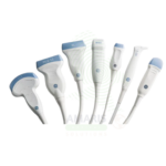
Phased Array Transducer: Dedicated cardiac probe with small footprint for intercostal imaging.
-
Doppler Capabilities: Color, Pulsed-wave, Continuous-wave Doppler.
-
Quantitative Software: Automated ejection fraction, strain imaging, 3D volume analysis.
-
ECG: Integrated ECG for cardiac cycle timing.
-
Stress Echo Package: Software for rest-stress comparison.
-
2. TECHNICAL & PERFORMANCE PROPERTIES
-
Transducer Frequency: Phased array: 1-5 MHz for adult cardiac; higher frequency for pediatric.
-
Doppler: Pulsed-wave (PW), Continuous-wave (CW), Color Doppler.
-
Spectral Analysis: Real-time spectral display; automated calculations.
-
2D Frame Rate: High frame rate for capturing rapid cardiac motion.
-
Harmonic Imaging: Tissue harmonic imaging for improved endocardial border definition.
-
Strain Imaging: Speckle tracking for myocardial deformation analysis.
-
3D/4D Capability: Real-time 3D imaging for volumetric assessment.
3. PHYSICAL & OPERATIONAL PROPERTIES
-
Construction: Cart-based with large high-resolution monitor; portable options available.
-
ECG Integration: Wired or wireless ECG for gating.
-
Controls: Dedicated cardiac presets; touchscreen interface.
-
Image Storage: Digital storage; DICOM export; vendor-neutral format support.
4. SAFETY & COMPLIANCE ATTRIBUTES
-
Regulatory Status: Class II medical device regulated by FDA.
-
Acoustic Output: Regulated by FDA output display standards.
-
Electrical Safety: Compliant with IEC 60601-1.
-
Transducer Safety: Compatible with sterilization and disinfection protocols.
5. STORAGE & HANDLING ATTRIBUTES
-
Storage: Stored in an echocardiography laboratory or designated area.
-
Cleaning: Wipe console and transducers with hospital-grade disinfectants.
-
Transducer Care: Use appropriate disinfection level; handle carefully to avoid damage.
-
ECG Lead Care: Replace leads as needed; clean between patients.
6. LABORATORY & CLINICAL APPLICATIONS
-
Primary Application: Non-invasive assessment of cardiac structure, function, and hemodynamics.
-
Clinical Role: Essential imaging modality in cardiology, cardiac surgery, and critical care.
SAFETY HANDLING PRECAUTIONS
1. SAFETY PRECAUTIONS
-
Acoustic Output: Follow ALARA principles; use appropriate power settings.
-
Transducer Handling: Handle phased array transducers carefully; avoid drops.
-
ECG Safety: Use isolated ECG leads; follow electrical safety protocols.
-
Patient Positioning: Ensure patient comfort and safety during examination, especially during stress testing.
2. FIRST AID MEASURES
-
Patient Syncope: If patient faints during stress echo, discontinue stress; place in Trendelenburg; monitor vitals.
-
Electrical Shock: If shock occurs, disconnect power; seek medical attention.
3. FIRE FIGHTING MEASURES
-
Flammability: Plastic components are combustible; electrical components may pose fire risk.
-
Extinguishing Media: For electrical fire, use CO₂ or dry chemical extinguisher.
Related products
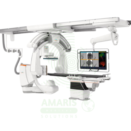
Angiographic Intervention System
An Angiographic Intervention System is a high-end fluoroscopic imaging system designed for guiding minimally invasive vascular and interventional procedures. Essential for cardiac catheterization laboratories, interventional radiology suites, and hybrid operating rooms, it provides real-time, high-resolution visualization of blood vessels, catheters, and devices during coronary interventions, peripheral vascular procedures, neurovascular interventions, and structural heart procedures. Advanced features include rotational angiography for 3D reconstruction, digital subtraction angiography, and dose reduction technologies, enabling precise treatment with minimal radiation exposure.
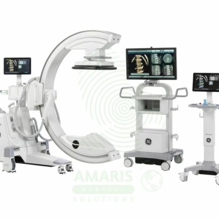
C-Arm Surgical System
A C-Arm Surgical System is a mobile fluoroscopic X-ray imaging device with a distinctive C-shaped arm connecting the X-ray tube and detector. It is an indispensable tool in modern operating rooms and interventional suites, providing real-time live imaging to guide complex procedures in orthopedics, spine surgery, pain management, and vascular interventions. Its mobility allows precise positioning around the patient, while features like pulsed fluoroscopy and dose monitoring are critical for radiation safety. Modern flat-panel systems offer high-resolution imaging and advanced capabilities like 3D Cone-Beam CT. Safe operation demands rigorous adherence to radiation protection protocols (ALARA) for both patients and the surgical team.
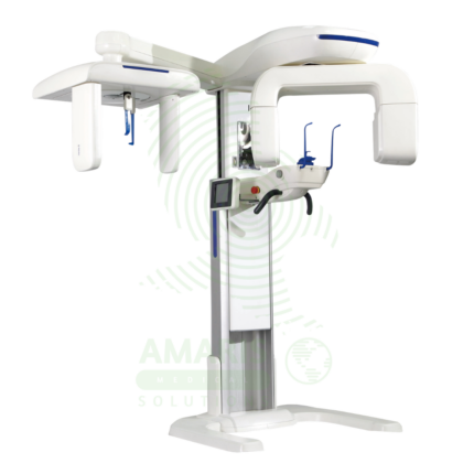
Dental X-ray Machine
A Dental X-ray Machine is a specialized radiographic system designed for imaging teeth, jaws, and facial structures. It encompasses intraoral units for detailed tooth-specific views, panoramic machines for wide screening shots, and advanced Cone Beam CT (CBCT) scanners for 3D surgical planning. Utilizing low-dose radiation and digital imaging technology, it is indispensable for diagnosing cavities, gum disease, infections, and planning treatments like implants, orthodontics, and oral surgery. Its safe operation requires strict adherence to radiation protection protocols, including the use of lead aprons, proper collimation, and operator training to ensure patient and staff safety while obtaining critical diagnostic information.
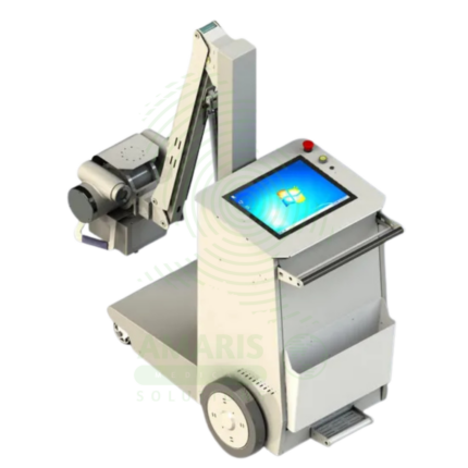
Digital & Analog X-ray Machine
A Digital & Analog X-ray Machine is a fundamental medical imaging device that uses a controlled beam of ionizing radiation to produce static or real-time images of the body's internal structures. It is indispensable for diagnosing fractures, lung diseases, dental issues, and many abdominal conditions. The transition from Analog (film-based) to Digital (CR or DR) technology has revolutionized the field, offering faster results, superior image manipulation, improved dose efficiency, and seamless integration into digital healthcare networks. Its operation demands strict adherence to radiation safety protocols (ALARA) to protect patients and staff, making it a cornerstone of safe, effective diagnostic medicine.
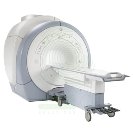
Magnetic Resonance Imaging
Magnetic Resonance Imaging (MRI) is a non-invasive diagnostic imaging modality that uses powerful magnetic fields and radiofrequency waves to produce detailed images of soft tissues, organs, and internal structures without ionizing radiation. It is the gold standard for imaging the brain, spinal cord, joints, muscles, and ligaments, and is essential for neurological, musculoskeletal, oncologic, and cardiovascular diagnosis. MRI provides exceptional soft tissue contrast, enabling precise anatomical characterization, tumor staging, and treatment planning. Strict safety protocols for ferromagnetic screening and contrast administration are essential for patient safety.
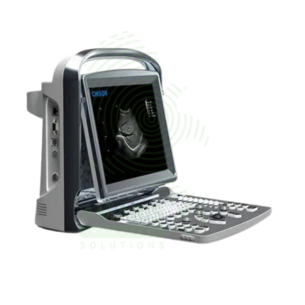
Portable Ultrasound Machine
A Portable Ultrasound Machine is a compact, battery-powered imaging device designed for point-of-care use, bringing diagnostic capability directly to the patient's bedside. It is essential for rapid triage in emergency and critical care settings, procedural guidance, and basic examinations in clinics and remote locations. While offering core imaging modes like B-mode and Color Doppler in a lightweight, durable package, its effective use requires clinician training to recognize both its diagnostic value and its limitations compared to comprehensive departmental systems. Proper cleaning, battery management, and data security are paramount for its safe and effective deployment.
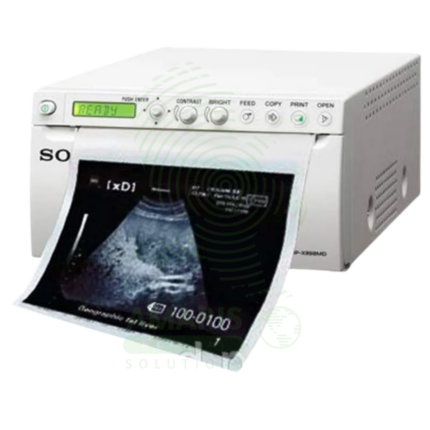
Sony Ultrasound Printer
A Sony Ultrasound Printer is a specialized medical-grade printer that produces high-quality hard copies of ultrasound images. Utilizing dry thermal, laser, or dye-sublimation technology, it creates grayscale or color prints on photographic paper or film, providing a physical record for patient charts, referrals, and consultations. While its role has diminished with the rise of fully digital PACS workflows, it remains essential in settings requiring immediate tangible output, patient education, or where digital infrastructure is limited. Its performance is defined by high spatial and contrast resolution to accurately reproduce the subtleties of sonographic images.