Dermatoscope and Magnifiers
 Diagnostic Kits
 Vital Signs Monitors
 Stethoscopes and Accessories
 Otoscopes, Ophthalmoscopes, and Retinoscopes
 Reflex Hammers and Neurological Tools
 Scales and Measuring Devices
 Spirometers and Pulmonary Function Tests
 Electrosurgical Units and Accessories
 Cutting Instruments
 Grasping and Holding Instruments
 Hemostatic Instruments
 Specialized Surgical Sets
 Single-Use Procedure Trays and Packs
 Surgical Drapes, Gowns, and Covers
 Tissue Unifying Instruments
 Radiation Protection
 X-Ray Machines and Accessories
 Ultrasound Systems and Probes
 MRI and CT Scanners
 Radiology Consumables
 Bone Densitometers
 Fluoroscopy Equipment
 Imaging Tables and Positioning Aids
 Microscopes and Accessories
 Centrifuges and Separators
 Analyzers
 Incubators and Ovens
 Pipettes, Dispensers, and Lab Glassware
 Refrigerators, Freezers, and Storage Units
 Lab Consumables
 Sterilizers and Autoclaves for Lab Use
 Multi-Parameter Monitors
 Ventilators and Respiratory Support Devices
 Defibrillators and AEDs
 Infusion Pumps and IV Systems
 Patient Warmers and Cooling Devices
 Central Monitoring Stations
 Accessories
 Anesthesia Machines and Workstations
 Oxygen Concentrators and Delivery Systems
 Nebulizers and Inhalers
 CPAP/BiPAP Machines
 Airway Management
 Anesthesia Masks, Circuits, and Bags
 Humidifiers and Heaters
 Respiratory Therapy Accessories
 First Aid Kits and Cabinets
 Emergency Resuscitation Equipment
 Trauma Supplies
 Emergency Carts and Crash Carts
 Burn Care Products
 Bleeding Control
 Automated External Defibrillators (AEDs)
 Transport and Evacuation
 Wheelchairs and Accessories
 Walkers, Crutches, and Canes
 Prosthetics and Orthotics
 Physical Therapy Equipment
 Transfer Devices
 Bathroom Safety
 Orthopedic Traction and Tables
 Hot/Cold Therapy Packs and Units
 Beds and Mattresses
 Chairs and Stools
 Tables
 Cabinets and Storage
 Privacy Screens & Curtains
 Stands and Racks
 Linens and Textiles
 Lighting
 Autoclaves and Sterilizers
 Ultrasonic Cleaners
 Disinfectant Solutions and Wipes
 Sterilization Pouches, Wraps, and Indicators
 Instrument Trays and Containers
 UV and Ozone Disinfection Devices
 Washer Disinfectors
 Wound Care
 Gloves
 Masks and Respirators
 Catheters and Tubing
 Swabs, Applicators, and Sponges
 Incontinence Products
 Personal Protective Equipment (PPE)
 Dental Chairs and Units
 Handpieces and Burs
 Instruments
 Consumables
 Sterilization for Dental Use
 Orthodontic Supplies
 Endodontic Tools
 Slit Lamps and Tonometers
 Lensometers and Phoropters
 Ophthalmic Surgical Instruments
 Eyewear Frames and Lenses
 Contact Lens Supplies
 Vision Testing Charts and Devices
 Eye Care Consumables
 Laser Systems for Eye Care
 ENT Exam Chairs and Tables
 Endoscopes
 Audiometers and Hearing Tests
 ENT Instruments
 Nasal and Throat Packs
 Hearing Aids and Accessories
 Otology Supplies
 Fetal Dopplers and Monitors
 Delivery Beds and Tables
 Gynecological Instruments
 Neonatal Incubators and Warmers
 Breast Pumps and Accessories
 Contraceptive Devices
 Maternity Supports and Pads
 Neonatal Consumables
 Cystoscopes and Urethroscopes
 Dialysis Machines and Supplies
 Urological Catheters and Bags
 Lithotripters
 Prostate Treatment Devices
 Urinary Incontinence Products
 Kidney Stone Management Tools
 Consumables & Disposables
 EEG and EMG Machines
 Neurosurgical Instruments
 Nerve Stimulators
 Headrests and Positioning Aids
 Lumbar Puncture Kits
 Seizure Monitoring Devices
 Consumables
 Rehabilitation for Neurological Conditions
 ECG Machines and Accessories
 Holter Monitors
 Stress Test Systems
 Pacemakers and Defibrillator Accessories
 Vascular Access Devices
 Cardiac Catheters and Guidewires
 Blood Flow Meters
 Consumables
 Orthopedic Instruments
 Casts, Splints, and Padding
 Joint Replacement Supplies
 Prosthetic Limbs and Components
 Bone Grafts and Substitutes
 Traction Devices
 Orthopedic Braces and Supports
 Rehabilitation Aids for Orthopedics
 Home Oxygen Therapy
 Hospital Beds for Home Use
 Mobility Aids
 Bathroom and Daily Living Aids
 Wound Care for Home
 Monitoring Devices
 Enteral Feeding Pumps and Tubes
 Hand Sanitizers and Dispensers
 Face Shields and Goggles
 Isolation Gowns and Suits
 Biohazard Waste Containers
 Air Purifiers and HEPA Filters
 Surface Disinfectants
 Sharps Containers
 Protective Barriers
 Cardiovascular & Endurance Training
 Strength Training & Weightlifting
 Functional Training & Core Conditioning
 Physical Therapy & Rehabilitation
 Sports & Outdoor Recreation
 Gym Flooring & Facility Equipment
 Fitness Monitoring & Accessories
 Kids & Novelties
Chest Bottle
WhatsApp Order
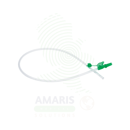
A Chest Bottle (Underwater Seal Drainage System) is a three-chamber disposable device used to drain air and fluid from the pleural space via a chest tube. Its critical component is a water seal chamber that acts as a one-way valve, preventing air from re-entering the chest while allowing drainage. It is used to treat pneumothorax, hemothorax, and pleural effusions, and is mandatory post-thoracic surgery. The system must always be kept upright and below the patient’s chest level. It provides essential clinical information through tidaling in the water seal (indicating tube patency and lung expansion) and bubbling (indicating an air leak), making it both a therapeutic and diagnostic tool.
Description
Chest Bottle
PRIMARY CLINICAL & DIAGNOSTIC USES
1. Underwater Seal Drainage of the Pleural Cavity
-
Primary Use: Provides a one-way valve for air and fluid (blood, effusion, chyle, empyema) draining from a patient’s pleural space via a chest tube, preventing atmospheric air from being sucked back into the chest during inspiration.
-
How it helps: For the surgeon, pulmonologist, or intensive care nurse, the underwater seal is a simple yet elegant physiological solution—it lets unwanted contents leave the chest but absolutely nothing return. For the patient with a collapsed lung or fluid around their lung, this mechanism ensures that as they breathe in, they draw air into their lungs, not back into the pleural space, allowing the lung to gradually re-expand and heal.
2. Treatment of Pneumothorax
-
Primary Use: Evacuates air from the pleural space, allowing a collapsed lung to re-expand, with bubbling in the water seal chamber indicating an ongoing air leak.
-
How it helps: For the clinician managing a pneumothorax, the chest bottle transforms an invisible physiological problem into a visible one—bubbles tell the story of air leaking from the lung. For the patient who arrived gasping with a collapsed lung, watching bubbles travel through the tube provides tangible evidence that the trapped air is escaping and their lung has room to breathe again.
3. Treatment of Hemothorax, Pleural Effusion, and Empyema
-
Primary Use: Allows for the controlled drainage of blood, serous fluid, or pus from the pleural cavity, monitoring the volume and rate of output.
-
How it helps: For the trauma surgeon or medical team, the graduated collection chamber provides continuous feedback—how much blood is this patient losing? Is the effusion draining? Is the infection clearing? For the patient, each milliliter drained is fluid that was compressing their lung, making every breath a little easier as the chest bottle slowly restores space for their lung to expand.
4. Post-Operative Thoracic Drainage
-
Primary Use: Standard following thoracic surgeries (lobectomy, pneumonectomy, cardiac surgery) to drain residual air and fluid, monitor for bleeding, and ensure lung re-expansion.
-
How it helps: For the cardiothoracic surgical team, the chest bottle is their window into the chest after they’ve closed it—watching for post-op bleeding, air leaks from suture lines, or proper lung re-expansion. For the patient waking from heart or lung surgery, this silent companion by the bedside is working continuously to clear the surgical site, prevent complications, and help them breathe deeply as they begin their recovery.
SECONDARY & SUPPORTIVE USES
1. Measurement of Output: For the nursing and medical team, the calibrated collection chamber allows precise measurement of fluid volume and character—distinguishing between expected post-op drainage, concerning hemorrhage, or signs of infection. For the patient, accurate output monitoring ensures that complications are caught early, before they become emergencies.
2. Assessment of Air Leak: For the respiratory therapist or surgeon, the water seal chamber provides a visual window into lung healing—continuous bubbling suggests a significant air leak from lung parenchyma, while intermittent bubbling with cough is normal. For the patient with a lung injury or recent surgery, the gradual decrease and cessation of bubbling over days signals that their lung is sealing and healing as it should.
3. Application of Controlled Suction: For the clinician managing complex drainage, the suction control chamber allows precise negative pressure application (typically -10 to -20 cm H₂O) to enhance drainage and lung expansion. For the patient with a persistent pneumothorax or large effusion, gentle suction actively encourages the lung to re-expand and hold against the chest wall, speeding recovery and reducing time with a chest tube in place.
KEY PRODUCT FEATURES
1. BASIC IDENTIFICATION ATTRIBUTES
-
Device Type: A disposable, rigid plastic, three-chamber collection system used in conjunction with a chest tube.
-
Common Names: Often called an "Underwater Seal Drainage System," "Pleurovac," or "Chest Drainage Unit."
-
Core Chambers & Function:
-
Collection Chamber: The first chamber. Receives fluid and air directly from the patient via the chest tube. It is calibrated to measure output (in mL).
-
Water Seal Chamber: The second and most critical chamber. Contains sterile water (typically filled to the 2cm mark). The tube from the collection chamber dips 2cm below the water surface, creating a one-way seal. This chamber shows tidaling (fluctuation with respiration) and bubbling (air leak).
-
Suction Control Chamber: The third chamber. Used when suction is applied. Filled with water to a level (e.g., -20 cm) that determines the maximum negative pressure applied to the pleural space. Bubbling in this chamber indicates suction is being applied.
-
2. TECHNICAL & PERFORMANCE PROPERTIES
-
Calibration: Clear, accurate mL markings on the collection chamber for measuring output.
-
Water Seal Depth: Standard 2 cm H2O. This is the minimum pressure needed to overcome the seal and allow air to escape from the pleural space.
-
Suction Control Setting: Adjustable by adding or removing water (e.g., -10, -15, -20 cm H2O). The suction source must be set high enough to cause gentle, continuous bubbling in this chamber.
-
High-Volume Capacity: Collection chambers can typically hold 2000-2500 mL of fluid before requiring emptying/replacement.
3. PHYSICAL & OPERATIONAL PROPERTIES
-
Connections: Standardized tubing connectors: a patient port (from chest tube), a suction port (to wall vacuum), and an atmospheric vent.
-
Stability: Designed with a wide base and often a hanger to be positioned below the patient's chest level (on the floor or bed rail) at all times to maintain the water seal.
-
Transparency: Made of clear plastic for continuous visual assessment of all chambers.
4. SAFETY & COMPLIANCE ATTRIBUTES
-
Regulatory Status: Classified as a Class II medical device.
-
Sterility: The system is supplied sterile, and the water added to the seal and suction chambers must be sterile.
-
One-Way Valve Integrity: The underwater seal is a passive, fail-safe mechanism. The system must remain upright and intact to maintain this seal.
5. STORAGE & HANDLING ATTRIBUTES
-
Storage: Store in a clean, dry area in its sealed package.
-
Setup: Must be set up and filled with sterile water according to strict aseptic technique before connection to the patient's chest tube.
-
Handling During Use: The unit must always be kept upright and below the level of the patient's chest. Tilting or raising it above the chest can cause fluid to flow back into the pleural space.
-
Disposal: Once disconnected from the patient, the entire unit is disposed of as biohazardous/clinical waste due to contamination with bodily fluids.
6. LABORATORY & CLINICAL APPLICATIONS
-
Primary Application: The standard of care for managing chest tubes in thoracic surgery, pulmonology, emergency medicine, and critical care.
-
Clinical Role: A critical monitoring and therapeutic device that provides both treatment (drainage) and continuous diagnostic information (air leak, output volume) for pleural space pathologies.
SAFETY HANDLING PRECAUTIONS
1. SAFETY PRECAUTIONS
-
Position is Paramount: The bottle must always be kept below chest level. Elevating it will cause siphoning of fluid back into the pleural cavity, risking infection or tamponade.
-
Maintain Water Seal: Never empty the water seal chamber while the system is connected to the patient. The water level must be checked and maintained at the 2cm mark.
-
Tube Patency: Ensure the chest tube and all connecting tubing are not kinked, compressed, or clogged. Monitor for tidaling in the water seal chamber; absence of tidaling may indicate tube obstruction, lung re-expansion, or system malfunction.
-
Clamping Chest Tubes: Clamp chest tubes only under specific, directed circumstances (e.g., to quickly assess for a persistent air leak, or to change the drainage system). Never clamp for transport or without a clear clinical reason, as it can rapidly cause a tension pneumothorax.
-
Sudden Cessation of Bubbling/Tidaling: This may indicate lung re-expansion (desired) or tube obstruction (dangerous). Assess the patient clinically (breath sounds, oxygenation) and the system immediately.
2. FIRST AID MEASURES
-
Accidental Disconnection at Patient Port: Immediately clamp the chest tube close to the patient's body (if a clamp is at hand), ask the patient to exhale and hold it if possible, and reconnect the tubing using aseptic technique. The exposed end of the chest tube can be temporarily submerged in a bottle of sterile water to create an emergency seal.
-
Bottle Tip-Over/Breakage: If the system is compromised, clamp the chest tube temporarily. Replace the entire drainage system as quickly as possible using a new, pre-set-up unit.
-
Tension Pneumothorax Suspected (Patient in Distress): This is a clinical emergency. If a tension pneumothorax is suspected due to system malfunction or occlusion, immediately decompress the chest with a needle thoracostomy if necessary, following emergency protocols. Do not wait.
3. FIRE FIGHTING MEASURES
-
Flammability: Plastic components are combustible.
-
Extinguishing Media: Use water, CO2, or foam as appropriate for the surrounding fire.
Related products
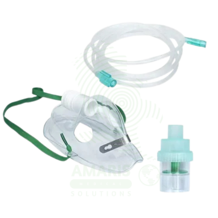
Adult & Pediatric Nebulizer Kit
An Adult & Pediatric Nebulizer Kit is a complete set of patient-contact components required for aerosol medication delivery, including a nebulizer cup, connecting tubing, and both an adult mouthpiece and a pediatric mask (or universal fit). Designed for use with a compressor, it provides the delivery circuit for treating asthma, COPD, and other respiratory conditions. Available in disposable (single-patient-use) formats for infection control in clinics or reusable formats for home care, its proper use, coupled with rigorous cleaning for reusable kits, is essential for effective therapy and preventing respiratory infections. The kit's universal connectors ensure compatibility with standard compressors.
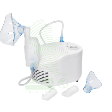
Compressor Nebulizer Machine
A Compressor Nebulizer Machine is a pneumatic device that transforms liquid medication into a breathable mist for treating respiratory conditions. Consisting of an electric air compressor, a nebulizer cup, tubing, and a mask or mouthpiece, it is particularly effective for infants, children, and patients with severe asthma, COPD, or cystic fibrosis who require reliable aerosol delivery. While offering robust performance for home and clinical use, its safety and efficacy depend critically on proper cleaning to prevent infection, correct assembly, and the use of prescribed nebulizer-compatible medications. It remains a fundamental tool for both acute intervention and chronic management of pulmonary diseases.
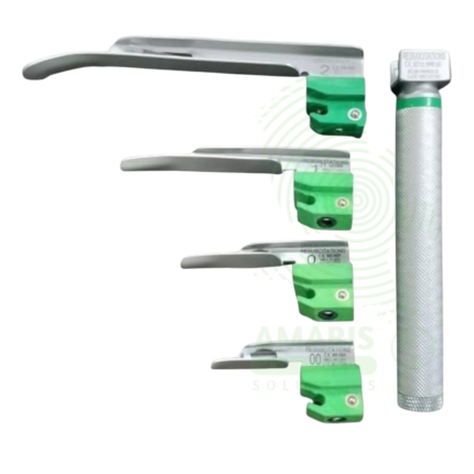
Miller Fiber Optic Laryngoscope
A Miller Fiber Optic Laryngoscope is a rigid laryngoscope with straight Miller blade (sizes 0-4, 70-160 mm) incorporating an integrated fiber optic light bundle that transmits bright, focused illumination (2,000-10,000+ Lux) from a handle-mounted LED or xenon bulb to the blade tip for enhanced visualization during tracheal intubation. The straight blade design allows direct elevation of the epiglottis rather than the indirect vallecula technique, making it particularly useful for pediatric/neonatal intubation, patients with floppy or prominent epiglottis, anterior airways, and difficult airways requiring direct epiglottic control. Features stainless steel reusable blades, ergonomic handles with knurled grip, ISO standard hook-on fittings, and steam autoclave compatibility. Primary clinical applications include direct epiglottis elevation for tracheal intubation (especially pediatric and neonatal), difficult airway management with fiber optic illumination, patients with prominent or floppy epiglottis, cervical spine precautions, anterior airway management, teaching and training (straight blade technique), and neonatal resuscitation. Class II medical device requiring FDA clearance. Critical safety considerations include pre-use light check (dark spots indicate broken fibers), appropriate blade size selection (especially critical in pediatrics), proper lifting technique (direct epiglottic lift, not levering on teeth), pediatric fragility awareness, battery verification, fiber optic care (avoid sharp bending), and backup device availability.
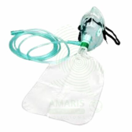
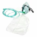
Non-Rebreathing Oxygen Mask
An Non-Rebreathing Oxygen Mask is a high-concentration oxygen delivery device designed for emergency use in critically hypoxic patients. It features a face mask with an attached reservoir bag and a system of one-way valves to prevent the rebreathing of exhaled carbon dioxide, allowing for the delivery of up to 95% FiO2. Its safe operation depends on a minimum oxygen flow of 10-15 L/min to keep the reservoir bag inflated. It is contraindicated where uncontrolled high-flow oxygen may be harmful (e.g., in some COPD patients) and carries a severe fire risk. It is a vital, immediate-intervention tool found in all emergency response settings.
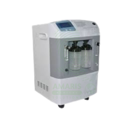
Oxygen Concentrator
An Oxygen Concentrator is a Class II medical device that delivers 90-95% pure supplemental oxygen to patients with chronic hypoxemia using pressure swing adsorption (PSA) technology to concentrate oxygen from room air. Available as stationary/home units (10-30 kg, 0.5-10 L/min continuous flow) for long-term oxygen therapy (LTOT) and portable/ambulatory units (1-10 kg, 0.5-3 L/min pulse-dose or continuous flow) for active patients requiring mobility. Features include oxygen purity monitoring with alarms, digital flow control, hour meters, washable filters, and (for portable units) rechargeable batteries, FAA approval for travel, and pulse-dose delivery to conserve oxygen. Primary clinical indications include COPD, pulmonary fibrosis, cystic fibrosis, and other chronic respiratory conditions with resting, exertional, or nocturnal hypoxemia (PaO2 ≤55 mmHg or SpO2 ≤88%). Essential for home-based oxygen therapy, enabling patients to maintain independence, reduce hospitalizations, and improve quality of life. Critical safety precautions include NO SMOKING in the oxygen environment, keeping away from heat sources and open flames, proper electrical safety, regular filter cleaning, and having backup oxygen for power outages.
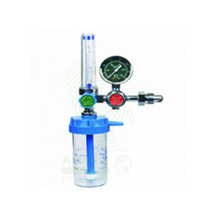
Oxygen Regulator Set
An Oxygen Regulator Set is a critical pressure control device that attaches to a medical oxygen cylinder, reducing its extremely high internal pressure to a safe, usable level and providing precise control over the flow rate delivered to the patient. Consisting of a pin-indexed yoke, high-pressure gauge, adjustable flowmeter, and safety outlet, it is the essential interface for safe oxygen delivery from cylinders in emergencies, during transport, and in home care. Its safe operation hinges on absolute adherence to oil-free handling procedures, correct connection techniques, and regular inspection for leaks or damage to prevent catastrophic oxygen-fuelled fires or equipment failure.
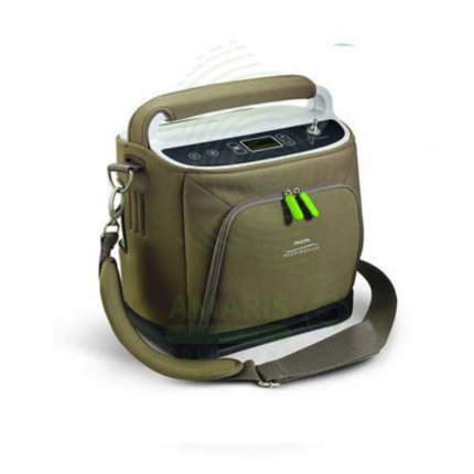
Portable Oxygen Concentrator
A Portable Oxygen Concentrator (POC) is a lightweight, battery-powered device that delivers oxygen via pulse dose technology, enabling active, mobile lifestyles for patients with chronic lung disease. By providing oxygen on-demand with each breath, it maximizes battery efficiency and portability, allowing users to travel, exercise, and socialize freely. It is a prescription-only device that requires careful titration to match a patient's needs during activity and is not a substitute for a stationary concentrator used at home and during sleep. Key considerations include FAA approval for air travel, battery life management, and understanding its specific use case as an ambulatory aid, not a primary oxygen source.