Dermatoscope and Magnifiers
 Diagnostic Kits
 Vital Signs Monitors
 Stethoscopes and Accessories
 Otoscopes, Ophthalmoscopes, and Retinoscopes
 Reflex Hammers and Neurological Tools
 Scales and Measuring Devices
 Spirometers and Pulmonary Function Tests
 Electrosurgical Units and Accessories
 Cutting Instruments
 Grasping and Holding Instruments
 Hemostatic Instruments
 Specialized Surgical Sets
 Single-Use Procedure Trays and Packs
 Surgical Drapes, Gowns, and Covers
 Tissue Unifying Instruments
 Radiation Protection
 X-Ray Machines and Accessories
 Ultrasound Systems and Probes
 MRI and CT Scanners
 Radiology Consumables
 Bone Densitometers
 Fluoroscopy Equipment
 Imaging Tables and Positioning Aids
 Microscopes and Accessories
 Centrifuges and Separators
 Analyzers
 Incubators and Ovens
 Pipettes, Dispensers, and Lab Glassware
 Refrigerators, Freezers, and Storage Units
 Lab Consumables
 Sterilizers and Autoclaves for Lab Use
 Multi-Parameter Monitors
 Ventilators and Respiratory Support Devices
 Defibrillators and AEDs
 Infusion Pumps and IV Systems
 Patient Warmers and Cooling Devices
 Central Monitoring Stations
 Accessories
 Anesthesia Machines and Workstations
 Oxygen Concentrators and Delivery Systems
 Nebulizers and Inhalers
 CPAP/BiPAP Machines
 Airway Management
 Anesthesia Masks, Circuits, and Bags
 Humidifiers and Heaters
 Respiratory Therapy Accessories
 First Aid Kits and Cabinets
 Emergency Resuscitation Equipment
 Trauma Supplies
 Emergency Carts and Crash Carts
 Burn Care Products
 Bleeding Control
 Automated External Defibrillators (AEDs)
 Transport and Evacuation
 Wheelchairs and Accessories
 Walkers, Crutches, and Canes
 Prosthetics and Orthotics
 Physical Therapy Equipment
 Transfer Devices
 Bathroom Safety
 Orthopedic Traction and Tables
 Hot/Cold Therapy Packs and Units
 Beds and Mattresses
 Chairs and Stools
 Tables
 Cabinets and Storage
 Privacy Screens & Curtains
 Stands and Racks
 Linens and Textiles
 Lighting
 Autoclaves and Sterilizers
 Ultrasonic Cleaners
 Disinfectant Solutions and Wipes
 Sterilization Pouches, Wraps, and Indicators
 Instrument Trays and Containers
 UV and Ozone Disinfection Devices
 Washer Disinfectors
 Wound Care
 Gloves
 Masks and Respirators
 Catheters and Tubing
 Swabs, Applicators, and Sponges
 Incontinence Products
 Personal Protective Equipment (PPE)
 Dental Chairs and Units
 Handpieces and Burs
 Instruments
 Consumables
 Sterilization for Dental Use
 Orthodontic Supplies
 Endodontic Tools
 Slit Lamps and Tonometers
 Lensometers and Phoropters
 Ophthalmic Surgical Instruments
 Eyewear Frames and Lenses
 Contact Lens Supplies
 Vision Testing Charts and Devices
 Eye Care Consumables
 Laser Systems for Eye Care
 ENT Exam Chairs and Tables
 Endoscopes
 Audiometers and Hearing Tests
 ENT Instruments
 Nasal and Throat Packs
 Hearing Aids and Accessories
 Otology Supplies
 Fetal Dopplers and Monitors
 Delivery Beds and Tables
 Gynecological Instruments
 Neonatal Incubators and Warmers
 Breast Pumps and Accessories
 Contraceptive Devices
 Maternity Supports and Pads
 Neonatal Consumables
 Cystoscopes and Urethroscopes
 Dialysis Machines and Supplies
 Urological Catheters and Bags
 Lithotripters
 Prostate Treatment Devices
 Urinary Incontinence Products
 Kidney Stone Management Tools
 Consumables & Disposables
 EEG and EMG Machines
 Neurosurgical Instruments
 Nerve Stimulators
 Headrests and Positioning Aids
 Lumbar Puncture Kits
 Seizure Monitoring Devices
 Consumables
 Rehabilitation for Neurological Conditions
 ECG Machines and Accessories
 Holter Monitors
 Stress Test Systems
 Pacemakers and Defibrillator Accessories
 Vascular Access Devices
 Cardiac Catheters and Guidewires
 Blood Flow Meters
 Consumables
 Orthopedic Instruments
 Casts, Splints, and Padding
 Joint Replacement Supplies
 Prosthetic Limbs and Components
 Bone Grafts and Substitutes
 Traction Devices
 Orthopedic Braces and Supports
 Rehabilitation Aids for Orthopedics
 Home Oxygen Therapy
 Hospital Beds for Home Use
 Mobility Aids
 Bathroom and Daily Living Aids
 Wound Care for Home
 Monitoring Devices
 Enteral Feeding Pumps and Tubes
 Hand Sanitizers and Dispensers
 Face Shields and Goggles
 Isolation Gowns and Suits
 Biohazard Waste Containers
 Air Purifiers and HEPA Filters
 Surface Disinfectants
 Sharps Containers
 Protective Barriers
 Cardiovascular & Endurance Training
 Strength Training & Weightlifting
 Functional Training & Core Conditioning
 Physical Therapy & Rehabilitation
 Sports & Outdoor Recreation
 Gym Flooring & Facility Equipment
 Fitness Monitoring & Accessories
 Kids & Novelties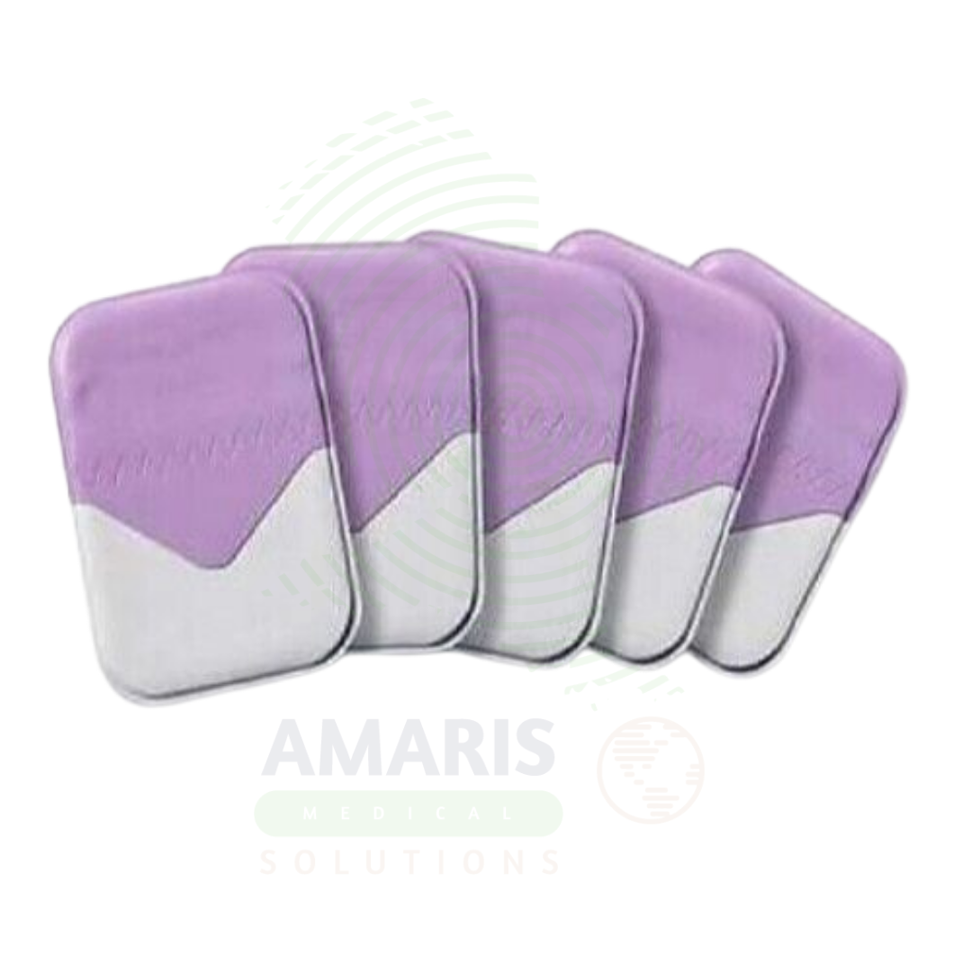

Dental X-ray Films
$1.00
WhatsApp Order
Dental X-ray Films are photosensitive silver halide-coated materials used as the image receptor in analog dental radiography. Supplied in intraoral packets or extraoral sheets, they capture the latent image from X-rays, which is then chemically processed to produce a permanent diagnostic radiograph. Essential for intraoral (periapical, bitewing) and extraoral (panoramic, cephalometric) views, they provide a tangible, archival record. Their use demands careful handling to avoid artifacts, proper storage to prevent fogging, and adherence to a labor-intensive wet chemical processing workflow, which has made them largely obsolete with the advent of digital radiography, though they remain in use in some practices.
Description
Dental X-ray Films
USES
PRIMARY CLINICAL & DIAGNESTIC USES
1. Capture and Recording of Diagnostic Radiographic Images: The primary use is to serve as the image receptor in analog dental radiography. It records the pattern of X-rays that pass through oral structures, creating a latent image that, when chemically processed, produces a permanent radiograph for diagnosis.
2. Intraoral Radiography: Used for detailed imaging of individual teeth and their immediate supporting structures. This includes:
-
-
Periapical Films: To view the entire tooth from crown to root tip and surrounding bone, diagnosing abscesses, cysts, fractures, and periapical pathology.
-
Bitewing Films: To visualize the crowns of upper and lower posterior teeth in one image, primarily for detecting interproximal caries (cavities between teeth) and monitoring bone levels in periodontal disease.
-
Occlusal Films: Larger films to show a broader segment of the jaw.
-
3. Extraoral Radiography: Used for broader imaging of the skull and jaws.
-
-
-
Panoramic Films: Long, curved films used in panoramic machines to capture the entire dentition, jaws, TMJs, and sinuses on a single image for screening and treatment planning.
-
Cephalometric Films: For orthodontic analysis of facial and jaw relationships.
-
-
4. Legal and Archival Medical Record: Provides a physical, long-term legal document of the radiographic examination, which can be stored in the patient’s chart.
5. Teaching and Consultation: Physical films are used for teaching students and for discussing cases with colleagues or specialists.
SECONDARY & SUPPORTIVE USES
1. Quality Assurance Testing: Used to create test exposures to verify the proper functioning and calibration of X-ray equipment and processors.
2. Forensic Dentistry: Used for dental identification in legal investigations.
3. Teleradiology (via digitization): While analog, the resulting radiograph can be digitized with a scanner for electronic transmission.
KEY PRODUCT FEATURES
1. BASIC IDENTIFICATION ATTRIBUTES
-
Type: A photosensitive material (film) used to record X-ray images in analog dental radiography.
-
Designation: Radiographic Film, Dental X-ray Film. Classified by use and packaging.
-
Common Variants:
-
Intraoral Film Packets: Individual, light-tight packets containing the film. They have a lead foil backing to reduce backscatter and a plastic/paper wrapper. Sizes include:
-
Size 0: Small child (for primary teeth)
-
Size 1: Narrow anterior adult
-
Size 2: Standard adult (most common for periapical and bitewing)
-
Size 3: Long, narrow for bitewings only
-
Size 4: Occlusal films
-
-
Extraoral Films: Larger, single sheets without individual packets, used in cassettes with intensifying screens (for panoramic, cephalometric).
-
Film Speed: Rated by ISO (International Standards Organization). D-speed (Ultraspeed) and E-speed (Ektaspeed) are common. Faster film (higher ISO) requires less radiation exposure but may have slightly lower resolution.
-
Single vs. Double Emulsion: Intraoral films are single-emulsion. Extraoral films are often double-emulsion for use with screens.
-
2. TECHNICAL & PERFORMANCE PROPERTIES
-
Film Construction: A transparent polyester base coated with a gelatin emulsion containing photosensitive silver halide crystals (primarily silver bromide).
-
Latent Image Formation: X-rays or light from intensifying screens cause a physico-chemical change in the silver halide crystals, forming the latent image.
-
Spectral Sensitivity: Must be matched to the color of light emitted by intensifying screens (for extraoral film) or to X-rays directly (for intraoral).
-
Contrast and Latitude: The ability to distinguish between different tissue densities (e.g., enamel vs. dentin vs. pulp). High-contrast films have a steeper characteristic curve.
3. PHYSICAL & OPERATIONAL PROPERTIES
-
Handling (Intraoral): Must be handled in normal operatory light before and after exposure but requires a darkroom for processing.
-
Processing: Requires chemical development (developer and fixer) in an automatic processor or manual tanks to convert the latent image into a permanent, visible one.
-
Orientation: Film packets have a raised identification dot to determine left/right orientation on the radiograph.
4. SAFETY & COMPLIANCE ATTRIBUTES
-
Regulatory Status: Class II medical device (as part of a radiographic system).
-
Radiation Safety: Using the fastest film speed acceptable (E-speed over D-speed) reduces the required patient radiation dose (ALARA principle).
-
Lead Foil: The backing reduces patient dose from backscatter.
5. STORAGE & HANDLING ATTRIBUTES
-
Storage (Unprocessed): Store in a cool, dry, radiation-free environment. Store on edge, not flat. Protect from exposure to light, chemicals, and fumes (especially mercury from amalgam).
-
Shelf Life: Has an expiration date. Outdated film has increased base fog, reducing image quality.
-
Darkroom Requirements: A dedicated light-tight space with safelights is required for opening packets and manual processing.
6. LABORATORY & CLINICAL APPLICATIONS
-
Primary Application: The image receptor in analog dental radiography. Found in dental practices, clinics, and schools that have not fully transitioned to digital sensors.
-
Workflow: Part of a multi-step chain: exposure → transport to darkroom → film removal from packet → chemical processing → drying → mounting/viewing.
SAFETY HANDLING PRECAUTIONS
1. SAFETY PRECAUTIONS
-
Film Fogging: Protect unexposed film from all sources of radiation (X-ray scatter, cosmic background) and light leaks. Store away from the X-ray unit.
-
Chemical Handling: Processing chemicals (developer, fixer) are hazardous and require proper handling, ventilation, and disposal. Silver must be recovered from a used fixer.
-
Film Identification: Mislabelling a processed film is a critical error. Use the identification dot and proper mounting techniques.
-
Artifact Prevention: Dust, fingerprints, scratches, or static electricity will cause permanent artifacts on the image. Handle films by the edges only.
2. FIRST AID MEASURES
-
Chemical Contact: From processing chemicals, not the film itself. Rinse affected area with copious water and seek medical attention if needed.
-
Film Ingestion/Aspiration: If a film packet is dropped in the mouth and swallowed or inhaled, it is a medical emergency due to risk of airway obstruction or GI tract blockage from the lead foil. Seek immediate medical help.
3. FIRE FIGHTING MEASURES
-
Flammability: The polyester base and paper/plastic packaging are combustible.
-
Extinguishing Media: Use water, foam, or CO₂.
Related products
Analogue Mobile X-ray Machine
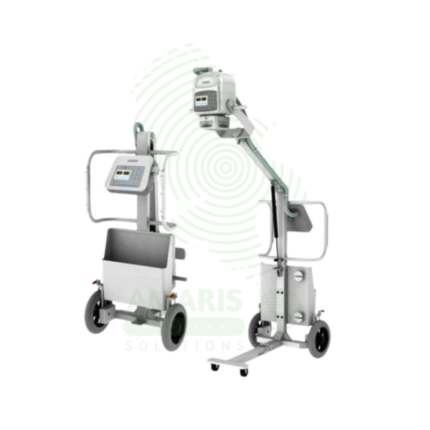
An Analogue Mobile X-ray Machine is a battery-powered, portable X-ray system using traditional film cassettes for bedside imaging in intensive care units, neonatal intensive care units, emergency departments, and operating rooms. The mobile unit enables chest, abdominal, and extremity imaging at the patient's bedside, eliminating the risks associated with transporting critically ill patients. Film cassettes are processed in darkroom facilities for image development. Used in hospitals without digital radiography, as backup for digital systems, and in resource-limited settings where digital infrastructure is not available.
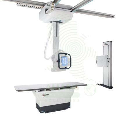
Digital Ceiling X-ray
A Digital Ceiling X-ray is a ceiling-mounted digital radiography system for general diagnostic imaging of the skeletal, chest, abdominal, and extremity anatomy. The ceiling-mounted tube assembly provides full room coverage for flexible patient positioning, while digital flat panel detectors produce immediate high-resolution images for rapid diagnosis. Integrated with PACS and RIS, it supports efficient digital workflow from image acquisition to interpretation. Used in radiology departments, emergency rooms, and outpatient imaging centers.
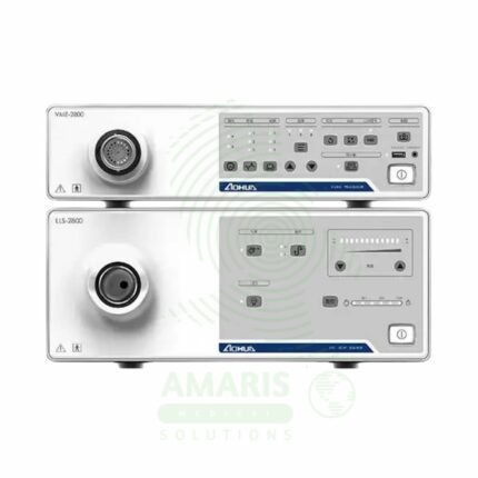
Endoscopy System
The Endoscopy System is a complete video processor and light source stack that forms the core of a modern digital endoscopy suite. It provides high-definition imaging for compatible Fujinon flexible video endoscopes, enabling diagnostic and therapeutic procedures in gastroenterology, pulmonology, and urology. Key features include advanced image processing and enhancement technologies like Narrow Band Imaging (NBI) for improved diagnostic accuracy. As a critical piece of capital equipment, it requires careful handling, the use of compatible accessories, and regular professional maintenance to ensure optimal performance and safety.
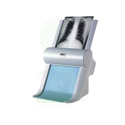
Film Digitizer
A Film Digitizer is a specialized scanner that converts analog radiographic films into high-fidelity digital images (DICOM format). It is the essential tool for migrating historical film archives into a modern digital PACS, enabling filmless workflow, remote access, and long-term preservation of diagnostic records. Its critical performance characteristics are a wide optical density range (to capture all film details) and high spatial resolution. By creating secure, accessible digital copies, it protects against the loss of physical films and integrates past patient history with current digital imaging, supporting comprehensive care and efficient radiology practice.
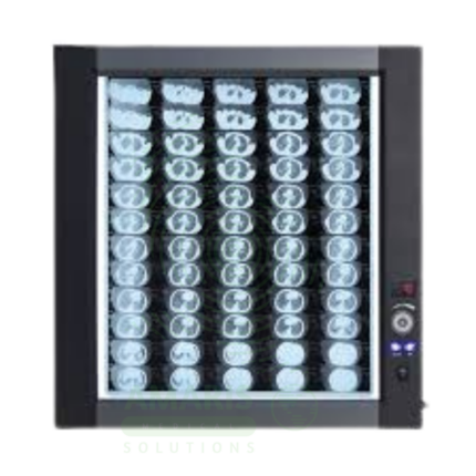
LED Medical Film Viewer
An LED Medical Film Viewer is a light box designed for viewing and interpreting analog X-ray films. Using LED backlight technology, it provides uniform, high-luminance illumination with instant on capability and long life. Available in single, dual, and multi-panel configurations, it supports side-by-side comparison of current and prior studies, pre-operative planning, and group teaching. Essential for radiology departments, orthopedic clinics, emergency departments, and operating rooms where film-based imaging is still used.
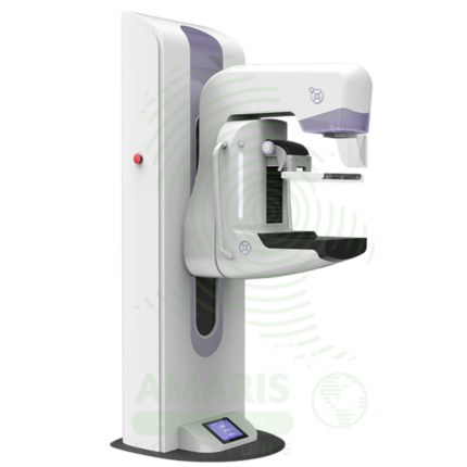
Mammography Machine
A Mammography Machine is a specialized, low-dose X-ray system designed exclusively for imaging the breast. It is the gold-standard tool for breast cancer screening and diagnostic evaluation, utilizing firm breast compression and high-resolution digital detectors to produce detailed images of breast tissue. Modern systems often incorporate Digital Breast Tomosynthesis (DBT or "3D mammography") to reduce tissue overlap and improve cancer detection. Its operation is highly regulated, requiring certified technologists, qualified interpreting physicians, and a rigorous quality assurance program to ensure patient safety, optimal image quality, and accurate early detection of breast cancer, which is vital for reducing mortality.
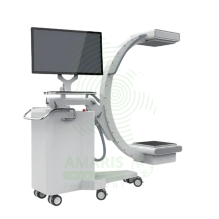
Mobile Film
Mobile Film is a battery-powered, portable X-ray system using traditional film cassettes for bedside imaging in intensive care units, neonatal intensive care units, emergency departments, and operating rooms. The mobile unit enables chest, abdominal, and extremity imaging at the patient's bedside, eliminating the risks associated with transporting critically ill patients. Film cassettes are processed in darkroom or daylight processors for image development. Used in hospitals without digital radiography capabilities or as backup for digital systems.