Dermatoscope and Magnifiers
 Diagnostic Kits
 Vital Signs Monitors
 Stethoscopes and Accessories
 Otoscopes, Ophthalmoscopes, and Retinoscopes
 Reflex Hammers and Neurological Tools
 Scales and Measuring Devices
 Spirometers and Pulmonary Function Tests
 Electrosurgical Units and Accessories
 Cutting Instruments
 Grasping and Holding Instruments
 Hemostatic Instruments
 Specialized Surgical Sets
 Single-Use Procedure Trays and Packs
 Surgical Drapes, Gowns, and Covers
 Tissue Unifying Instruments
 Radiation Protection
 X-Ray Machines and Accessories
 Ultrasound Systems and Probes
 MRI and CT Scanners
 Radiology Consumables
 Bone Densitometers
 Fluoroscopy Equipment
 Imaging Tables and Positioning Aids
 Microscopes and Accessories
 Centrifuges and Separators
 Analyzers
 Incubators and Ovens
 Pipettes, Dispensers, and Lab Glassware
 Refrigerators, Freezers, and Storage Units
 Lab Consumables
 Sterilizers and Autoclaves for Lab Use
 Multi-Parameter Monitors
 Ventilators and Respiratory Support Devices
 Defibrillators and AEDs
 Infusion Pumps and IV Systems
 Patient Warmers and Cooling Devices
 Central Monitoring Stations
 Accessories
 Anesthesia Machines and Workstations
 Oxygen Concentrators and Delivery Systems
 Nebulizers and Inhalers
 CPAP/BiPAP Machines
 Airway Management
 Anesthesia Masks, Circuits, and Bags
 Humidifiers and Heaters
 Respiratory Therapy Accessories
 First Aid Kits and Cabinets
 Emergency Resuscitation Equipment
 Trauma Supplies
 Emergency Carts and Crash Carts
 Burn Care Products
 Bleeding Control
 Automated External Defibrillators (AEDs)
 Transport and Evacuation
 Wheelchairs and Accessories
 Walkers, Crutches, and Canes
 Prosthetics and Orthotics
 Physical Therapy Equipment
 Transfer Devices
 Bathroom Safety
 Orthopedic Traction and Tables
 Hot/Cold Therapy Packs and Units
 Beds and Mattresses
 Chairs and Stools
 Tables
 Cabinets and Storage
 Privacy Screens & Curtains
 Stands and Racks
 Linens and Textiles
 Lighting
 Autoclaves and Sterilizers
 Ultrasonic Cleaners
 Disinfectant Solutions and Wipes
 Sterilization Pouches, Wraps, and Indicators
 Instrument Trays and Containers
 UV and Ozone Disinfection Devices
 Washer Disinfectors
 Wound Care
 Gloves
 Masks and Respirators
 Catheters and Tubing
 Swabs, Applicators, and Sponges
 Incontinence Products
 Personal Protective Equipment (PPE)
 Dental Chairs and Units
 Handpieces and Burs
 Instruments
 Consumables
 Sterilization for Dental Use
 Orthodontic Supplies
 Endodontic Tools
 Slit Lamps and Tonometers
 Lensometers and Phoropters
 Ophthalmic Surgical Instruments
 Eyewear Frames and Lenses
 Contact Lens Supplies
 Vision Testing Charts and Devices
 Eye Care Consumables
 Laser Systems for Eye Care
 ENT Exam Chairs and Tables
 Endoscopes
 Audiometers and Hearing Tests
 ENT Instruments
 Nasal and Throat Packs
 Hearing Aids and Accessories
 Otology Supplies
 Fetal Dopplers and Monitors
 Delivery Beds and Tables
 Gynecological Instruments
 Neonatal Incubators and Warmers
 Breast Pumps and Accessories
 Contraceptive Devices
 Maternity Supports and Pads
 Neonatal Consumables
 Cystoscopes and Urethroscopes
 Dialysis Machines and Supplies
 Urological Catheters and Bags
 Lithotripters
 Prostate Treatment Devices
 Urinary Incontinence Products
 Kidney Stone Management Tools
 Consumables & Disposables
 EEG and EMG Machines
 Neurosurgical Instruments
 Nerve Stimulators
 Headrests and Positioning Aids
 Lumbar Puncture Kits
 Seizure Monitoring Devices
 Consumables
 Rehabilitation for Neurological Conditions
 ECG Machines and Accessories
 Holter Monitors
 Stress Test Systems
 Pacemakers and Defibrillator Accessories
 Vascular Access Devices
 Cardiac Catheters and Guidewires
 Blood Flow Meters
 Consumables
 Orthopedic Instruments
 Casts, Splints, and Padding
 Joint Replacement Supplies
 Prosthetic Limbs and Components
 Bone Grafts and Substitutes
 Traction Devices
 Orthopedic Braces and Supports
 Rehabilitation Aids for Orthopedics
 Home Oxygen Therapy
 Hospital Beds for Home Use
 Mobility Aids
 Bathroom and Daily Living Aids
 Wound Care for Home
 Monitoring Devices
 Enteral Feeding Pumps and Tubes
 Hand Sanitizers and Dispensers
 Face Shields and Goggles
 Isolation Gowns and Suits
 Biohazard Waste Containers
 Air Purifiers and HEPA Filters
 Surface Disinfectants
 Sharps Containers
 Protective Barriers
 Cardiovascular & Endurance Training
 Strength Training & Weightlifting
 Functional Training & Core Conditioning
 Physical Therapy & Rehabilitation
 Sports & Outdoor Recreation
 Gym Flooring & Facility Equipment
 Fitness Monitoring & Accessories
 Kids & Novelties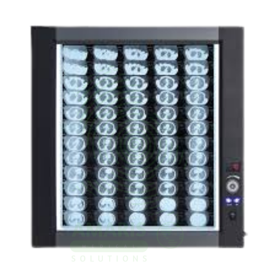
LED Medical Film Viewer
$1.00
WhatsApp Order
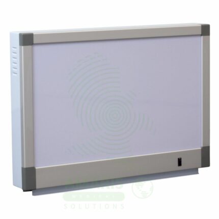
An LED Medical Film Viewer is a light box designed for viewing and interpreting analog X-ray films. Using LED backlight technology, it provides uniform, high-luminance illumination with instant on capability and long life. Available in single, dual, and multi-panel configurations, it supports side-by-side comparison of current and prior studies, pre-operative planning, and group teaching. Essential for radiology departments, orthopedic clinics, emergency departments, and operating rooms where film-based imaging is still used.
Description
LED Medical Film Viewer
PRIMARY CLINICAL & DIAGNOSTIC USES
1. Viewing and Interpreting Radiographic Films
-
Primary Use: Provides a uniformly illuminated, high-luminance light source for viewing and interpreting analog X-ray films. The LED backlight delivers consistent brightness across the viewing surface, allowing physicians, radiologists, and surgeons to visualize the captured image with optimal contrast and clarity for accurate diagnosis.
-
How it helps: For the radiologist and clinician, the LED film viewer transforms a sheet of film into a diagnostic tool—illuminating the subtle shades of gray that reveal fractures, tumors, and infections, and providing the contrast needed to distinguish normal anatomy from pathology. For the patient, proper film viewing ensures that their X-ray films can be properly interpreted, leading to accurate diagnosis and appropriate treatment.
2. Comparison of Current and Prior Studies
-
Primary Use: Multi-panel film viewers allow side-by-side comparison of new and old X-ray films to assess disease progression, healing of fractures, or changes in tumor size over time. The even illumination across all panels ensures consistent viewing conditions for accurate comparison.
-
How it helps: For the radiologist and treating physician, the ability to place current and prior films side by side on a multi-panel viewer provides invaluable perspective—showing whether a fracture is healing, a tumor is growing, or pneumonia is resolving, and guiding decisions about continuing, changing, or stopping treatment. For the patient, this comparison ensures that treatment decisions are based on objective evidence of how their condition is evolving over time.
3. Surgical and Procedural Planning
-
Primary Use: Used by surgeons to review pre-operative X-rays in the operating room or clinic to plan surgical approaches, select appropriate implants, or visualize fracture patterns before reduction and fixation. The bright, uniform illumination ensures all details are visible.
-
How it helps: For the orthopedic surgeon and operating room team, having a film viewer in the surgical suite means the patient’s films are available for immediate reference during the procedure—confirming fracture patterns, guiding implant selection, and ensuring that the surgical approach matches the pre-operative plan. For the patient undergoing surgery, this ready access to their images means the surgeon can refer back to the films throughout the procedure, ensuring accuracy and optimal outcomes.
4. Diagnosis of Medical Conditions from Film
-
Primary Use: Essential for diagnosing fractures, dislocations, lung diseases, abdominal conditions, and dental issues by providing the necessary contrast and clarity to see anatomical details and pathologies captured on the film. LED technology provides consistent brightness with no flicker or warm-up time.
-
How it helps: For the emergency physician, orthopedist, and pulmonologist, the LED film viewer brings diagnostic images to life—revealing the hairline fracture that might otherwise be missed, showing the infiltrate that confirms pneumonia, and demonstrating the obstruction that explains a patient’s abdominal pain. For the patient, proper film viewing means that their condition is accurately diagnosed and appropriately treated.
5. Teaching and Case Discussion
-
Primary Use: Provides a communal viewing platform for medical students, residents, and healthcare teams to discuss radiographic findings, anatomy, and pathology during teaching conferences or clinical rounds. The bright, uniform illumination supports group viewing.
-
How it helps: For the medical educator and trainees, the film viewer creates a shared space for learning—allowing groups to gather around illuminated films, point out findings, discuss differential diagnoses, and build the interpretive skills essential for clinical practice. For the future patients who will be cared for by these trainees, every teaching session on a film viewer contributes to their diagnostic accuracy and clinical competence.
SECONDARY & SUPPORTIVE USES
1. Quality Control of Film Processing: Used in radiology departments to check the density, contrast, and overall quality of films produced by automated processors, ensuring diagnostic adequacy.
2. Review of Other Analog Imaging Films: Can be used to view other types of analog medical imaging films, such as those from older CT scanners, mammography units, or nuclear medicine studies.
3. Forensic and Veterinary Radiology: Used in non-human settings, such as forensic pathology or veterinary clinics, to examine skeletal remains or animal radiographs.
4. Display of Photographic Negatives: In non-medical contexts, the uniform light box is suitable for viewing photographic negatives or slides.
5. Archival Film Review: Used to review archived film studies for comparison with current digital images.
6. Multidisciplinary Case Review: Supports case review conferences across multiple specialties with shared viewing capability.
KEY PRODUCT FEATURES
1. BASIC IDENTIFICATION ATTRIBUTES
-
Product Type: A light box with LED backlighting for viewing and interpreting analog X-ray films.
-
Designation: LED Medical Film Viewer, X-ray Film Viewer, Light Box, Illuminator, Film Viewing Box.
-
Configurations:
-
Single Panel: One viewing panel for individual film review.
-
Dual Panel: Two panels side by side for comparison viewing.
-
Multi-Panel: Three or more panels for multi-film comparison.
-
Wall-Mounted: Fixed installation on wall.
-
Desktop: Portable unit for table placement.
-
-
Key Components:
-
LED Backlight: Array of LEDs for uniform illumination.
-
Diffuser: Evenly distributes light across viewing surface.
-
Viewing Surface: Translucent acrylic or glass panel.
-
Frame: Metal or plastic housing.
-
Brightness Control: Adjustable intensity (some models).
-
Film Clips: Hold films in place during viewing.
-
2. TECHNICAL & PERFORMANCE PROPERTIES
-
Luminance: Typically 2,000-4,000 cd/m²; high-brightness models up to 6,000 cd/m².
-
Color Temperature: 4,000-6,500 K (cool white) for accurate film interpretation.
-
Uniformity: >90% uniformity across viewing surface.
-
LED Life: 50,000-100,000 hours.
-
Warm-Up Time: Instant on; no warm-up required.
-
Power Consumption: Low compared to fluorescent models.
3. PHYSICAL & OPERATIONAL PROPERTIES
-
Construction: Metal or plastic housing with acrylic viewing surface.
-
Mounting: Wall-mounted or desktop options.
-
Panel Size: Single: 14x17 inches; multiple sizes available.
-
Brightness Control: Optional dimming control for different film densities.
-
Cooling: Passive cooling; no fans.
4. SAFETY & COMPLIANCE ATTRIBUTES
-
Regulatory Status: Class I medical device (when used for medical film viewing).
-
Electrical Safety: Compliant with medical electrical equipment standards.
-
LED Safety: No UV emission; safe for prolonged viewing.
-
Heat Output: Minimal compared to fluorescent units.
5. STORAGE & HANDLING ATTRIBUTES
-
Storage: Wall-mounted units remain installed; desktop units stored on the work surface.
-
Cleaning: Wipe viewing surface with soft, non-abrasive cloth; use mild cleaner.
-
Maintenance: LED units require no bulb replacement; occasional cleaning of surface.
6. LABORATORY & CLINICAL APPLICATIONS
-
Primary Application: Viewing and interpreting analog X-ray films in radiology, orthopedics, emergency medicine, and surgical settings.
-
Clinical Role: Essential equipment in radiology departments, orthopedic clinics, emergency departments, and operating rooms.
SAFETY HANDLING PRECAUTIONS
1. SAFETY PRECAUTIONS
-
Electrical Safety: Keep away from water; unplug during cleaning.
-
Surface Care: Use soft cloth; avoid abrasive cleaners that may scratch the viewing surface.
-
Mounting: Ensure wall-mounted units are securely attached.
-
Brightness: Adjust brightness to appropriate level for film density.
2. FIRST AID MEASURES
-
Electrical Shock: If shock occurs, disconnect power; seek medical attention if needed.
-
Broken Surface: If viewing surface breaks, disconnect power; carefully collect fragments; dispose safely.
3. FIRE FIGHTING MEASURES
-
Flammability: Plastic components are combustible; electrical components may pose fire risk.
-
Extinguishing Media: For electrical fire, use CO₂ or dry chemical extinguisher.
Related products
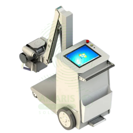
Digital & Analog X-ray Machine
A Digital & Analog X-ray Machine is a fundamental medical imaging device that uses a controlled beam of ionizing radiation to produce static or real-time images of the body's internal structures. It is indispensable for diagnosing fractures, lung diseases, dental issues, and many abdominal conditions. The transition from Analog (film-based) to Digital (CR or DR) technology has revolutionized the field, offering faster results, superior image manipulation, improved dose efficiency, and seamless integration into digital healthcare networks. Its operation demands strict adherence to radiation safety protocols (ALARA) to protect patients and staff, making it a cornerstone of safe, effective diagnostic medicine.
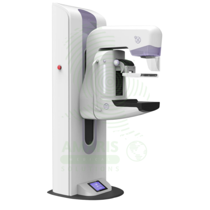
Digital Mammography System
A Digital Mammography System is a specialized X-ray imaging system designed for breast cancer screening and diagnosis. Using high-resolution digital detectors, it provides superior image quality with lower radiation dose compared to film mammography. Advanced systems offer tomosynthesis (3D) imaging, which improves cancer detection rates and reduces false positives. Used in breast imaging centers, radiology departments, and women's health facilities, it is the gold standard for early detection of breast cancer.
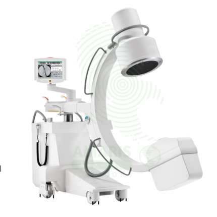
Mobile C-arm Surgical System
A Mobile C-arm Surgical System is a portable fluoroscopic imaging device used for real-time intraoperative guidance during orthopedic, spinal, vascular, and pain management procedures. The C-shaped arm allows flexible positioning around the patient, providing AP, lateral, and oblique views to verify instrument placement, fracture reduction, and device deployment. Essential for minimally invasive surgery, it enables surgeons to achieve precision and accuracy while reducing operative time and improving patient outcomes.
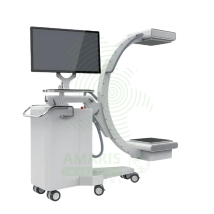
Mobile Film
Mobile Film is a battery-powered, portable X-ray system using traditional film cassettes for bedside imaging in intensive care units, neonatal intensive care units, emergency departments, and operating rooms. The mobile unit enables chest, abdominal, and extremity imaging at the patient's bedside, eliminating the risks associated with transporting critically ill patients. Film cassettes are processed in darkroom or daylight processors for image development. Used in hospitals without digital radiography capabilities or as backup for digital systems.
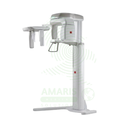
Panoramic X-ray Dental Machine
A Panoramic X-ray Dental Machine is a rotating extraoral radiographic system that produces a single, broad 2D image of the entire jaws, teeth, TMJs, and sinuses. By focusing on a curved "focal trough," it provides an efficient screening tool for wisdom teeth evaluation, orthodontic planning, and detecting large jaw pathologies. While offering a valuable overview with a relatively low radiation dose, its diagnostic utility is entirely dependent on precise patient positioning to avoid blurring and distortion. It is a cornerstone of modern dental diagnostic imaging, serving as a crucial first step in comprehensive oral assessment and treatment planning.
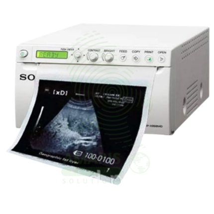
Sony Ultrasound Printer
A Sony Ultrasound Printer is a specialized medical-grade printer that produces high-quality hard copies of ultrasound images. Utilizing dry thermal, laser, or dye-sublimation technology, it creates grayscale or color prints on photographic paper or film, providing a physical record for patient charts, referrals, and consultations. While its role has diminished with the rise of fully digital PACS workflows, it remains essential in settings requiring immediate tangible output, patient education, or where digital infrastructure is limited. Its performance is defined by high spatial and contrast resolution to accurately reproduce the subtleties of sonographic images.
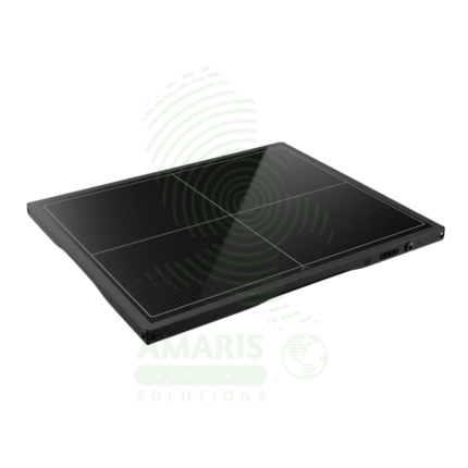
Wire & Wireless Flat Panel Detector
A Wire & Wireless Flat Panel Detector is a digital X-ray image receptor that converts X-ray energy into high-quality digital images for general radiography. Available in wired and wireless configurations, these detectors replace traditional film cassettes, providing immediate image preview, eliminating film processing, and enabling seamless digital workflow integration. Wireless detectors offer flexibility for bedside imaging in ICUs, emergency departments, and operating rooms, while wired detectors provide reliable connectivity for fixed systems. High detective quantum efficiency enables dose reduction while maintaining diagnostic image quality.