Dermatoscope and Magnifiers
 Diagnostic Kits
 Vital Signs Monitors
 Stethoscopes and Accessories
 Otoscopes, Ophthalmoscopes, and Retinoscopes
 Reflex Hammers and Neurological Tools
 Scales and Measuring Devices
 Spirometers and Pulmonary Function Tests
 Electrosurgical Units and Accessories
 Cutting Instruments
 Grasping and Holding Instruments
 Hemostatic Instruments
 Specialized Surgical Sets
 Single-Use Procedure Trays and Packs
 Surgical Drapes, Gowns, and Covers
 Tissue Unifying Instruments
 Radiation Protection
 X-Ray Machines and Accessories
 Ultrasound Systems and Probes
 MRI and CT Scanners
 Radiology Consumables
 Bone Densitometers
 Fluoroscopy Equipment
 Imaging Tables and Positioning Aids
 Microscopes and Accessories
 Centrifuges and Separators
 Analyzers
 Incubators and Ovens
 Pipettes, Dispensers, and Lab Glassware
 Refrigerators, Freezers, and Storage Units
 Lab Consumables
 Sterilizers and Autoclaves for Lab Use
 Multi-Parameter Monitors
 Ventilators and Respiratory Support Devices
 Defibrillators and AEDs
 Infusion Pumps and IV Systems
 Patient Warmers and Cooling Devices
 Central Monitoring Stations
 Accessories
 Anesthesia Machines and Workstations
 Oxygen Concentrators and Delivery Systems
 Nebulizers and Inhalers
 CPAP/BiPAP Machines
 Airway Management
 Anesthesia Masks, Circuits, and Bags
 Humidifiers and Heaters
 Respiratory Therapy Accessories
 First Aid Kits and Cabinets
 Emergency Resuscitation Equipment
 Trauma Supplies
 Emergency Carts and Crash Carts
 Burn Care Products
 Bleeding Control
 Automated External Defibrillators (AEDs)
 Transport and Evacuation
 Wheelchairs and Accessories
 Walkers, Crutches, and Canes
 Prosthetics and Orthotics
 Physical Therapy Equipment
 Transfer Devices
 Bathroom Safety
 Orthopedic Traction and Tables
 Hot/Cold Therapy Packs and Units
 Beds and Mattresses
 Chairs and Stools
 Tables
 Cabinets and Storage
 Privacy Screens & Curtains
 Stands and Racks
 Linens and Textiles
 Lighting
 Autoclaves and Sterilizers
 Ultrasonic Cleaners
 Disinfectant Solutions and Wipes
 Sterilization Pouches, Wraps, and Indicators
 Instrument Trays and Containers
 UV and Ozone Disinfection Devices
 Washer Disinfectors
 Wound Care
 Gloves
 Masks and Respirators
 Catheters and Tubing
 Swabs, Applicators, and Sponges
 Incontinence Products
 Personal Protective Equipment (PPE)
 Dental Chairs and Units
 Handpieces and Burs
 Instruments
 Consumables
 Sterilization for Dental Use
 Orthodontic Supplies
 Endodontic Tools
 Slit Lamps and Tonometers
 Lensometers and Phoropters
 Ophthalmic Surgical Instruments
 Eyewear Frames and Lenses
 Contact Lens Supplies
 Vision Testing Charts and Devices
 Eye Care Consumables
 Laser Systems for Eye Care
 ENT Exam Chairs and Tables
 Endoscopes
 Audiometers and Hearing Tests
 ENT Instruments
 Nasal and Throat Packs
 Hearing Aids and Accessories
 Otology Supplies
 Fetal Dopplers and Monitors
 Delivery Beds and Tables
 Gynecological Instruments
 Neonatal Incubators and Warmers
 Breast Pumps and Accessories
 Contraceptive Devices
 Maternity Supports and Pads
 Neonatal Consumables
 Cystoscopes and Urethroscopes
 Dialysis Machines and Supplies
 Urological Catheters and Bags
 Lithotripters
 Prostate Treatment Devices
 Urinary Incontinence Products
 Kidney Stone Management Tools
 Consumables & Disposables
 EEG and EMG Machines
 Neurosurgical Instruments
 Nerve Stimulators
 Headrests and Positioning Aids
 Lumbar Puncture Kits
 Seizure Monitoring Devices
 Consumables
 Rehabilitation for Neurological Conditions
 ECG Machines and Accessories
 Holter Monitors
 Stress Test Systems
 Pacemakers and Defibrillator Accessories
 Vascular Access Devices
 Cardiac Catheters and Guidewires
 Blood Flow Meters
 Consumables
 Orthopedic Instruments
 Casts, Splints, and Padding
 Joint Replacement Supplies
 Prosthetic Limbs and Components
 Bone Grafts and Substitutes
 Traction Devices
 Orthopedic Braces and Supports
 Rehabilitation Aids for Orthopedics
 Home Oxygen Therapy
 Hospital Beds for Home Use
 Mobility Aids
 Bathroom and Daily Living Aids
 Wound Care for Home
 Monitoring Devices
 Enteral Feeding Pumps and Tubes
 Hand Sanitizers and Dispensers
 Face Shields and Goggles
 Isolation Gowns and Suits
 Biohazard Waste Containers
 Air Purifiers and HEPA Filters
 Surface Disinfectants
 Sharps Containers
 Protective Barriers
 Cardiovascular & Endurance Training
 Strength Training & Weightlifting
 Functional Training & Core Conditioning
 Physical Therapy & Rehabilitation
 Sports & Outdoor Recreation
 Gym Flooring & Facility Equipment
 Fitness Monitoring & Accessories
 Kids & Novelties
Endobronchial Tubes
WhatsApp Order
Endobronchial Tubes are specialized double-lumen endotracheal tubes designed for selective lung ventilation and isolation during thoracic surgery, management of massive hemoptysis, and bronchopleural fistulas. The dual-lumen design allows independent ventilation of each lung, enabling surgical access, infection control, and airway protection. Essential for thoracic surgery, anesthesiology, and critical care, they provide safe, effective single-lung ventilation with proper placement confirmed by fiberoptic bronchoscopy.
Description
Endobronchial Tubes
PRIMARY CLINICAL & DIAGNOSTIC USES
1. Selective Lung Ventilation for Thoracic Surgery
-
Primary Use: Provides selective ventilation of one lung while allowing the other lung to be collapsed during thoracic surgical procedures including lobectomy, pneumonectomy, esophagectomy, and video-assisted thoracoscopic surgery. The double-lumen tube design allows independent ventilation of the left and right lungs.
-
How it helps: For the thoracic surgeon and anesthesiologist, endobronchial tubes enable optimal surgical access by selectively deflating the lung on the operative side while maintaining ventilation to the contralateral lung—providing a clear surgical field and protecting the healthy lung from contamination. For the patient undergoing thoracic surgery, this technology allows for minimally invasive approaches, reduces operative time, and minimizes the risk of complications associated with single-lung ventilation.
2. Isolation of the Lung for Infection Control
-
Primary Use: Isolates a lung affected by infection, hemorrhage, or massive abscess to prevent spillage of infectious material or blood into the healthy lung during surgical or therapeutic procedures.
-
How it helps: For the thoracic surgeon and anesthesiologist, endobronchial tubes provide a critical barrier that contains infectious or hemorrhagic material within the diseased lung—protecting the healthy lung from contamination and preventing life-threatening aspiration. For the patient with severe pulmonary infection or massive hemoptysis, this isolation can be life-saving, allowing for definitive treatment while preserving respiratory function.
3. Management of Bronchopleural Fistula
-
Primary Use: Used to manage bronchopleural fistulas by selectively isolating the affected bronchus, preventing air leak into the pleural space and maintaining adequate ventilation.
-
How it helps: For the critical care team and thoracic surgeon, endobronchial tubes provide a means of managing this challenging complication—sealing off the fistulous communication and allowing the pleural space to heal. For the patient with a bronchopleural fistula, proper tube placement can prevent respiratory compromise and facilitate healing without additional surgical intervention.
4. Facilitation of Bronchoscopic Procedures
-
Primary Use: Provides a conduit for passage of fiberoptic bronchoscopes for diagnostic and therapeutic procedures, including lavage, biopsy, and laser therapy, while maintaining ventilation.
-
How it helps: For the interventional pulmonologist, endobronchial tubes allow simultaneous access for bronchoscopic instruments and maintenance of ventilation—enabling complex airway interventions without interrupting gas exchange. For the patient undergoing bronchoscopic procedures, this means safer, more efficient interventions with continuous respiratory support.
5. Management of Massive Hemoptysis
-
Primary Use: Used in the emergency management of massive hemoptysis to isolate the bleeding lung and prevent aspiration of blood into the contralateral airway.
-
How it helps: For the emergency physician and critical care team, endobronchial tube placement provides immediate airway protection in life-threatening hemoptysis—containing the bleeding source and maintaining a patent airway for ventilation. For the patient with massive hemoptysis, this intervention can be life-saving, preventing asphyxiation and allowing time for definitive treatment.
SECONDARY & SUPPORTIVE USES
1. Difficult Airway Management: Used in complex airway scenarios where conventional endotracheal intubation is challenging or where selective ventilation is required for airway control.
2. Tracheobronchial Trauma: Provides airway stabilization and selective ventilation in patients with traumatic injury to the tracheobronchial tree.
3. Lung Transplantation: Used during lung transplantation procedures to selectively ventilate the transplanted lung while the native lung is managed.
4. Pulmonary Lavage: Facilitates whole-lung lavage in patients with pulmonary alveolar proteinosis by allowing sequential lavage of one lung while the other is ventilated.
5. Thoracic Trauma: Used in the acute management of thoracic trauma where lung isolation is required to prevent contamination or manage air leaks.
6. Pediatric Applications: Smaller endobronchial tubes are available for pediatric thoracic surgery and airway management in children.
KEY PRODUCT FEATURES
1. BASIC IDENTIFICATION ATTRIBUTES
-
Device Type: A specialized double-lumen endotracheal tube designed for selective lung ventilation and isolation.
-
Designation: Endobronchial Tube, Double-Lumen Tube, DLT, Bronchial Catheter, Carlens Tube, Robertshaw Tube.
-
Key Components:
-
Dual Lumens: Separate channels for ventilation of each lung (tracheal lumen and bronchial lumen).
-
Bronchial Cuff: Inflatable cuff that seals the bronchus of the ventilated lung.
-
Tracheal Cuff: Inflatable cuff that seals the trachea, preventing air leak and aspiration.
-
Bronchial Tip: Extended tip designed to enter the main bronchus (left or right specific).
-
Pilot Balloons: Color-coded balloons for monitoring cuff inflation.
-
Radiopaque Markers: Positioned to confirm correct placement under fluoroscopy or X-ray.
-
Connectors: Standard 15 mm connectors for ventilator circuits.
-
2. TECHNICAL & PERFORMANCE PROPERTIES
-
Sizes: 26 Fr to 41 Fr for adults; pediatric sizes available.
-
Lumen Diameter: Varies by size; sufficient for passage of fiberoptic bronchoscope.
-
Cuff Types: High-volume, low-pressure cuffs to minimize tracheal and bronchial injury.
-
Material: Medical-grade polyvinyl chloride (PVC) or silicone.
-
Radiopacity: Embedded markers for radiographic confirmation.
-
Direction: Left-sided or right-sided configuration based on intended placement.
-
Sterility: Ethylene oxide or gamma irradiation sterilized.
3. PHYSICAL & OPERATIONAL PROPERTIES
-
Construction: Flexible but kink-resistant tubing; smooth surface for atraumatic insertion.
-
Color Coding: Blue for tracheal cuff, white or clear for bronchial cuff.
-
Length: 28-40 cm depending on size.
-
Markings: Depth markings at 1 cm intervals for accurate placement.
-
Packaging: Sterile, single-use; individually packaged.
4. SAFETY & COMPLIANCE ATTRIBUTES
-
Regulatory Status: Class II medical device regulated by FDA.
-
Biocompatibility: Materials safe for airway contact.
-
Cuff Safety: High-volume, low-pressure cuffs reduce risk of mucosal ischemia.
-
Latex-Free: Manufactured without natural rubber latex.
5. STORAGE & HANDLING ATTRIBUTES
-
Storage: Store in a clean, dry location at room temperature.
-
Sterility Maintenance: Do not use it if the package is opened, damaged, or wet.
-
Expiration: Check expiration date before use; do not use after expiration.
-
Single-Use Only: Intended for single patient use only; do not resterilize or reuse.
6. LABORATORY & CLINICAL APPLICATIONS
-
Primary Application: Selective lung ventilation and isolation for thoracic surgery, massive hemoptysis, and bronchopleural fistula management.
-
Clinical Role: Essential equipment in thoracic surgery, anesthesiology, and critical care settings.
SAFETY HANDLING PRECAUTIONS
1. SAFETY PRECAUTIONS
-
Confirm Placement: Verify correct position with fiberoptic bronchoscopy after intubation.
-
Cuff Inflation: Inflate cuffs to minimal occluding volume; monitor cuff pressures.
-
Tube Security: Secure tube after confirming placement to prevent dislodgement.
-
Position Monitoring: Monitor for signs of tube migration, obstruction, or malposition.
-
Suctioning: Suction both lumens as needed to maintain patency.
-
Deflation Prior to Removal: Deflate cuffs completely before extubation.
2. FIRST AID MEASURES
-
Tube Displacement: If tube is displaced, manually ventilate patient; remove tube; reintubate as needed.
-
Cuff Rupture: If cuff ruptures, consider tube replacement if the seal cannot be maintained.
-
Obstruction: If lumen is obstructed, attempt to suction; consider tube replacement if patency cannot be restored.
3. FIRE FIGHTING MEASURES
-
Flammability: Plastic components are combustible; an oxygen-enriched environment increases fire risk.
-
Extinguishing Media: For electrical fire, use CO₂ or dry chemical extinguisher.
Related products
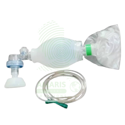
Breath Resuscitation Bag
A Breath Resuscitation Bag (manual resuscitator, bag-valve-mask) is a Class II medical device used to deliver positive-pressure ventilation to patients in respiratory arrest, cardiac arrest, or respiratory failure. Available in adult (1,500-2,000 mL), pediatric (450-750 mL), and infant/neonatal (200-350 mL) sizes with self-inflating silicone or PVC bags, one-way non-rebreathing valves, transparent cushioned masks, and oxygen reservoirs for high FiO2 delivery (90-100% with reservoir). Oxygen inlet allows connection to flow meter (10-15 L/min recommended). Pediatric/neonatal models include pressure-limiting valves (35-45 cmH2O) to prevent barotrauma. Essential for CPR (AHA guidelines: adult 10-12 breaths/min, child 12-20, infant 20-30), preoxygenation before intubation, transport ventilation, and recovery from anesthesia. Critical safety considerations include avoiding excessive ventilation pressure (prevents gastric insufflation), maintaining proper mask seal, monitoring chest rise, and having backup equipment readily available. Indispensable emergency airway equipment in hospitals, ambulances, crash carts, and emergency response kits worldwide.
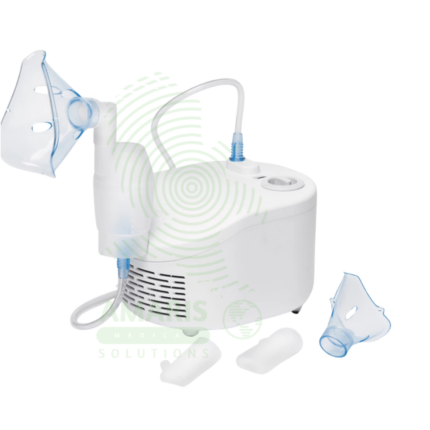
Compressor Nebulizer Machine
A Compressor Nebulizer Machine is a pneumatic device that transforms liquid medication into a breathable mist for treating respiratory conditions. Consisting of an electric air compressor, a nebulizer cup, tubing, and a mask or mouthpiece, it is particularly effective for infants, children, and patients with severe asthma, COPD, or cystic fibrosis who require reliable aerosol delivery. While offering robust performance for home and clinical use, its safety and efficacy depend critically on proper cleaning to prevent infection, correct assembly, and the use of prescribed nebulizer-compatible medications. It remains a fundamental tool for both acute intervention and chronic management of pulmonary diseases.
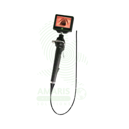
Flexible Fiber Optic Laryngoscope
A Flexible Fiber Optic Laryngoscope is a flexible endoscope (2-5 mm diameter, 30-60 cm working length) with fiber optic image transmission and steerable tip (120-180° angulation) for visualization of the upper airway and facilitation of difficult intubations. Features include control handle with angulation lever, working channel (1-2 mm) for suction or oxygen, external light source (halogen/xenon/LED), and optional camera for video display. Primary clinical applications include awake intubation in difficult airway management (limited mouth opening, cervical spine instability, obstructing pathology), nasotracheal intubation for oral surgery or maxillofacial trauma, intubation with cervical spine precautions (minimal neck movement), diagnostic airway assessment (stridor, hoarseness, vocal cord dysfunction, masses), double-lumen tube placement for thoracic surgery, pediatric difficult airway management, and tracheostomy tube placement guidance. Class II medical device requiring FDA clearance. Critical safety considerations include mandatory leak testing before immersion, antifog preparation, gentle insertion technique, airway maintenance with oxygen, topical anesthesia for patient comfort, suction availability, backup airway device, and strict infection control with validated reprocessing protocols.
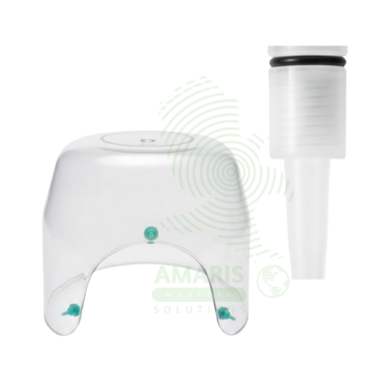
Infant Oxygen Hood
An Infant Oxygen Hood is a Class II medical device used to deliver controlled concentrations of supplemental oxygen to spontaneously breathing newborns and infants. Made of transparent medical-grade acrylic or polycarbonate, the hood fits over the infant's head with a soft foam or rubber neck seal, creating an oxygen-enriched environment while allowing easy access for monitoring and care. Available in premature/neonatal, infant, and older infant sizes with gas inlet ports (22 mm) for connection to air/oxygen blenders (FiO2 21-100%), access ports for monitoring leads and IV lines, and outlet vents to prevent CO2 accumulation. Requires minimum flow rate of 5 L/min to ensure adequate CO2 washout. Used with heated humidifiers for prolonged therapy to prevent airway drying. Primary clinical applications include management of respiratory distress syndrome (RDS) in premature infants, neonatal pneumonia, post-extubation oxygen support, congenital heart disease, and palliative care. Critical safety considerations include maintaining adequate flow rate to prevent rebreathing, monitoring FiO2 at hood level, ensuring proper neck seal without constriction, and preventing hyperoxia to reduce retinopathy of prematurity (ROP) risk. Essential equipment in NICUs and special care nurseries for controlled oxygen delivery to vulnerable infants.
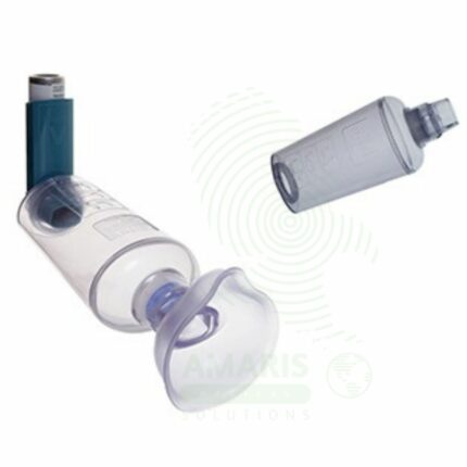
Inhaler Spacer with Child Mask
An Inhaler Spacer with Child Mask is a valved holding chamber designed to optimize aerosol medication delivery from metered-dose inhalers to infants and young children. The spacer captures the medication spray, allowing the child to inhale over multiple breaths, while the soft mask creates a seal over the nose and mouth. This improves pulmonary drug deposition, reduces oropharyngeal side effects, and eliminates the need for complex breath coordination. Essential for pediatric asthma management, the spacer enables effective inhaler use in children as young as infancy, supporting home management of reactive airway disease.
Macintosh Bulb Laryngoscope
A Macintosh Bulb Laryngoscope is a rigid laryngoscope with curved Macintosh blade (sizes 0-4, 70-160 mm) featuring a distal incandescent (xenon, krypton, halogen) or LED bulb at the blade tip for direct illumination during tracheal intubation. The curved blade design allows indirect epiglottis elevation by placing the tip in the vallecula, requiring less force and neck extension than straight blades. Features stainless steel reusable blades (or disposable plastic), ergonomic handles with knurled grip, ISO standard hook-on fittings, and autoclavable options. Light output 500-3,000 Lux depending on bulb type and battery condition. Primary clinical applications include routine and emergency tracheal intubation during general anesthesia, difficult airway management, cervical spine precautions (minimal neck movement), rapid sequence intubation, neonatal and pediatric intubation (sizes 0-2), teaching and training, and use in resource-limited settings. Class II medical device requiring FDA clearance. Critical safety considerations include pre-use light check (brightness, bulb security), appropriate blade size selection, proper lifting technique (not levering on teeth), battery verification, bulb obstruction risk from secretions, backup device availability, and infection control (sterilization or disposable blades).
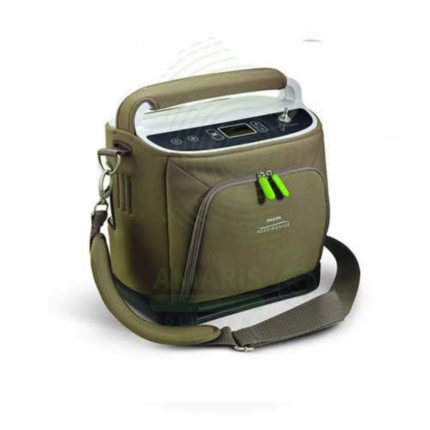
Portable Oxygen Concentrator
A Portable Oxygen Concentrator (POC) is a lightweight, battery-powered device that delivers oxygen via pulse dose technology, enabling active, mobile lifestyles for patients with chronic lung disease. By providing oxygen on-demand with each breath, it maximizes battery efficiency and portability, allowing users to travel, exercise, and socialize freely. It is a prescription-only device that requires careful titration to match a patient's needs during activity and is not a substitute for a stationary concentrator used at home and during sleep. Key considerations include FAA approval for air travel, battery life management, and understanding its specific use case as an ambulatory aid, not a primary oxygen source.