Dermatoscope and Magnifiers
 Diagnostic Kits
 Vital Signs Monitors
 Stethoscopes and Accessories
 Otoscopes, Ophthalmoscopes, and Retinoscopes
 Reflex Hammers and Neurological Tools
 Scales and Measuring Devices
 Spirometers and Pulmonary Function Tests
 Electrosurgical Units and Accessories
 Cutting Instruments
 Grasping and Holding Instruments
 Hemostatic Instruments
 Specialized Surgical Sets
 Single-Use Procedure Trays and Packs
 Surgical Drapes, Gowns, and Covers
 Tissue Unifying Instruments
 Radiation Protection
 X-Ray Machines and Accessories
 Ultrasound Systems and Probes
 MRI and CT Scanners
 Radiology Consumables
 Bone Densitometers
 Fluoroscopy Equipment
 Imaging Tables and Positioning Aids
 Microscopes and Accessories
 Centrifuges and Separators
 Analyzers
 Incubators and Ovens
 Pipettes, Dispensers, and Lab Glassware
 Refrigerators, Freezers, and Storage Units
 Lab Consumables
 Sterilizers and Autoclaves for Lab Use
 Multi-Parameter Monitors
 Ventilators and Respiratory Support Devices
 Defibrillators and AEDs
 Infusion Pumps and IV Systems
 Patient Warmers and Cooling Devices
 Central Monitoring Stations
 Accessories
 Anesthesia Machines and Workstations
 Oxygen Concentrators and Delivery Systems
 Nebulizers and Inhalers
 CPAP/BiPAP Machines
 Airway Management
 Anesthesia Masks, Circuits, and Bags
 Humidifiers and Heaters
 Respiratory Therapy Accessories
 First Aid Kits and Cabinets
 Emergency Resuscitation Equipment
 Trauma Supplies
 Emergency Carts and Crash Carts
 Burn Care Products
 Bleeding Control
 Automated External Defibrillators (AEDs)
 Transport and Evacuation
 Wheelchairs and Accessories
 Walkers, Crutches, and Canes
 Prosthetics and Orthotics
 Physical Therapy Equipment
 Transfer Devices
 Bathroom Safety
 Orthopedic Traction and Tables
 Hot/Cold Therapy Packs and Units
 Beds and Mattresses
 Chairs and Stools
 Tables
 Cabinets and Storage
 Privacy Screens & Curtains
 Stands and Racks
 Linens and Textiles
 Lighting
 Autoclaves and Sterilizers
 Ultrasonic Cleaners
 Disinfectant Solutions and Wipes
 Sterilization Pouches, Wraps, and Indicators
 Instrument Trays and Containers
 UV and Ozone Disinfection Devices
 Washer Disinfectors
 Wound Care
 Gloves
 Masks and Respirators
 Catheters and Tubing
 Swabs, Applicators, and Sponges
 Incontinence Products
 Personal Protective Equipment (PPE)
 Dental Chairs and Units
 Handpieces and Burs
 Instruments
 Consumables
 Sterilization for Dental Use
 Orthodontic Supplies
 Endodontic Tools
 Slit Lamps and Tonometers
 Lensometers and Phoropters
 Ophthalmic Surgical Instruments
 Eyewear Frames and Lenses
 Contact Lens Supplies
 Vision Testing Charts and Devices
 Eye Care Consumables
 Laser Systems for Eye Care
 ENT Exam Chairs and Tables
 Endoscopes
 Audiometers and Hearing Tests
 ENT Instruments
 Nasal and Throat Packs
 Hearing Aids and Accessories
 Otology Supplies
 Fetal Dopplers and Monitors
 Delivery Beds and Tables
 Gynecological Instruments
 Neonatal Incubators and Warmers
 Breast Pumps and Accessories
 Contraceptive Devices
 Maternity Supports and Pads
 Neonatal Consumables
 Cystoscopes and Urethroscopes
 Dialysis Machines and Supplies
 Urological Catheters and Bags
 Lithotripters
 Prostate Treatment Devices
 Urinary Incontinence Products
 Kidney Stone Management Tools
 Consumables & Disposables
 EEG and EMG Machines
 Neurosurgical Instruments
 Nerve Stimulators
 Headrests and Positioning Aids
 Lumbar Puncture Kits
 Seizure Monitoring Devices
 Consumables
 Rehabilitation for Neurological Conditions
 ECG Machines and Accessories
 Holter Monitors
 Stress Test Systems
 Pacemakers and Defibrillator Accessories
 Vascular Access Devices
 Cardiac Catheters and Guidewires
 Blood Flow Meters
 Consumables
 Orthopedic Instruments
 Casts, Splints, and Padding
 Joint Replacement Supplies
 Prosthetic Limbs and Components
 Bone Grafts and Substitutes
 Traction Devices
 Orthopedic Braces and Supports
 Rehabilitation Aids for Orthopedics
 Home Oxygen Therapy
 Hospital Beds for Home Use
 Mobility Aids
 Bathroom and Daily Living Aids
 Wound Care for Home
 Monitoring Devices
 Enteral Feeding Pumps and Tubes
 Hand Sanitizers and Dispensers
 Face Shields and Goggles
 Isolation Gowns and Suits
 Biohazard Waste Containers
 Air Purifiers and HEPA Filters
 Surface Disinfectants
 Sharps Containers
 Protective Barriers
 Cardiovascular & Endurance Training
 Strength Training & Weightlifting
 Functional Training & Core Conditioning
 Physical Therapy & Rehabilitation
 Sports & Outdoor Recreation
 Gym Flooring & Facility Equipment
 Fitness Monitoring & Accessories
 Kids & Novelties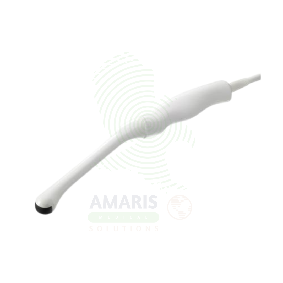
Endocavity Probes
$1.00
WhatsApp Order
Endocavity Probes are specialized ultrasound transducers designed for insertion into the vagina or rectum to provide high-resolution imaging of pelvic structures, the uterus, ovaries, and prostate gland. Transvaginal probes are essential for early pregnancy assessment, gynecologic evaluation, and infertility treatment, while transrectal probes are critical for prostate evaluation and biopsy guidance. Used with sterile probe covers and high-level disinfection between patients, they provide superior image quality for accurate diagnosis and procedure guidance.
Description
Endocavity Probes
PRIMARY CLINICAL & DIAGNOSTIC USES
1. Transvaginal Ultrasound for Pelvic Imaging
-
Primary Use: Provides high-resolution imaging of the uterus, endometrium, ovaries, and adnexa through the vaginal approach. Transvaginal probes offer superior image quality compared to transabdominal imaging for pelvic structures due to closer proximity to the organs of interest and the ability to use higher frequencies.
-
How it helps: For the gynecologist, obstetrician, and reproductive endocrinologist, transvaginal ultrasound provides the detailed imaging needed to evaluate the endometrium, detect ovarian cysts, diagnose ectopic pregnancy, and assess early fetal development. For the patient, transvaginal imaging offers a more comfortable experience than a full bladder for transabdominal imaging and provides superior diagnostic information.
2. Early Pregnancy Assessment
-
Primary Use: Essential for early pregnancy evaluation, including confirmation of intrauterine pregnancy, detection of fetal heartbeat, measurement of gestational sac and crown-rump length, and diagnosis of ectopic pregnancy or pregnancy failure.
-
How it helps: For the obstetrician and emergency physician, transvaginal ultrasound provides the ability to diagnose pregnancy location and viability weeks before transabdominal imaging is possible—guiding management of early pregnancy complications and providing reassurance to patients with viable pregnancies. For the patient with early pregnancy concerns, transvaginal imaging provides answers when they are most needed.
3. Transrectal Ultrasound for Prostate Evaluation
-
Primary Use: Endocavity probes with transrectal capability provide detailed imaging of the prostate gland for evaluation of prostate volume, detection of nodules, guidance for prostate biopsy, and assessment of seminal vesicles.
-
How it helps: For the urologist, transrectal ultrasound provides the imaging needed to evaluate prostate pathology, guide biopsy for suspected prostate cancer, and assess prostate volume for benign prostatic hyperplasia treatment planning. For the patient undergoing prostate evaluation, transrectal ultrasound enables targeted biopsy of suspicious lesions, improving cancer detection rates.
4. Endometrial Assessment and Biopsy Guidance
-
Primary Use: High-resolution imaging of the endometrium for evaluation of thickness, echogenicity, and focal abnormalities. Endocavity probes can guide endometrial biopsy and aspiration procedures.
-
How it helps: For the gynecologist managing patients with abnormal uterine bleeding, transvaginal ultrasound provides the detailed endometrial assessment needed to guide the need for biopsy and to localize focal abnormalities such as polyps or fibroids. For the patient, accurate imaging helps avoid unnecessary invasive procedures and guides appropriate treatment.
5. Pelvic Floor and Urogynecologic Assessment
-
Primary Use: Used to evaluate pelvic floor anatomy, assess for cystocele, rectocele, and uterine prolapse, and to assess pelvic floor muscle function. Dynamic imaging during Valsalva and contraction maneuvers provides functional assessment.
-
How it helps: For the urogynecologist and pelvic floor physical therapist, endocavity probes provide dynamic assessment of pelvic floor structures—evaluating pelvic organ prolapse, assessing pelvic floor muscle function, and guiding treatment planning. For the patient with pelvic floor disorders, accurate imaging helps identify the underlying cause of symptoms and guide appropriate therapy.
SECONDARY & SUPPORTIVE USES
1. Infertility Assessment and Treatment: Used for monitoring follicle development, endometrial thickness, and guiding oocyte retrieval and embryo transfer.
2. Ectopic Pregnancy Diagnosis: Essential for differentiating intrauterine from ectopic pregnancy in patients with pain or bleeding.
3. Postmenopausal Bleeding Evaluation: Assessment of endometrial thickness and morphology in patients with postmenopausal bleeding.
4. Uterine Anomaly Assessment: Evaluation of congenital uterine anomalies, intrauterine adhesions, and fibroid location.
5. Rectal Cancer Staging: Transrectal ultrasound for local staging of rectal cancer.
6. Interventional Guidance: Guidance for transvaginal oocyte retrieval, cyst aspiration, and drainage procedures.
KEY PRODUCT FEATURES
1. BASIC IDENTIFICATION ATTRIBUTES
-
Device Type: Specialized ultrasound transducers designed for insertion into body cavities (vagina or rectum) for imaging pelvic and prostate structures.
-
Designation: Endocavity Probes, Transvaginal Probes, Transrectal Probes, Intracavitary Ultrasound Transducers.
-
Types:
-
Transvaginal Probes: Designed for vaginal insertion for pelvic imaging.
-
Transrectal Probes: Designed for rectal insertion for prostate imaging.
-
Endocavitary Biplane Probes: Provide both longitudinal and transverse imaging planes.
-
-
Key Components:
-
Elongated Insertion Shaft: Designed for comfortable insertion.
-
High-Frequency Transducer: 5-9 MHz for optimal resolution.
-
Biplane Capability: Some probes offer dual-plane imaging.
-
Ergonomic Handle: Designed for comfortable operator grip.
-
Sterile Sheath Compatibility: Designed for use with sterile probe covers.
-
2. TECHNICAL & PERFORMANCE PROPERTIES
-
Frequency Range: 5-9 MHz for optimal penetration and resolution.
-
Field of View: Wide field of view for comprehensive assessment.
-
Biplane Imaging: Some probes offer simultaneous longitudinal and transverse imaging.
-
Doppler Capabilities: Color and Spectral Doppler for vascular assessment.
-
3D/4D Capability: Some endocavity probes support 3D and 4D imaging.
-
Resolution: High resolution for detailed assessment of small structures.
3. PHYSICAL & OPERATIONAL PROPERTIES
-
Construction: Ergonomic design with smooth, cleanable surfaces.
-
Insertion Shaft: Narrow diameter for patient comfort.
-
Handle: Designed for precise manipulation and control.
-
Cable: Flexible, durable cable with strain relief.
-
Sterility: Used with sterile probe covers; probe itself is high-level disinfected.
4. SAFETY & COMPLIANCE ATTRIBUTES
-
Regulatory Status: Class II medical device regulated by FDA.
-
Acoustic Output: Regulated by FDA output display standards.
-
Biocompatibility: Materials safe for mucous membrane contact.
-
Sterile Sheath: Must be used with sterile probe covers for each patient.
-
High-Level Disinfection: Probe must undergo high-level disinfection between patients.
5. STORAGE & HANDLING ATTRIBUTES
-
Storage: Store in protective holder or case.
-
Cleaning: Thorough cleaning after each use; enzymatic cleaner recommended.
-
Disinfection: High-level disinfection required between patients.
-
Inspection: Regular inspection for damage, cracks, or fluid ingress.
-
Sheath Use: Always use sterile probe cover for each patient.
6. LABORATORY & CLINICAL APPLICATIONS
-
Primary Application: Pelvic imaging for gynecology, obstetrics, and reproductive medicine; prostate imaging for urology.
-
Clinical Role: Essential equipment for obstetrics, gynecology, reproductive endocrinology, urology, and pelvic floor assessment.
SAFETY HANDLING PRECAUTIONS
1. SAFETY PRECAUTIONS
-
Sterile Sheath: Always use a sterile probe cover for each patient to prevent cross-contamination.
-
High-Level Disinfection: Perform high-level disinfection between patients per facility protocol.
-
Insertion Technique: Insert with care; use gentle pressure; do not force.
-
Patient Positioning: Ensure the patient is properly positioned and comfortable.
-
Informed Consent: Explain procedure to patient; obtain consent before examination.
2. FIRST AID MEASURES
-
Patient Discomfort: If patient experiences significant discomfort, stop procedure; reassess technique.
-
Sheath Breakage: If sheath breaks during procedure, remove probe; assess for contamination; follow infection control protocol.
3. FIRE FIGHTING MEASURES
-
Flammability: Plastic components are combustible.
-
Extinguishing Media: Use water, foam, or CO₂ as appropriate for surrounding materials.
Related products
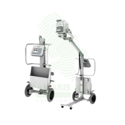
Analogue Mobile X-ray Machine
An Analogue Mobile X-ray Machine is a battery-powered, portable X-ray system using traditional film cassettes for bedside imaging in intensive care units, neonatal intensive care units, emergency departments, and operating rooms. The mobile unit enables chest, abdominal, and extremity imaging at the patient's bedside, eliminating the risks associated with transporting critically ill patients. Film cassettes are processed in darkroom facilities for image development. Used in hospitals without digital radiography, as backup for digital systems, and in resource-limited settings where digital infrastructure is not available.
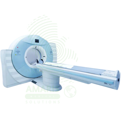
Computed Tomography (CT)
Computed Tomography (CT) is a diagnostic imaging modality that uses X-rays and computer processing to create detailed cross-sectional images of the body. Essential for trauma evaluation, cancer diagnosis, vascular imaging, and surgical planning, CT provides rapid, high-resolution images that guide life-saving decisions in emergency medicine, oncology, and surgery. Advanced multi-slice systems enable whole-body scanning in seconds with sub-millimeter resolution. Radiation dose optimization and contrast safety protocols are essential for patient safety.
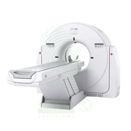
CT Scanner
A CT Scanner is an advanced diagnostic imaging device that uses a rotating X-ray source and detector array to create detailed cross-sectional images ("slices") of the body. By combining these slices, it generates comprehensive 2D and 3D views of bones, organs, and blood vessels, making it indispensable for trauma evaluation, cancer staging, vascular assessment, and guiding complex procedures. While offering unparalleled diagnostic clarity, its operation requires strict adherence to radiation safety principles (ALARA) and protocols for the safe use of contrast agents to maximize patient benefit and minimize risk.
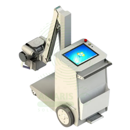
Digital & Analog X-ray Machine
A Digital & Analog X-ray Machine is a fundamental medical imaging device that uses a controlled beam of ionizing radiation to produce static or real-time images of the body's internal structures. It is indispensable for diagnosing fractures, lung diseases, dental issues, and many abdominal conditions. The transition from Analog (film-based) to Digital (CR or DR) technology has revolutionized the field, offering faster results, superior image manipulation, improved dose efficiency, and seamless integration into digital healthcare networks. Its operation demands strict adherence to radiation safety protocols (ALARA) to protect patients and staff, making it a cornerstone of safe, effective diagnostic medicine.
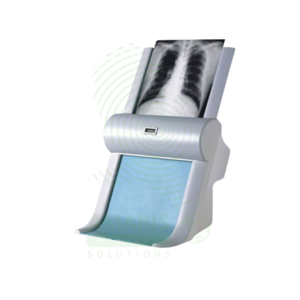
Film Digitizer
A Film Digitizer is a specialized scanner that converts analog radiographic films into high-fidelity digital images (DICOM format). It is the essential tool for migrating historical film archives into a modern digital PACS, enabling filmless workflow, remote access, and long-term preservation of diagnostic records. Its critical performance characteristics are a wide optical density range (to capture all film details) and high spatial resolution. By creating secure, accessible digital copies, it protects against the loss of physical films and integrates past patient history with current digital imaging, supporting comprehensive care and efficient radiology practice.
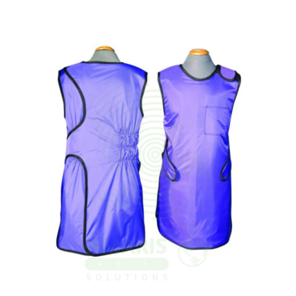
Lead Apron
A Lead Apron is a protective garment worn by healthcare workers to shield against scatter radiation during fluoroscopic procedures, X-ray examinations, and interventional radiology. Made of lead-impregnated material, it attenuates scatter radiation to the thyroid, chest, and reproductive organs, ensuring occupational radiation exposure remains within safe limits. Available in frontal, wrap-around, and two-piece designs with lead equivalence ranging from 0.25 mm to 0.5 mm, proper storage, annual inspection, and use of thyroid shields are essential for effective radiation protection.
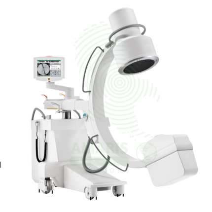
Mobile C-arm Surgical System
A Mobile C-arm Surgical System is a portable fluoroscopic imaging device used for real-time intraoperative guidance during orthopedic, spinal, vascular, and pain management procedures. The C-shaped arm allows flexible positioning around the patient, providing AP, lateral, and oblique views to verify instrument placement, fracture reduction, and device deployment. Essential for minimally invasive surgery, it enables surgeons to achieve precision and accuracy while reducing operative time and improving patient outcomes.