Dermatoscope and Magnifiers
 Diagnostic Kits
 Vital Signs Monitors
 Stethoscopes and Accessories
 Otoscopes, Ophthalmoscopes, and Retinoscopes
 Reflex Hammers and Neurological Tools
 Scales and Measuring Devices
 Spirometers and Pulmonary Function Tests
 Electrosurgical Units and Accessories
 Cutting Instruments
 Grasping and Holding Instruments
 Hemostatic Instruments
 Specialized Surgical Sets
 Single-Use Procedure Trays and Packs
 Surgical Drapes, Gowns, and Covers
 Tissue Unifying Instruments
 Radiation Protection
 X-Ray Machines and Accessories
 Ultrasound Systems and Probes
 MRI and CT Scanners
 Radiology Consumables
 Bone Densitometers
 Fluoroscopy Equipment
 Imaging Tables and Positioning Aids
 Microscopes and Accessories
 Centrifuges and Separators
 Analyzers
 Incubators and Ovens
 Pipettes, Dispensers, and Lab Glassware
 Refrigerators, Freezers, and Storage Units
 Lab Consumables
 Sterilizers and Autoclaves for Lab Use
 Multi-Parameter Monitors
 Ventilators and Respiratory Support Devices
 Defibrillators and AEDs
 Infusion Pumps and IV Systems
 Patient Warmers and Cooling Devices
 Central Monitoring Stations
 Accessories
 Anesthesia Machines and Workstations
 Oxygen Concentrators and Delivery Systems
 Nebulizers and Inhalers
 CPAP/BiPAP Machines
 Airway Management
 Anesthesia Masks, Circuits, and Bags
 Humidifiers and Heaters
 Respiratory Therapy Accessories
 First Aid Kits and Cabinets
 Emergency Resuscitation Equipment
 Trauma Supplies
 Emergency Carts and Crash Carts
 Burn Care Products
 Bleeding Control
 Automated External Defibrillators (AEDs)
 Transport and Evacuation
 Wheelchairs and Accessories
 Walkers, Crutches, and Canes
 Prosthetics and Orthotics
 Physical Therapy Equipment
 Transfer Devices
 Bathroom Safety
 Orthopedic Traction and Tables
 Hot/Cold Therapy Packs and Units
 Beds and Mattresses
 Chairs and Stools
 Tables
 Cabinets and Storage
 Privacy Screens & Curtains
 Stands and Racks
 Linens and Textiles
 Lighting
 Autoclaves and Sterilizers
 Ultrasonic Cleaners
 Disinfectant Solutions and Wipes
 Sterilization Pouches, Wraps, and Indicators
 Instrument Trays and Containers
 UV and Ozone Disinfection Devices
 Washer Disinfectors
 Wound Care
 Gloves
 Masks and Respirators
 Catheters and Tubing
 Swabs, Applicators, and Sponges
 Incontinence Products
 Personal Protective Equipment (PPE)
 Dental Chairs and Units
 Handpieces and Burs
 Instruments
 Consumables
 Sterilization for Dental Use
 Orthodontic Supplies
 Endodontic Tools
 Slit Lamps and Tonometers
 Lensometers and Phoropters
 Ophthalmic Surgical Instruments
 Eyewear Frames and Lenses
 Contact Lens Supplies
 Vision Testing Charts and Devices
 Eye Care Consumables
 Laser Systems for Eye Care
 ENT Exam Chairs and Tables
 Endoscopes
 Audiometers and Hearing Tests
 ENT Instruments
 Nasal and Throat Packs
 Hearing Aids and Accessories
 Otology Supplies
 Fetal Dopplers and Monitors
 Delivery Beds and Tables
 Gynecological Instruments
 Neonatal Incubators and Warmers
 Breast Pumps and Accessories
 Contraceptive Devices
 Maternity Supports and Pads
 Neonatal Consumables
 Cystoscopes and Urethroscopes
 Dialysis Machines and Supplies
 Urological Catheters and Bags
 Lithotripters
 Prostate Treatment Devices
 Urinary Incontinence Products
 Kidney Stone Management Tools
 Consumables & Disposables
 EEG and EMG Machines
 Neurosurgical Instruments
 Nerve Stimulators
 Headrests and Positioning Aids
 Lumbar Puncture Kits
 Seizure Monitoring Devices
 Consumables
 Rehabilitation for Neurological Conditions
 ECG Machines and Accessories
 Holter Monitors
 Stress Test Systems
 Pacemakers and Defibrillator Accessories
 Vascular Access Devices
 Cardiac Catheters and Guidewires
 Blood Flow Meters
 Consumables
 Orthopedic Instruments
 Casts, Splints, and Padding
 Joint Replacement Supplies
 Prosthetic Limbs and Components
 Bone Grafts and Substitutes
 Traction Devices
 Orthopedic Braces and Supports
 Rehabilitation Aids for Orthopedics
 Home Oxygen Therapy
 Hospital Beds for Home Use
 Mobility Aids
 Bathroom and Daily Living Aids
 Wound Care for Home
 Monitoring Devices
 Enteral Feeding Pumps and Tubes
 Hand Sanitizers and Dispensers
 Face Shields and Goggles
 Isolation Gowns and Suits
 Biohazard Waste Containers
 Air Purifiers and HEPA Filters
 Surface Disinfectants
 Sharps Containers
 Protective Barriers
 Cardiovascular & Endurance Training
 Strength Training & Weightlifting
 Functional Training & Core Conditioning
 Physical Therapy & Rehabilitation
 Sports & Outdoor Recreation
 Gym Flooring & Facility Equipment
 Fitness Monitoring & Accessories
 Kids & Novelties
Near Probes
$1.00
WhatsApp Order
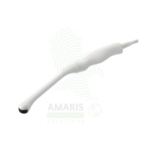
Near Probes are high-frequency linear array ultrasound transducers designed for imaging superficial structures including the thyroid, breast, testicles, muscles, tendons, ligaments, and superficial vessels. Operating at frequencies up to 18 MHz, they provide exceptional resolution for visualizing fine detail, enabling accurate characterization of small lesions, assessment of tendon integrity, and guidance for interventional procedures. Essential for radiology, breast imaging, musculoskeletal imaging, and vascular access, they deliver the image quality needed for precise diagnosis and treatment.
Description
Near Probes
PRIMARY CLINICAL & DIAGNOSTIC USES
1. High-Resolution Imaging of Superficial Structures
-
Primary Use: Near probes (high-frequency linear transducers) provide exceptional resolution for imaging superficial structures including the thyroid, breast, testicles, muscles, tendons, ligaments, and superficial vessels. The high frequency allows visualization of fine detail not visible with lower frequency probes.
-
How it helps: For the radiologist, sonographer, and musculoskeletal specialist, near probes deliver the resolution needed to evaluate superficial structures with exceptional clarity—revealing subtle tendon tears, small thyroid nodules, and early breast lesions that might be missed with lower frequency probes. For the patient with a palpable lump, joint pain, or thyroid nodule, high-frequency imaging provides the detailed characterization needed for accurate diagnosis.
2. Thyroid and Parathyroid Imaging
-
Primary Use: Near probes are essential for evaluating thyroid nodules, diffuse thyroid disease, and parathyroid adenomas. High-frequency imaging allows detailed assessment of nodule characteristics including margins, echogenicity, calcifications, and vascularity.
-
How it helps: For the endocrinologist and thyroid surgeon, near probes provide the resolution needed to characterize thyroid nodules, guide fine-needle aspiration, and localize parathyroid adenomas preoperatively. For the patient with a thyroid nodule, high-quality ultrasound helps distinguish benign from suspicious lesions, guiding the need for biopsy or surgical intervention.
3. Breast Imaging
-
Primary Use: Used for evaluation of palpable breast masses, characterization of mammographic findings, and guidance for breast biopsies. Near probes provide detailed imaging of breast parenchyma, cysts, solid masses, and ductal structures.
-
How it helps: For the breast imager and surgeon, near probes provide the detail needed to differentiate cystic from solid masses, characterize suspicious lesions, and guide percutaneous biopsies. For the patient with a breast lump, high-resolution ultrasound provides reassurance for benign findings and accurate targeting for biopsy when needed.
4. Musculoskeletal Imaging
-
Primary Use: Near probes are the primary tool for evaluating muscles, tendons, ligaments, joints, and peripheral nerves. Dynamic imaging allows assessment of tendon gliding, joint effusions, and nerve compression syndromes.
-
How it helps: For the orthopedic surgeon, sports medicine physician, and rheumatologist, near probes provide dynamic assessment of musculoskeletal structures—showing tendon tears with movement, revealing inflammation in real time, and guiding precise placement of therapeutic injections. For the patient with shoulder pain, tennis elbow, or carpal tunnel syndrome, high-resolution ultrasound provides accurate diagnosis and guides treatment.
5. Vascular Access and Superficial Vessel Imaging
-
Primary Use: Near probes are used to image superficial veins for peripheral IV access, PICC line placement, and arterial line insertion. High-frequency imaging allows visualization of small vessels and guides needle placement.
-
How it helps: For the anesthesiologist, emergency physician, and vascular access team, near probes provide the resolution needed to visualize superficial vessels, assess patency, and guide needle placement—improving success rates and reducing complications. For the patient with difficult venous access, ultrasound guidance with a near probe means fewer needle sticks and successful IV placement.
SECONDARY & SUPPORTIVE USES
1. Pediatric Imaging: High-frequency probes for imaging infants and children, where structures are smaller and closer to the surface.
2. Scrotal Imaging: Detailed evaluation of testicular pathology including masses, torsion, varicocele, and epididymitis.
3. Salivary Gland Imaging: Evaluation of parotid, submandibular, and sublingual glands for stones, masses, and inflammatory conditions.
4. Dermatologic Ultrasound: High-resolution imaging of skin lesions, nail abnormalities, and superficial soft tissue masses.
5. Foreign Body Localization: Detecting and localizing superficial foreign bodies such as glass, metal, or wood splinters.
6. Regional Anesthesia: Guidance for peripheral nerve blocks and superficial nerve blocks.
KEY PRODUCT FEATURES
1. BASIC IDENTIFICATION ATTRIBUTES
-
Device Type: High-frequency linear array ultrasound transducers designed for imaging superficial structures.
-
Designation: Near Probes, High-Frequency Linear Probes, Superficial Ultrasound Transducers, Linear Array Probes.
-
Key Components:
-
Linear Array: Arrangement of piezoelectric elements in a straight line.
-
Frequency Range: 5-18 MHz depending on application.
-
Footprint: Long, narrow contact surface for optimal skin coupling.
-
Acoustic Lens: Focuses beam for optimal resolution at superficial depths.
-
Controls: Integrated buttons for image capture and mode selection.
-
2. TECHNICAL & PERFORMANCE PROPERTIES
-
Frequency Range: 5-18 MHz; higher frequencies provide better resolution but less penetration.
-
Field of View: Wide field of view for comprehensive evaluation of superficial structures.
-
Resolution: High spatial resolution for visualization of fine detail.
-
Penetration: Optimal for depths up to 6-8 cm.
-
Harmonic Imaging: Tissue harmonic imaging for improved image quality.
-
Doppler Capabilities: Color, Power, and Spectral Doppler for vascular assessment.
3. PHYSICAL & OPERATIONAL PROPERTIES
-
Construction: Ergonomic design for comfortable handling; lightweight.
-
Footprint: Long, narrow shape for optimal contact with superficial structures.
-
Cable: Flexible, durable cable with strain relief.
-
Sterilization: Compatible with high-level disinfection for semi-critical applications.
4. SAFETY & COMPLIANCE ATTRIBUTES
-
Regulatory Status: Class II medical device regulated by FDA.
-
Acoustic Output: Regulated by FDA output display standards.
-
Biocompatibility: Materials safe for skin contact.
-
Disinfection: Compatible with high-level disinfection for endocavitary use; intermediate-level for external use.
5. STORAGE & HANDLING ATTRIBUTES
-
Storage: Store in protective holder or case.
-
Cleaning: Wipe with hospital-grade disinfectants; follow manufacturer guidelines.
-
Disinfection: High-level disinfection for procedures involving mucous membranes; intermediate-level for intact skin.
-
Handling: Handle carefully; protect from drops and impacts.
6. LABORATORY & CLINICAL APPLICATIONS
-
Primary Application: High-resolution imaging of superficial structures including thyroid, breast, musculoskeletal, and vascular structures.
-
Clinical Role: Essential equipment for radiology, breast imaging, musculoskeletal imaging, vascular access, and interventional procedures.
SAFETY HANDLING PRECAUTIONS
1. SAFETY PRECAUTIONS
-
Probe Handling: Handle carefully; high-frequency probes are delicate.
-
Disinfection: Follow appropriate disinfection protocols based on application.
-
Gel Use: Use appropriate ultrasound gel for acoustic coupling.
-
Patient Comfort: Ensure adequate gel for comfortable scanning; avoid excessive pressure.
2. FIRST AID MEASURES
-
Probe Damage: If probe is dropped or damaged, removed from service; contact service provider.
-
Patient Discomfort: If a patient experiences discomfort, adjust scanning technique; use adequate gel.
3. FIRE FIGHTING MEASURES
-
Flammability: Plastic components are combustible.
-
Extinguishing Media: Use water, foam, or CO₂ as appropriate for surrounding materials.
Related products
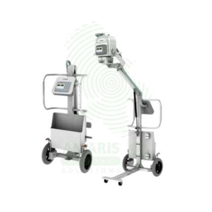
Analogue Mobile X-ray Machine
An Analogue Mobile X-ray Machine is a battery-powered, portable X-ray system using traditional film cassettes for bedside imaging in intensive care units, neonatal intensive care units, emergency departments, and operating rooms. The mobile unit enables chest, abdominal, and extremity imaging at the patient's bedside, eliminating the risks associated with transporting critically ill patients. Film cassettes are processed in darkroom facilities for image development. Used in hospitals without digital radiography, as backup for digital systems, and in resource-limited settings where digital infrastructure is not available.
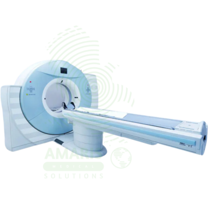
Computed Tomography (CT)
Computed Tomography (CT) is a diagnostic imaging modality that uses X-rays and computer processing to create detailed cross-sectional images of the body. Essential for trauma evaluation, cancer diagnosis, vascular imaging, and surgical planning, CT provides rapid, high-resolution images that guide life-saving decisions in emergency medicine, oncology, and surgery. Advanced multi-slice systems enable whole-body scanning in seconds with sub-millimeter resolution. Radiation dose optimization and contrast safety protocols are essential for patient safety.
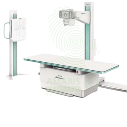
Digital Fixed X-ray
A Digital Fixed X-ray is a permanent installation digital radiography system designed for high-volume general imaging in radiology departments and outpatient imaging centers. Featuring digital flat panel detectors, ceiling-mounted tube assemblies, and tilting tables, it provides high-resolution images for skeletal, chest, abdominal, and extremity examinations. Integrated with PACS and RIS, it supports efficient digital workflow from image acquisition to interpretation, enabling rapid diagnosis and treatment planning.
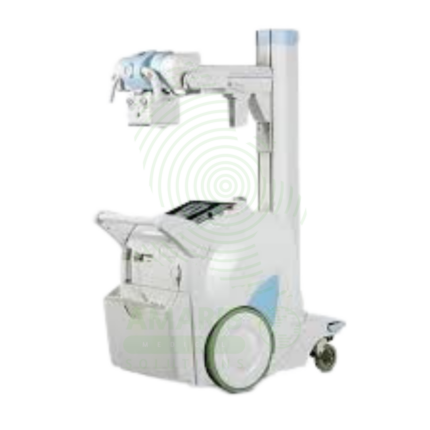
Digital Mobile X-ray
A Digital Mobile X-ray is a battery-powered, portable digital radiography system designed for bedside imaging in intensive care units, neonatal intensive care units, emergency departments, and operating rooms. The mobile unit enables high-quality chest, abdominal, and extremity imaging at the patient's bedside, eliminating the risks associated with transporting critically ill patients. Wireless digital detectors provide immediate image capture and transmission to PACS, supporting rapid diagnosis and treatment decisions.
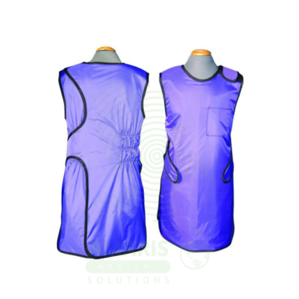
Lead Apron
A Lead Apron is a protective garment worn by healthcare workers to shield against scatter radiation during fluoroscopic procedures, X-ray examinations, and interventional radiology. Made of lead-impregnated material, it attenuates scatter radiation to the thyroid, chest, and reproductive organs, ensuring occupational radiation exposure remains within safe limits. Available in frontal, wrap-around, and two-piece designs with lead equivalence ranging from 0.25 mm to 0.5 mm, proper storage, annual inspection, and use of thyroid shields are essential for effective radiation protection.
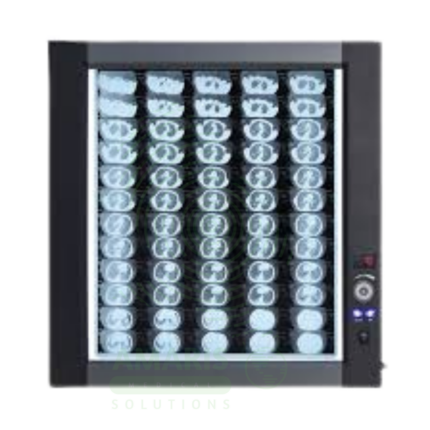
LED Medical Film Viewer
An LED Medical Film Viewer is a light box designed for viewing and interpreting analog X-ray films. Using LED backlight technology, it provides uniform, high-luminance illumination with instant on capability and long life. Available in single, dual, and multi-panel configurations, it supports side-by-side comparison of current and prior studies, pre-operative planning, and group teaching. Essential for radiology departments, orthopedic clinics, emergency departments, and operating rooms where film-based imaging is still used.
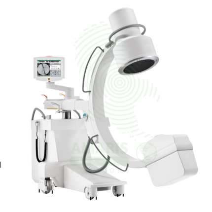
Mobile C-arm Surgical System
A Mobile C-arm Surgical System is a portable fluoroscopic imaging device used for real-time intraoperative guidance during orthopedic, spinal, vascular, and pain management procedures. The C-shaped arm allows flexible positioning around the patient, providing AP, lateral, and oblique views to verify instrument placement, fracture reduction, and device deployment. Essential for minimally invasive surgery, it enables surgeons to achieve precision and accuracy while reducing operative time and improving patient outcomes.