Dermatoscope and Magnifiers
 Diagnostic Kits
 Vital Signs Monitors
 Stethoscopes and Accessories
 Otoscopes, Ophthalmoscopes, and Retinoscopes
 Reflex Hammers and Neurological Tools
 Scales and Measuring Devices
 Spirometers and Pulmonary Function Tests
 Electrosurgical Units and Accessories
 Cutting Instruments
 Grasping and Holding Instruments
 Hemostatic Instruments
 Specialized Surgical Sets
 Single-Use Procedure Trays and Packs
 Surgical Drapes, Gowns, and Covers
 Tissue Unifying Instruments
 Radiation Protection
 X-Ray Machines and Accessories
 Ultrasound Systems and Probes
 MRI and CT Scanners
 Radiology Consumables
 Bone Densitometers
 Fluoroscopy Equipment
 Imaging Tables and Positioning Aids
 Microscopes and Accessories
 Centrifuges and Separators
 Analyzers
 Incubators and Ovens
 Pipettes, Dispensers, and Lab Glassware
 Refrigerators, Freezers, and Storage Units
 Lab Consumables
 Sterilizers and Autoclaves for Lab Use
 Multi-Parameter Monitors
 Ventilators and Respiratory Support Devices
 Defibrillators and AEDs
 Infusion Pumps and IV Systems
 Patient Warmers and Cooling Devices
 Central Monitoring Stations
 Accessories
 Anesthesia Machines and Workstations
 Oxygen Concentrators and Delivery Systems
 Nebulizers and Inhalers
 CPAP/BiPAP Machines
 Airway Management
 Anesthesia Masks, Circuits, and Bags
 Humidifiers and Heaters
 Respiratory Therapy Accessories
 First Aid Kits and Cabinets
 Emergency Resuscitation Equipment
 Trauma Supplies
 Emergency Carts and Crash Carts
 Burn Care Products
 Bleeding Control
 Automated External Defibrillators (AEDs)
 Transport and Evacuation
 Wheelchairs and Accessories
 Walkers, Crutches, and Canes
 Prosthetics and Orthotics
 Physical Therapy Equipment
 Transfer Devices
 Bathroom Safety
 Orthopedic Traction and Tables
 Hot/Cold Therapy Packs and Units
 Beds and Mattresses
 Chairs and Stools
 Tables
 Cabinets and Storage
 Privacy Screens & Curtains
 Stands and Racks
 Linens and Textiles
 Lighting
 Autoclaves and Sterilizers
 Ultrasonic Cleaners
 Disinfectant Solutions and Wipes
 Sterilization Pouches, Wraps, and Indicators
 Instrument Trays and Containers
 UV and Ozone Disinfection Devices
 Washer Disinfectors
 Wound Care
 Gloves
 Masks and Respirators
 Catheters and Tubing
 Swabs, Applicators, and Sponges
 Incontinence Products
 Personal Protective Equipment (PPE)
 Dental Chairs and Units
 Handpieces and Burs
 Instruments
 Consumables
 Sterilization for Dental Use
 Orthodontic Supplies
 Endodontic Tools
 Slit Lamps and Tonometers
 Lensometers and Phoropters
 Ophthalmic Surgical Instruments
 Eyewear Frames and Lenses
 Contact Lens Supplies
 Vision Testing Charts and Devices
 Eye Care Consumables
 Laser Systems for Eye Care
 ENT Exam Chairs and Tables
 Endoscopes
 Audiometers and Hearing Tests
 ENT Instruments
 Nasal and Throat Packs
 Hearing Aids and Accessories
 Otology Supplies
 Fetal Dopplers and Monitors
 Delivery Beds and Tables
 Gynecological Instruments
 Neonatal Incubators and Warmers
 Breast Pumps and Accessories
 Contraceptive Devices
 Maternity Supports and Pads
 Neonatal Consumables
 Cystoscopes and Urethroscopes
 Dialysis Machines and Supplies
 Urological Catheters and Bags
 Lithotripters
 Prostate Treatment Devices
 Urinary Incontinence Products
 Kidney Stone Management Tools
 Consumables & Disposables
 EEG and EMG Machines
 Neurosurgical Instruments
 Nerve Stimulators
 Headrests and Positioning Aids
 Lumbar Puncture Kits
 Seizure Monitoring Devices
 Consumables
 Rehabilitation for Neurological Conditions
 ECG Machines and Accessories
 Holter Monitors
 Stress Test Systems
 Pacemakers and Defibrillator Accessories
 Vascular Access Devices
 Cardiac Catheters and Guidewires
 Blood Flow Meters
 Consumables
 Orthopedic Instruments
 Casts, Splints, and Padding
 Joint Replacement Supplies
 Prosthetic Limbs and Components
 Bone Grafts and Substitutes
 Traction Devices
 Orthopedic Braces and Supports
 Rehabilitation Aids for Orthopedics
 Home Oxygen Therapy
 Hospital Beds for Home Use
 Mobility Aids
 Bathroom and Daily Living Aids
 Wound Care for Home
 Monitoring Devices
 Enteral Feeding Pumps and Tubes
 Hand Sanitizers and Dispensers
 Face Shields and Goggles
 Isolation Gowns and Suits
 Biohazard Waste Containers
 Air Purifiers and HEPA Filters
 Surface Disinfectants
 Sharps Containers
 Protective Barriers
 Cardiovascular & Endurance Training
 Strength Training & Weightlifting
 Functional Training & Core Conditioning
 Physical Therapy & Rehabilitation
 Sports & Outdoor Recreation
 Gym Flooring & Facility Equipment
 Fitness Monitoring & Accessories
 Kids & Novelties
Polypectomy Snare
WhatsApp Order
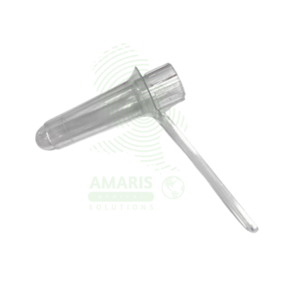
A Polypectomy Snare is a sterile, single-use Class II medical device (FDA-cleared, CE-marked) used during endoscopy (colonoscopy, gastroscopy, enteroscopy) for resecting polyps and mucosal lesions from the gastrointestinal tract. The device consists of a flexible stainless steel braided wire loop (10-40 mm diameter) that opens and closes via an ergonomic handle, delivered through an insulated PTFE or PEEK sheath compatible with standard endoscopes (working channel 2.0-2.8 mm). Available in various shapes (oval, round, crescent, hexagonal) for different polyp types (pedunculated, sessile, flat). Used with electrosurgical generators for hot snare polypectomy (cautery-assisted) or without for cold snare polypectomy (small polyps). Primary clinical applications include removal of colorectal polyps to prevent colorectal cancer, resection of gastric and esophageal polyps, treatment of bleeding polyps, debulking of large sessile polyps via piecemeal technique, and surveillance in hereditary polyposis syndromes (FAP, Lynch). Critical safety precautions include single-use only, proper electrosurgical technique, ensuring complete polyp capture before current application, submucosal injection for sessile lesions, and post-procedure monitoring for bleeding or perforation. Essential instrument for therapeutic endoscopy and colorectal cancer prevention.
Description
Polypectomy Snare
PRIMARY CLINICAL & DIAGNOSTIC USES
1. Endoscopic Removal of Colorectal Polyps:
-
Primary Use: The polypectomy snare is the primary instrument used during colonoscopy to remove polyps from the colon and rectum, preventing progression to colorectal cancer by excising precancerous adenomatous polyps and enabling histopathological examination.
-
How it helps: Removes the precursors to colorectal cancer before they have a chance to become malignant, offering patients one of the most effective cancer prevention interventions available in modern medicine.
2. Resection of Gastric Polyps:
-
Primary Use: Used during upper gastrointestinal endoscopy (esophagogastroduodenoscopy) to remove polyps from the stomach, aiding in diagnosis and treatment of gastric lesions and reducing risk of malignant transformation.
-
How it helps: Allows gastroenterologists to clear the stomach of potentially dangerous growths, preventing them from developing into gastric cancer while providing tissue for pathological analysis.
3. Excision of Esophageal Polyps:
-
Primary Use: Employed in endoscopic procedures to remove polyps and other mucosal lesions from the esophagus, alleviating symptoms such as dysphagia and bleeding while providing tissue for pathological analysis.
-
How it helps: Restores comfortable swallowing and stops bleeding by removing growths that obstruct the esophagus, improving quality of life and ruling out malignancy.
4. Removal of Small Intestinal Polyps:
-
Primary Use: Used during enteroscopy to resect polyps in the small intestine, particularly in patients with hereditary polyposis syndromes such as Familial Adenomatous Polyposis (FAP) or Peutz-Jeghers syndrome.
-
How it helps: Provides life-saving surveillance and intervention for patients born with genetic conditions that cause hundreds of polyps to form throughout their intestines, reducing their risk of developing cancer at a young age.
5. Treatment of Bleeding Polyps:
-
Primary Use: Utilized to remove polyps that are actively bleeding or at high risk of bleeding, achieving hemostasis while definitively treating the underlying lesion.
-
How it helps: Stops active bleeding from polyps that are causing blood loss, anemia, and weakness, removing the source of bleeding in one therapeutic procedure.
6. Debulking of Large or Sessile Polyps:
-
Primary Use: Used in piecemeal polypectomy techniques to remove large or flat (sessile) polyps that cannot be resected in a single pass, often combined with submucosal injection to lift the lesion and reduce perforation risk.
-
How it helps: Tackles challenging polyps that would otherwise require surgery, removing them piece by piece through the endoscope and sparing patients from more invasive procedures.
7. Surveillance and Management of Hereditary Polyposis Syndromes:
-
Primary Use: Essential for regular surveillance and polypectomy in patients with FAP, Lynch syndrome, and other hereditary conditions predisposing to multiple colorectal polyps and cancer.
-
How it helps: Offers patients with genetic predispositions to colon cancer a way to manage their risk through regular surveillance and polyp removal, dramatically reducing their chances of developing cancer.
SECONDARY & SUPPORTIVE USES
1. Obtaining Tissue for Histopathological Diagnosis: Provides tissue samples for pathological examination to determine polyp type (adenomatous, hyperplastic, sessile serrated), grade of dysplasia, and presence of malignancy, guiding further management.
2. Therapeutic Resection of Submucosal Lesions: Used with submucosal injection techniques to remove selected submucosal tumors and lesions after appropriate lifting, expanding the range of conditions that can be treated endoscopically.
3. Management of Post-Polypectomy Bleeding: Can be used to apply cautery or mechanical pressure to bleeding sites following polypectomy, providing an immediate solution for procedural complications.
4. Training and Education: Used in endoscopic simulation and training programs to teach polypectomy techniques to gastroenterology fellows and endoscopy nurses, training the next generation of endoscopists.
5. Research and Clinical Trials: Employed in studies investigating new polypectomy techniques, devices, and outcomes in polyp management, advancing the field of therapeutic endoscopy.
6. Removal of Polypoid Lesions in Other Gastrointestinal Sites: Occasionally used for polyp removal in the duodenum, ampulla, or other accessible gastrointestinal locations, extending the utility of this versatile tool.
7. Treatment of Polypoid Dysplasia in Inflammatory Bowel Disease: Used to resect polypoid dysplastic lesions in patients with ulcerative colitis or Crohn’s disease undergoing colonoscopic surveillance, managing cancer risk in this high-risk population.
KEY PRODUCT FEATURES
1. BASIC IDENTIFICATION ATTRIBUTES
-
Product Type: Endoscopic surgical instrument for resection of polyps and mucosal lesions.
-
Common Names: Polypectomy Snare, Endoscopic Snare, Colonoscopy Snare, Electrosurgical Snare, Diathermy Snare.
-
Design Variations:
-
Rigid Snare: Fixed shape, non-retractable.
-
Semi-Rigid Snare: Maintains shape but has some flexibility.
-
Flexible Snare: Retractable into sheath; most common for therapeutic endoscopy.
-
-
Shape Configurations:
-
Oval/Round: Standard for pedunculated polyps.
-
Crescent/Half-Moon: For sessile or flat polyps.
-
Dual-Loop/Hexagonal: For large polyps or piecemeal resection.
-
Barbed/Serrated: Enhances grip on polyp tissue.
-
-
Sizes: Loop diameter ranging from 10 mm to 30 mm (standard); mini snares for small polyps (5-10 mm); large snares for bulky lesions (up to 40 mm).
-
Sheath Diameter: 2.0-2.8 mm (compatible with standard endoscope working channels).
-
Working Length: 160-230 cm for colonoscopy; 230-260 cm for enteroscopy.
-
Material: Stainless steel braided wire loop; PTFE or polyether ether ketone (PEEK) sheath.
-
Sterility: Sterile, single-use device.
-
Packaging: Individually wrapped sterile peel pouch.
2. TECHNICAL & PERFORMANCE PROPERTIES
-
Electrosurgical Compatibility: Designed for use with electrosurgical generators (monopolar mode) for cautery-assisted resection.
-
Cutting Mechanism: Mechanical cutting with optional electrosurgical current for hemostasis and clean resection.
-
Conductivity: Stainless steel wire conducts electrosurgical current to cutting site.
-
Insulation: Sheath is insulated to prevent thermal injury to surrounding tissue.
-
Radial Expansion: Snare loop opens to predetermined diameter; maintains shape during resection.
-
Retraction Force: Smooth retraction mechanism for precise control during polyp capture.
-
Rotatability: Some models allow rotation of the snare loop for optimal positioning.
-
Marker Bands: Radiopaque markers for visualization under fluoroscopy.
-
Tensile Strength: Withstands tension during polyp retraction without breakage.
3. PHYSICAL & OPERATIONAL PROPERTIES
-
Handle Design: Ergonomic handle with sliding mechanism for opening/closing snare loop.
-
Handle Features: May include rotating mechanism, locking mechanism, and electrosurgical connection port.
-
Sheath Flexibility: Flexible but kink-resistant for easy passage through the endoscope channel.
-
Loop Visibility: Enhanced visibility under endoscopic light; some models have color-coded loops.
-
Color Coding: Some manufacturers color-code sheath or handle by snare size or type.
-
Packaging: Sterile peel pouch with Tyvek backing; may include introduction aid.
-
Shelf Life: 3-5 years from manufacture date.
4. SAFETY & COMPLIANCE ATTRIBUTES
-
Regulatory Status: Class II medical device requiring FDA 510(k) clearance; CE marked.
-
Quality Standards: Manufactured under ISO 13485.
-
Sterility: Sterile; SAL 10⁻⁶; ethylene oxide or gamma sterilized.
-
Biocompatibility: Materials meet ISO 10993 for tissue contact.
-
Latex-Free: All components latex-free.
-
Electrosurgical Safety: Compatible with standard electrosurgical generators; insulated to prevent unintended burns.
-
Single-Use: Strictly single-use; never resterilize or reuse.
-
Packaging Integrity: Maintains sterility until opened.
5. STORAGE & HANDLING ATTRIBUTES
-
Storage: Store in cool, dry places at room temperature; protect from direct sunlight and extreme temperatures.
-
Inspection: Before use, check packaging integrity; do not use if the package is damaged or compromised.
-
Preparation: Remove from sterile packaging using aseptic technique.
-
Priming: Flush sheath with sterile water or saline to remove air and ensure smooth movement.
-
Handle Check: Verify smooth opening and closing of snare loop before insertion.
-
Electrosurgical Connection: Connect to electrosurgical generator as per manufacturer instructions.
-
Disposal: Dispose of used snare as biohazardous sharps waste.
6. LABORATORY & CLINICAL APPLICATIONS
-
Primary Application: Endoscopic resection of polyps and mucosal lesions throughout the gastrointestinal tract.
-
Polypectomy Techniques:
-
Hot Snare: Electrosurgical current applied during resection for hemostasis; used for most polyps >5 mm.
-
Cold Snare: Mechanical resection without cautery; used for small polyps (<5-10 mm) to reduce thermal injury risk.
-
Piecemeal Snare: Resection of large polyps in multiple pieces; often with submucosal injection.
-
Endoscopic Mucosal Resection (EMR): Submucosal injection followed by snare resection for sessile or flat lesions.
-
-
Polyp Selection Criteria:
-
Pedunculated Polyps: Snare placed around stalk; hot snare preferred.
-
Sessile Polyps (5-20 mm): May be resected with snare after submucosal injection.
-
Large Sessile Polyps (>20 mm): Piecemeal EMR with specialized snares.
-
-
Complications: Bleeding (immediate or delayed), perforation, post-polypectomy syndrome.
-
Success Rate: >95% for complete resection of appropriate polyps.
SAFETY HANDLING PRECAUTIONS
1. SAFETY PRECAUTIONS
-
Single-Use Only: Never reuse a polypectomy snare; reuse risks cross-contamination, device failure, and patient injury.
-
Electrosurgical Safety: Ensure proper grounding of patient; test electrosurgical unit before use.
-
Tissue Capture: Ensure polyp is fully ensnared before applying current; incomplete capture risks incomplete resection or perforation.
-
Tension Control: Avoid excessive tension during snare closure; may cause mechanical transection before cautery or tissue trauma.
-
Insulation Integrity: Do not use if sheath insulation is damaged; risk of thermal injury.
-
Submucosal Injection: Consider sessile polyps to lift lesions and reduce perforation risk.
-
Visualization: Maintain clear endoscopic view throughout procedure; insufflate adequately.
-
Training: Polypectomy should be performed by trained endoscopists familiar with techniques and complications.
-
Post-Procedure Monitoring: Observe for signs of bleeding or perforation; provide appropriate post-procedure instructions.
2. FIRST AID MEASURES
-
Intraprocedural Bleeding: Endoscopic hemostasis with clips, cautery, or injection; monitor hemodynamically.
-
Perforation: Suspect if patient experiences severe pain, abdominal distension, or fever; obtain imaging; surgical consultation.
-
Device Malfunction: If snare fails to open or close properly during procedure, remove and replace with new device.
-
Electrosurgical Injury: Assess for burns; document and manage per institutional protocol.
3. FIRE FIGHTING MEASURES
-
Flammability: Plastic components are combustible; metal components non-combustible.
-
Extinguishing Media: For electrical fire, use CO₂ or dry chemical (Class C) extinguisher.
-
Electrosurgical Unit Fire Risk: Follow electrosurgical safety protocols; avoid flammable prep solutions.
Related products
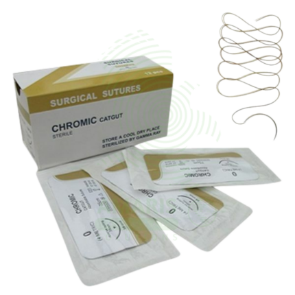
Chromic Sutures
Chromic Sutures are absorbable, monofilament surgical sutures derived from natural collagen and treated with chromic salts to delay absorption. Used for internal tissue approximation in gynecologic, urologic, gastrointestinal, and subcutaneous procedures, they provide temporary wound support during critical healing while being completely absorbed over time without requiring removal. Chromic treatment extends absorption compared to plain catgut, providing 10-14 days of tensile strength retention. Essential for procedures where suture removal would be impractical or where temporary wound support is required.
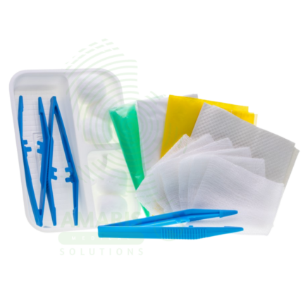
Disposable Dressing Set
A Disposable Dressing Set is a sterile, single-use convenience kit containing all components necessary for sterile wound dressing changes and minor wound care procedures. The set typically includes sterile drapes, gauze sponges, sterile gloves, antiseptic solution, cotton applicators, tape, and disposal bag. Used in hospitals, clinics, emergency departments, and home healthcare settings, it ensures consistent aseptic technique for post-operative wound care, central line dressing changes, and management of chronic wounds. Proper sterile technique and disposal are essential for safe and effective use.
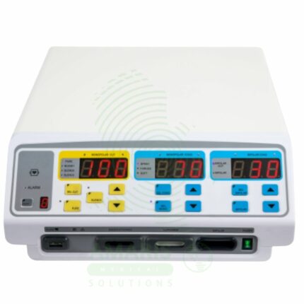
Electrosurgical Unit
An Electrosurgical Unit (ESU) is a radiofrequency generator that provides electrical energy for surgical cutting and coagulation. The ESU is a professional-grade system featuring monopolar and bipolar outputs, with safety-critical Return Electrode Monitoring (REM) for monopolar procedures. It is the universal surgical tool for precise tissue dissection with simultaneous hemostasis, used in open, laparoscopic, and endoscopic procedures across all specialties. Safe operation hinges on correct patient return pad placement, awareness of fire risks (especially with oxygen and flammable prep solutions), and adherence to protocols for patients with implanted electronic devices.

Green towel
A Green Towel is a sterile, absorbent surgical towel used in operating rooms and procedure areas for draping, fluid absorption, and creating sterile fields. Made from high-absorbency cotton or cotton-blend fabric, the green color reduces glare under operating lights and provides visual contrast against blood and tissue. Essential for establishing sterile fields, protecting instruments, and managing fluids during surgical procedures, it is a fundamental component of aseptic technique in surgery, emergency care, and other invasive procedures.
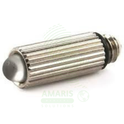
Macintosh Bulb Laryngoscope
A Macintosh Bulb Laryngoscope is a rigid laryngoscope with curved Macintosh blade (sizes 0-4, 70-160 mm) featuring a distal incandescent (xenon, krypton, halogen) or LED bulb at the blade tip for direct illumination during tracheal intubation. The curved blade design allows indirect epiglottis elevation by placing the tip in the vallecula, requiring less force and neck extension than straight blades. Features stainless steel reusable blades (or disposable plastic), ergonomic handles with knurled grip, ISO standard hook-on fittings, and autoclavable options. Light output 500-3,000 Lux depending on bulb type and battery condition. Primary clinical applications include routine and emergency tracheal intubation during general anesthesia, difficult airway management, cervical spine precautions (minimal neck movement), rapid sequence intubation, neonatal and pediatric intubation (sizes 0-2), teaching and training, and use in resource-limited settings. Class II medical device requiring FDA clearance. Critical safety considerations include pre-use light check (brightness, bulb security), appropriate blade size selection, proper lifting technique (not levering on teeth), battery verification, bulb obstruction risk from secretions, backup device availability, and infection control (sterilization or disposable blades).
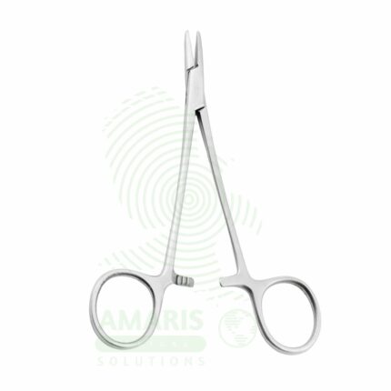
Needle Holders
Needle Holders are surgical instruments used to grasp, hold, and guide suture needles during wound closure and tissue approximation. Featuring serrated jaws with a locking ratchet mechanism, they provide secure needle control while allowing the surgeon to focus on precise suture placement. Available in various sizes and configurations including Mayo-Hegar for general surgery, Castroviejo for microsurgery, and Olsen-Hegar with integrated scissors. Made from surgical-grade stainless steel, often with tungsten carbide inserts for enhanced durability, they are essential instruments in all surgical specialties. Proper selection, handling, and regular inspection are critical for safe and effective use.
Proctoscope
A Proctoscope is a rigid, straight-tube endoscope specifically designed for the examination and treatment of the anal canal and distal rectum. It enables direct visualization to diagnose conditions like hemorrhoids, fissures, and proctitis, and serves as a conduit for therapeutic procedures such as band ligation. Available in reusable (autoclavable metal) or disposable (single-use plastic) formats, it is a fundamental tool in colorectal practice. Its effective use requires proper patient preparation, gentle insertion technique, and stringent adherence to sterilization protocols to ensure patient safety and diagnostic accuracy.