Dermatoscope and Magnifiers
 Diagnostic Kits
 Vital Signs Monitors
 Stethoscopes and Accessories
 Otoscopes, Ophthalmoscopes, and Retinoscopes
 Reflex Hammers and Neurological Tools
 Scales and Measuring Devices
 Spirometers and Pulmonary Function Tests
 Electrosurgical Units and Accessories
 Cutting Instruments
 Grasping and Holding Instruments
 Hemostatic Instruments
 Specialized Surgical Sets
 Single-Use Procedure Trays and Packs
 Surgical Drapes, Gowns, and Covers
 Tissue Unifying Instruments
 Radiation Protection
 X-Ray Machines and Accessories
 Ultrasound Systems and Probes
 MRI and CT Scanners
 Radiology Consumables
 Bone Densitometers
 Fluoroscopy Equipment
 Imaging Tables and Positioning Aids
 Microscopes and Accessories
 Centrifuges and Separators
 Analyzers
 Incubators and Ovens
 Pipettes, Dispensers, and Lab Glassware
 Refrigerators, Freezers, and Storage Units
 Lab Consumables
 Sterilizers and Autoclaves for Lab Use
 Multi-Parameter Monitors
 Ventilators and Respiratory Support Devices
 Defibrillators and AEDs
 Infusion Pumps and IV Systems
 Patient Warmers and Cooling Devices
 Central Monitoring Stations
 Accessories
 Anesthesia Machines and Workstations
 Oxygen Concentrators and Delivery Systems
 Nebulizers and Inhalers
 CPAP/BiPAP Machines
 Airway Management
 Anesthesia Masks, Circuits, and Bags
 Humidifiers and Heaters
 Respiratory Therapy Accessories
 First Aid Kits and Cabinets
 Emergency Resuscitation Equipment
 Trauma Supplies
 Emergency Carts and Crash Carts
 Burn Care Products
 Bleeding Control
 Automated External Defibrillators (AEDs)
 Transport and Evacuation
 Wheelchairs and Accessories
 Walkers, Crutches, and Canes
 Prosthetics and Orthotics
 Physical Therapy Equipment
 Transfer Devices
 Bathroom Safety
 Orthopedic Traction and Tables
 Hot/Cold Therapy Packs and Units
 Beds and Mattresses
 Chairs and Stools
 Tables
 Cabinets and Storage
 Privacy Screens & Curtains
 Stands and Racks
 Linens and Textiles
 Lighting
 Autoclaves and Sterilizers
 Ultrasonic Cleaners
 Disinfectant Solutions and Wipes
 Sterilization Pouches, Wraps, and Indicators
 Instrument Trays and Containers
 UV and Ozone Disinfection Devices
 Washer Disinfectors
 Wound Care
 Gloves
 Masks and Respirators
 Catheters and Tubing
 Swabs, Applicators, and Sponges
 Incontinence Products
 Personal Protective Equipment (PPE)
 Dental Chairs and Units
 Handpieces and Burs
 Instruments
 Consumables
 Sterilization for Dental Use
 Orthodontic Supplies
 Endodontic Tools
 Slit Lamps and Tonometers
 Lensometers and Phoropters
 Ophthalmic Surgical Instruments
 Eyewear Frames and Lenses
 Contact Lens Supplies
 Vision Testing Charts and Devices
 Eye Care Consumables
 Laser Systems for Eye Care
 ENT Exam Chairs and Tables
 Endoscopes
 Audiometers and Hearing Tests
 ENT Instruments
 Nasal and Throat Packs
 Hearing Aids and Accessories
 Otology Supplies
 Fetal Dopplers and Monitors
 Delivery Beds and Tables
 Gynecological Instruments
 Neonatal Incubators and Warmers
 Breast Pumps and Accessories
 Contraceptive Devices
 Maternity Supports and Pads
 Neonatal Consumables
 Cystoscopes and Urethroscopes
 Dialysis Machines and Supplies
 Urological Catheters and Bags
 Lithotripters
 Prostate Treatment Devices
 Urinary Incontinence Products
 Kidney Stone Management Tools
 Consumables & Disposables
 EEG and EMG Machines
 Neurosurgical Instruments
 Nerve Stimulators
 Headrests and Positioning Aids
 Lumbar Puncture Kits
 Seizure Monitoring Devices
 Consumables
 Rehabilitation for Neurological Conditions
 ECG Machines and Accessories
 Holter Monitors
 Stress Test Systems
 Pacemakers and Defibrillator Accessories
 Vascular Access Devices
 Cardiac Catheters and Guidewires
 Blood Flow Meters
 Consumables
 Orthopedic Instruments
 Casts, Splints, and Padding
 Joint Replacement Supplies
 Prosthetic Limbs and Components
 Bone Grafts and Substitutes
 Traction Devices
 Orthopedic Braces and Supports
 Rehabilitation Aids for Orthopedics
 Home Oxygen Therapy
 Hospital Beds for Home Use
 Mobility Aids
 Bathroom and Daily Living Aids
 Wound Care for Home
 Monitoring Devices
 Enteral Feeding Pumps and Tubes
 Hand Sanitizers and Dispensers
 Face Shields and Goggles
 Isolation Gowns and Suits
 Biohazard Waste Containers
 Air Purifiers and HEPA Filters
 Surface Disinfectants
 Sharps Containers
 Protective Barriers
 Cardiovascular & Endurance Training
 Strength Training & Weightlifting
 Functional Training & Core Conditioning
 Physical Therapy & Rehabilitation
 Sports & Outdoor Recreation
 Gym Flooring & Facility Equipment
 Fitness Monitoring & Accessories
 Kids & Novelties

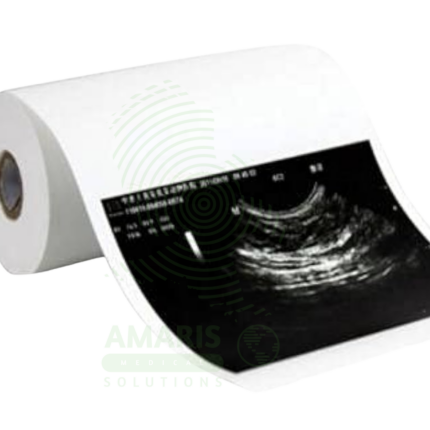
Ultrasound Machine
WhatsApp Order
The Ultrasound Machine is a diagnostic imaging system that uses high-frequency sound waves to produce real-time, dynamic images of internal body structures, including soft tissues, organs, blood vessels, and developing fetuses. It provides non-invasive, radiation-free visualization using multiple transducer probes for abdominal, cardiac, obstetric, gynecological, vascular, and musculoskeletal examinations. With core features like B-mode grayscale imaging, M-mode for motion assessment, and comprehensive Doppler capabilities (Color, Power, Spectral), it offers essential diagnostic functionality for evaluating anatomy, physiology, and blood flow. Its portability, digital image storage, and user-friendly interface make it a practical tool for hospitals, clinics, emergency departments, and point-of-care settings across virtually all medical specialties, requiring proper operator training and adherence to probe disinfection protocols for safe and effective use.
Description
Ultrasound Machine
PRIMARY CLINICAL & DIAGNOSTIC USES
1. Real-Time Anatomical and Physiological Imaging
-
Primary Use: Generates dynamic, real-time images of soft tissue structures and organs using high-frequency sound waves, providing a versatile, non-invasive, and radiation-free tool for visualizing muscles, tendons, internal organs, blood vessels, and developing fetuses.
-
How it helps: For the radiologist, sonographer, and clinician, the ultrasound machine transforms sound waves into living images of the human body—revealing organs in motion, blood flowing through vessels, and developing life in the womb, all without exposing the patient to radiation. For the patient, an ultrasound examination means their internal anatomy can be visualized safely, painlessly, and in real time, providing immediate answers and often the first glimpse of an unborn child.
2. Obstetrics and Gynecology
-
Primary Use: Essential for monitoring fetal development, evaluating the uterus and ovaries for conditions like fibroids and cysts, and guiding gynecological procedures.
-
How it helps: For the obstetrician and maternal-fetal medicine specialist, ultrasound provides a window into the womb—dating pregnancies, assessing fetal anatomy for anomalies, monitoring growth, and evaluating placental position, all critical for ensuring healthy outcomes. For the expectant parent, ultrasound offers the first images of their baby, providing reassurance of normal development and creating a profound early connection. For the gynecology patient, ultrasound reveals the cause of pelvic pain or abnormal bleeding, guiding diagnosis and treatment.
3. Abdominal and Pelvic Imaging
-
Primary Use: Used to assess organs such as the liver, gallbladder, kidneys, pancreas, spleen, bladder, and prostate for conditions like gallstones, cysts, tumors, ascites, and hydronephrosis.
-
How it helps: For the gastroenterologist, urologist, and primary care physician, abdominal ultrasound provides a rapid, non-invasive assessment of solid organs—revealing gallstones causing pain, hydronephrosis from obstruction, or tumors requiring further investigation. For the patient with abdominal pain, an ultrasound can often provide an immediate diagnosis, guiding treatment without the need for more invasive or radiation-intensive studies.
4. Vascular and Cardiac Evaluation
-
Primary Use: With Doppler ultrasound capabilities, it evaluates blood flow velocity and direction, used for diagnosing deep vein thrombosis, arterial stenosis, varicose veins, and for basic cardiac assessment.
-
How it helps: For the vascular surgeon and cardiologist, Doppler ultrasound makes blood flow visible—revealing clots in deep veins, blockages in arteries, and the direction and velocity of flow through vessels. For the patient with leg swelling, the test can rule out life-threatening DVT; for the patient with peripheral artery disease, it documents the location and severity of blockages.
5. Musculoskeletal Imaging
-
Primary Use: High-resolution linear probes allow detailed imaging of muscles, tendons, ligaments, and joints for diagnosing tears, sprains, inflammation, and guiding interventions like injections or aspirations.
-
How it helps: For the orthopedic surgeon, sports medicine physician, and rheumatologist, musculoskeletal ultrasound provides dynamic assessment of soft tissues—showing tendon tears with movement, revealing inflammation in real time, and guiding precise placement of therapeutic injections. For the athlete with a suspected tendon tear or the patient with chronic joint pain, ultrasound provides answers and guides treatment, often without the need for MRI.
SECONDARY & SUPPORTIVE USES
1. Point-of-Care Ultrasound: Its portability and ease of use make it ideal for rapid bedside assessment in emergency medicine, critical care, and anesthesiology. For the trauma patient, the FAST exam can reveal internal bleeding in seconds; for the critically ill, bedside ultrasound guides fluid resuscitation and vascular access.
2. Small Parts Imaging: Evaluates superficial structures like the thyroid, breast, testicles, and superficial soft tissue lumps. For the patient with a thyroid nodule, breast lump, or scrotal mass, ultrasound provides detailed characterization without radiation exposure.
3. Procedural Guidance: Provides real-time visualization for accurate needle placement during biopsies, fluid drainages, joint injections, and nerve blocks. For the patient undergoing a biopsy or injection, ultrasound guidance means the procedure is more accurate, safer, and less painful.
4. Pediatric Imaging: A preferred first-line imaging modality for children due to the lack of radiation, used for abdominal, hip, and cranial exams. For the child with suspected developmental dysplasia of the hip or abdominal pain, ultrasound provides answers while protecting developing tissues from radiation.
5. Urological Imaging: Assesses the kidneys, bladder, and scrotum, providing information about obstruction, residual urine volume, and testicular pathology. For the patient with urinary symptoms or scrotal pain, ultrasound provides rapid, non-invasive diagnosis.
KEY PRODUCT FEATURES
1. BASIC IDENTIFICATION ATTRIBUTES
-
Type: A diagnostic medical ultrasound system.
-
Common System Configuration:
-
Console: Cart-based system with an integrated computer, user interface (keyboard, trackball), and high-resolution LCD monitor.
-
Probes (Transducers): Supports multiple active array probes. A typical package includes:
-
Convex Array Probe (e.g., 3.5MHz): For general abdominal and obstetric imaging.
-
Linear Array Probe (e.g., 7.5MHz): For vascular, small parts, and musculoskeletal imaging.
-
Phased Array Probe (e.g., 2.5MHz): For cardiac and thoracic imaging.
-
Endocavitary Probe (optional): For gynecological and prostate imaging.
-
-
2. TECHNICAL & PERFORMANCE PROPERTIES
-
Imaging Principle: Uses a transducer probe to emit high-frequency sound waves into the body. The waves reflect off tissues of different densities, and the echoes are received and processed to create a real-time image (sonogram).
-
Key Imaging Modes:
-
B-Mode (Brightness Mode): Standard 2D grayscale imaging.
-
M-Mode (Motion Mode): Shows motion of structures over time along a single line, used in cardiac imaging.
-
Doppler Modes: Color Doppler maps blood flow direction and velocity; Pulsed-Wave (PW) Doppler measures flow velocity at a specific point; Continuous-Wave (CW) Doppler measures high velocities along a line.
-
Harmonic Imaging: Improves image clarity by using tissue-generated harmonic frequencies, reducing artefacts.
-
-
Connectivity: DICOM 3.0 standard for sending images to a PACS (Picture Archiving and Communication System) and hospital network. USB ports for data export.
3. PHYSICAL & OPERATIONAL PROPERTIES
-
Portability: A cart-based system with lockable wheels, designed for mobility within a department (e.g., from radiology to patient bedside).
-
User Interface: Features an intuitive control panel, customizable user presets, and ergonomic design for operator comfort during prolonged scanning.
-
Monitor: A high-resolution, adjustable color display for optimal image viewing.
4. SAFETY & COMPLIANCE ATTRIBUTES
-
Regulatory Status: Class II medical device.
-
Safety Standard: Complies with international standards (IEC 60601) for electrical and thermal safety. Ultrasound is considered very safe with no known ionizing radiation risk when used diagnostically.
-
ALARA Principle (As Low As Reasonably Achievable): Applies to acoustic output. Operators should use the lowest power output and shortest scan time necessary to obtain diagnostic information, especially during obstetric scanning.
5. STORAGE & HANDLING ATTRIBUTES
-
Storage: Store in a clean, dry, temperature-controlled environment. The cart should be parked securely with brakes engaged.
-
Cleaning & Disinfection:
-
Probes: Wipe down with a hospital-grade disinfectant wipe after each patient. Critical: Follow manufacturer guidelines for disinfecting endocavitary probes (high-level disinfection or sterilization required).
-
Console and Cart: Wipe surfaces regularly with a mild disinfectant. Avoid liquid ingress into controls or ports.
-
-
Probe Care: Handle probes carefully. Store in designated holders. Never immerse non-waterproof connector ends. Check routinely for cable wear or transducer face damage.
6. LABORATORY & CLINICAL APPLICATIONS
-
Primary Application: A versatile, foundational imaging tool used across hospital radiology departments, OB/GYN clinics, vascular labs, emergency rooms, outpatient imaging centers, and sports medicine facilities.
-
Operator-Dependent: Image quality and diagnostic accuracy are highly dependent on the skill and training of the sonographer or physician operator.
SAFETY HANDLING PRECAUTIONS
1. SAFETY PRECAUTIONS
-
Infection Control: Strict adherence to probe cleaning protocols between patients is mandatory to prevent cross-contamination.
-
Thermal and Mechanical Index Monitoring: The system displays Thermal Index (TI) and Mechanical Index (MI) values. Operators should be aware of these, particularly in obstetric scanning, to avoid potential bio-effects from excessive energy output.
-
Electrical Safety: Ensure the power cord and connections are intact. Do not use it in the presence of flammable anesthetics.
-
Ergonomics: Sonographers should practice proper posture and scanning technique to prevent repetitive strain injuries.
2. FIRST AID MEASURES
-
Electrical Shock: Immediately disconnect power. Do not touch the patient or equipment. Call for emergency medical help. Begin CPR if trained and safe to do so.
-
Probe Damage/Liquid Ingress: Turn off the system. Unplug the damaged probe. Tag it for service. Do not attempt to use it.
3. FIRE FIGHTING MEASURES
-
Flammability: The plastic housing, monitor, and internal electronics are combustible.
-
Extinguishing Media: For electrical fires, use a CO₂ or dry chemical extinguisher. Evacuate and alert the fire department.
Related products
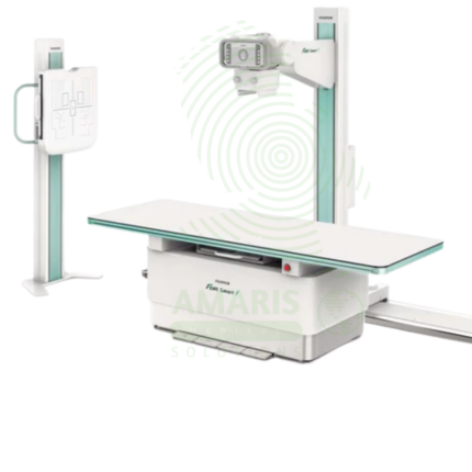
Digital Fixed X-ray
A Digital Fixed X-ray is a permanent installation digital radiography system designed for high-volume general imaging in radiology departments and outpatient imaging centers. Featuring digital flat panel detectors, ceiling-mounted tube assemblies, and tilting tables, it provides high-resolution images for skeletal, chest, abdominal, and extremity examinations. Integrated with PACS and RIS, it supports efficient digital workflow from image acquisition to interpretation, enabling rapid diagnosis and treatment planning.
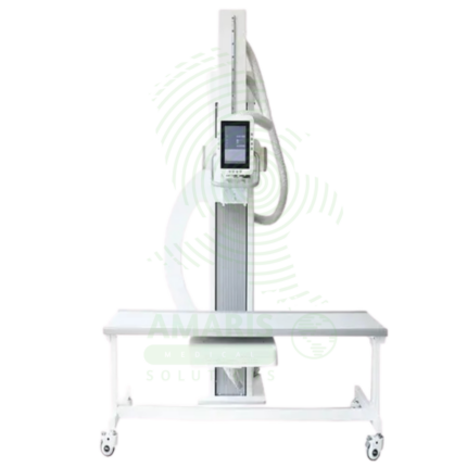
Digital U-arm X-ray
A Digital U-arm X-ray is a versatile digital radiography system designed for emergency departments, urgent care centers, and outpatient clinics. The U-arm configuration provides flexible positioning for chest, abdominal, skeletal, and extremity imaging with easy patient access for stretcher and wheelchair patients. Digital detectors produce immediate high-resolution images for rapid diagnosis, while the compact footprint allows installation in space-constrained settings. Essential for rapid, high-quality imaging in emergency and ambulatory care environments.

Lead Glass
Lead Glass is a transparent radiation shielding material used in X-ray rooms, CT suites, fluoroscopy suites, and radiation therapy control areas. Impregnated with lead oxide, it provides radiation attenuation equivalent to lead sheet while allowing direct visual observation of patients during procedures. Used for observation windows in control booths and procedure rooms, lead glass maintains the integrity of the radiation shielding envelope while enabling staff to monitor patient positioning, movement, and comfort. Proper installation with lead-lined frames and seals is essential for continuous radiation protection.
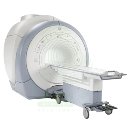
Magnetic Resonance Imaging
Magnetic Resonance Imaging (MRI) is a non-invasive diagnostic imaging modality that uses powerful magnetic fields and radiofrequency waves to produce detailed images of soft tissues, organs, and internal structures without ionizing radiation. It is the gold standard for imaging the brain, spinal cord, joints, muscles, and ligaments, and is essential for neurological, musculoskeletal, oncologic, and cardiovascular diagnosis. MRI provides exceptional soft tissue contrast, enabling precise anatomical characterization, tumor staging, and treatment planning. Strict safety protocols for ferromagnetic screening and contrast administration are essential for patient safety.
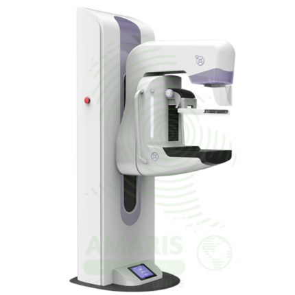
Mammography Machine
A Mammography Machine is a specialized, low-dose X-ray system designed exclusively for imaging the breast. It is the gold-standard tool for breast cancer screening and diagnostic evaluation, utilizing firm breast compression and high-resolution digital detectors to produce detailed images of breast tissue. Modern systems often incorporate Digital Breast Tomosynthesis (DBT or "3D mammography") to reduce tissue overlap and improve cancer detection. Its operation is highly regulated, requiring certified technologists, qualified interpreting physicians, and a rigorous quality assurance program to ensure patient safety, optimal image quality, and accurate early detection of breast cancer, which is vital for reducing mortality.
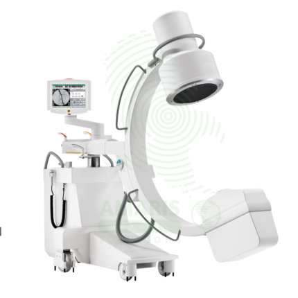
Mobile C-arm Surgical System
A Mobile C-arm Surgical System is a portable fluoroscopic imaging device used for real-time intraoperative guidance during orthopedic, spinal, vascular, and pain management procedures. The C-shaped arm allows flexible positioning around the patient, providing AP, lateral, and oblique views to verify instrument placement, fracture reduction, and device deployment. Essential for minimally invasive surgery, it enables surgeons to achieve precision and accuracy while reducing operative time and improving patient outcomes.
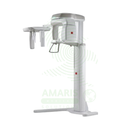
Panoramic X-ray Dental Machine
A Panoramic X-ray Dental Machine is a rotating extraoral radiographic system that produces a single, broad 2D image of the entire jaws, teeth, TMJs, and sinuses. By focusing on a curved "focal trough," it provides an efficient screening tool for wisdom teeth evaluation, orthodontic planning, and detecting large jaw pathologies. While offering a valuable overview with a relatively low radiation dose, its diagnostic utility is entirely dependent on precise patient positioning to avoid blurring and distortion. It is a cornerstone of modern dental diagnostic imaging, serving as a crucial first step in comprehensive oral assessment and treatment planning.