Dermatoscope and Magnifiers
 Diagnostic Kits
 Vital Signs Monitors
 Stethoscopes and Accessories
 Otoscopes, Ophthalmoscopes, and Retinoscopes
 Reflex Hammers and Neurological Tools
 Scales and Measuring Devices
 Spirometers and Pulmonary Function Tests
 Electrosurgical Units and Accessories
 Cutting Instruments
 Grasping and Holding Instruments
 Hemostatic Instruments
 Specialized Surgical Sets
 Single-Use Procedure Trays and Packs
 Surgical Drapes, Gowns, and Covers
 Tissue Unifying Instruments
 Radiation Protection
 X-Ray Machines and Accessories
 Ultrasound Systems and Probes
 MRI and CT Scanners
 Radiology Consumables
 Bone Densitometers
 Fluoroscopy Equipment
 Imaging Tables and Positioning Aids
 Microscopes and Accessories
 Centrifuges and Separators
 Analyzers
 Incubators and Ovens
 Pipettes, Dispensers, and Lab Glassware
 Refrigerators, Freezers, and Storage Units
 Lab Consumables
 Sterilizers and Autoclaves for Lab Use
 Multi-Parameter Monitors
 Ventilators and Respiratory Support Devices
 Defibrillators and AEDs
 Infusion Pumps and IV Systems
 Patient Warmers and Cooling Devices
 Central Monitoring Stations
 Accessories
 Anesthesia Machines and Workstations
 Oxygen Concentrators and Delivery Systems
 Nebulizers and Inhalers
 CPAP/BiPAP Machines
 Airway Management
 Anesthesia Masks, Circuits, and Bags
 Humidifiers and Heaters
 Respiratory Therapy Accessories
 First Aid Kits and Cabinets
 Emergency Resuscitation Equipment
 Trauma Supplies
 Emergency Carts and Crash Carts
 Burn Care Products
 Bleeding Control
 Automated External Defibrillators (AEDs)
 Transport and Evacuation
 Wheelchairs and Accessories
 Walkers, Crutches, and Canes
 Prosthetics and Orthotics
 Physical Therapy Equipment
 Transfer Devices
 Bathroom Safety
 Orthopedic Traction and Tables
 Hot/Cold Therapy Packs and Units
 Beds and Mattresses
 Chairs and Stools
 Tables
 Cabinets and Storage
 Privacy Screens & Curtains
 Stands and Racks
 Linens and Textiles
 Lighting
 Autoclaves and Sterilizers
 Ultrasonic Cleaners
 Disinfectant Solutions and Wipes
 Sterilization Pouches, Wraps, and Indicators
 Instrument Trays and Containers
 UV and Ozone Disinfection Devices
 Washer Disinfectors
 Wound Care
 Gloves
 Masks and Respirators
 Catheters and Tubing
 Swabs, Applicators, and Sponges
 Incontinence Products
 Personal Protective Equipment (PPE)
 Dental Chairs and Units
 Handpieces and Burs
 Instruments
 Consumables
 Sterilization for Dental Use
 Orthodontic Supplies
 Endodontic Tools
 Slit Lamps and Tonometers
 Lensometers and Phoropters
 Ophthalmic Surgical Instruments
 Eyewear Frames and Lenses
 Contact Lens Supplies
 Vision Testing Charts and Devices
 Eye Care Consumables
 Laser Systems for Eye Care
 ENT Exam Chairs and Tables
 Endoscopes
 Audiometers and Hearing Tests
 ENT Instruments
 Nasal and Throat Packs
 Hearing Aids and Accessories
 Otology Supplies
 Fetal Dopplers and Monitors
 Delivery Beds and Tables
 Gynecological Instruments
 Neonatal Incubators and Warmers
 Breast Pumps and Accessories
 Contraceptive Devices
 Maternity Supports and Pads
 Neonatal Consumables
 Cystoscopes and Urethroscopes
 Dialysis Machines and Supplies
 Urological Catheters and Bags
 Lithotripters
 Prostate Treatment Devices
 Urinary Incontinence Products
 Kidney Stone Management Tools
 Consumables & Disposables
 EEG and EMG Machines
 Neurosurgical Instruments
 Nerve Stimulators
 Headrests and Positioning Aids
 Lumbar Puncture Kits
 Seizure Monitoring Devices
 Consumables
 Rehabilitation for Neurological Conditions
 ECG Machines and Accessories
 Holter Monitors
 Stress Test Systems
 Pacemakers and Defibrillator Accessories
 Vascular Access Devices
 Cardiac Catheters and Guidewires
 Blood Flow Meters
 Consumables
 Orthopedic Instruments
 Casts, Splints, and Padding
 Joint Replacement Supplies
 Prosthetic Limbs and Components
 Bone Grafts and Substitutes
 Traction Devices
 Orthopedic Braces and Supports
 Rehabilitation Aids for Orthopedics
 Home Oxygen Therapy
 Hospital Beds for Home Use
 Mobility Aids
 Bathroom and Daily Living Aids
 Wound Care for Home
 Monitoring Devices
 Enteral Feeding Pumps and Tubes
 Hand Sanitizers and Dispensers
 Face Shields and Goggles
 Isolation Gowns and Suits
 Biohazard Waste Containers
 Air Purifiers and HEPA Filters
 Surface Disinfectants
 Sharps Containers
 Protective Barriers
 Cardiovascular & Endurance Training
 Strength Training & Weightlifting
 Functional Training & Core Conditioning
 Physical Therapy & Rehabilitation
 Sports & Outdoor Recreation
 Gym Flooring & Facility Equipment
 Fitness Monitoring & Accessories
 Kids & Novelties
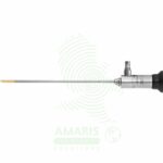
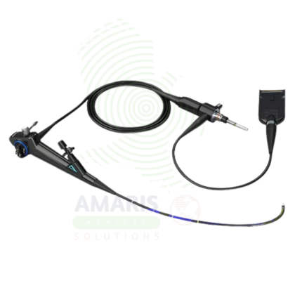
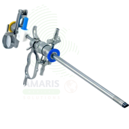
Urology Endoscopes
WhatsApp Order
Urology Endoscopes are specialized instruments for visualizing and operating within the urinary tract. They include rigid and flexible cystoscopes for the bladder, semi-rigid and flexible ureteroscopes for the ureters and kidneys, and nephroscopes for percutaneous kidney surgery. These devices enable critical procedures like TURP, TURBT, and laser lithotripsy for stones. Flexible scopes, essential for complex cases, require meticulous high-level disinfection and careful handling to prevent damage. Safe use hinges on managing irrigation fluid absorption, preventing perforation, and protecting the delicate optics during energy-based treatments, making them fundamental yet sophisticated tools in urological practice.
Description
Urology Endoscopes
PRIMARY CLINICAL & DIAGNOSTIC USES
1. Cystoscopy
-
Primary Use: Provides direct visualization of the urethra, bladder neck, and bladder mucosa for diagnostic evaluation of hematuria, recurrent urinary tract infections, lower urinary tract symptoms, urinary incontinence, and suspicion of bladder tumors or stones.
-
How it helps: For the urologist, the cystoscope transforms the bladder from a hidden, inaccessible organ into a clearly visualized cavity—revealing tumors that bleed, stones that obstruct, inflamed mucosa that causes symptoms, and anatomical abnormalities that explain incontinence. For the patient with blood in their urine, recurrent infections, or bothersome voiding symptoms, cystoscopy provides answers that imaging alone cannot, guiding diagnosis and treatment with certainty.
2. Ureteroscopy (URS)
-
Primary Use: Allows access to and visualization of the ureter and renal pelvis for the diagnosis and treatment of conditions such as ureteral stones, ureteral strictures, and upper tract urothelial carcinoma.
-
How it helps: For the urologist managing stone disease, the ureteroscope navigates the narrow, delicate ureter to reach stones where they lodge—fragmenting them with laser energy, retrieving fragments, and relieving obstruction. For the patient in excruciating pain from a lodged ureteral stone, ureteroscopy offers definitive treatment through natural urinary passages, avoiding open surgery and providing rapid relief.
3. Nephroscopy
-
Primary Use: Performed percutaneously to access and visualize the renal collecting system directly for the treatment of large or complex kidney stones.
-
How it helps: For the endourologist managing large or complex kidney stones, the nephroscope provides direct access to the renal pelvis through a small flank incision—allowing fragmentation and removal of stones too large for ureteroscopy or shock wave lithotripsy. For the patient with a staghorn calculus or massive stone burden, percutaneous nephroscopy offers the best chance for complete stone clearance and preservation of renal function.
4. Transurethral Resection of Bladder Tumor (TURBT)
-
Primary Use: The standard procedure for the diagnosis, staging, and therapeutic resection of bladder tumors using a resectoscope.
-
How it helps: For the urologic oncologist, the resectoscope allows simultaneous visualization and resection of bladder tumors—cutting away abnormal tissue while cauterizing the base, providing tissue for pathological staging, and treating the disease in a single procedure. For the patient with bladder cancer, TURBT offers both diagnosis and initial treatment, often through the same minimally invasive approach, with rapid recovery and preservation of bladder function.
5. Transurethral Resection of the Prostate (TURP)
-
Primary Use: The classic surgical treatment for benign prostatic hyperplasia, using a resectoscope to remove obstructive prostate tissue.
-
How it helps: For the urologist treating men with symptomatic BPH, the resectoscope provides a channel through the obstructing prostate—removing the tissue that blocks urinary flow, relieves straining, and causes incomplete emptying. For the aging man whose quality of life is diminished by frequent urination, weak stream, and nighttime awakening, TURP offers durable relief and return to normal voiding function.
SECONDARY & SUPPORTIVE USES
1. Ureteral Stent Placement and Removal: Urology endoscopes guide the insertion of double-J stents for ureteral obstruction and enable subsequent endoscopic removal. For the patient with a obstructing stone, tumor, or stricture, stent placement relieves pain, preserves renal function, and buys time for definitive treatment.
2. Treatment of Ureteral Strictures: Endoscopic techniques like balloon dilation or laser incision treat ureteral narrowing that would otherwise require open reconstruction. For the patient with a ureteral stricture causing pain and hydronephrosis, endoscopic management offers a minimally invasive alternative to major surgery.
3. Foreign Body Removal: Endoscopes enable extraction of objects from the bladder or urethra—forgotten stents, migrated devices, or self-inserted foreign bodies. For the patient with a retained foreign body causing pain, infection, or obstruction, endoscopic removal resolves the problem without open surgery.
4. Evaluation of Congenital Anomalies: In pediatric urology, endoscopes allow evaluation of conditions like posterior urethral valves in infants and children. For the youngest patients with congenital urologic abnormalities, endoscopic diagnosis and treatment can preserve renal function and improve quality of life from the earliest ages.
5. Prostate Procedures: Modified cystoscopes and continuous-flow resectoscopes enable laser vaporization and enucleation of the prostate (HoLEP, GreenLight PVP) for BPH. For the patient with an enlarged prostate, these modern endoscopic techniques offer the efficacy of TURP with potentially less bleeding and faster recovery.
6. Fulguration of Bleeding Points: Urology endoscopes allow precise cauterization of bleeding points within the bladder or prostatic fossa. For the patient with visible hematuria or post-procedure bleeding, endoscopic fulguration provides targeted control without the morbidity of open exploration.
KEY PRODUCT FEATURES
1. BASIC IDENTIFICATION ATTRIBUTES
-
Device Type: A family of rigid, semi-rigid, and flexible optical instruments designed for endoscopic access to the urinary tract.
-
Designation by Type and Access Route:
-
Cystoscope: For bladder and urethra. Can be rigid (metal) or flexible (fiberoptic/video).
-
Ureteroscope: For ureter and kidney. Can be semi-rigid (for lower ureter) or flexible (for entire ureter and intrarenal access).
-
Nephroscope: A large, rigid scope used through a percutaneous tract into the kidney (for PCNL).
-
Resectoscope: A specialized rigid cystoscope with an integrated cutting/coagulating loop for TURP and TURBT.
-
-
Core Components:
-
Sheath/Obturator (Rigid): The outer metal tube that maintains the urethral channel, with ports for irrigation inflow/outflow and instrument passage.
-
Telescope/Bridge: The optical component (rod-lens telescope) that inserts into the sheath. Bridges hold working instruments.
-
Working Channel: Present in flexible and some rigid scopes for passing lasers, fibers, baskets, graspers, and biopsy forceps.
-
Deflection Mechanism (Flexible): A lever on the handle that controls up/down (and sometimes left/right) deflection of the distal tip to navigate anatomy.
-
2. TECHNICAL & PERFORMANCE PROPERTIES
-
Diameter (French/Charr): Critical for access and patient comfort. Cystoscopes range from 16Fr-22Fr. Flexible ureteroscopes are typically 6Fr-9Fr at the tip.
-
Length: Varies by application (cystoscopes ~30-40cm, ureteroscopes 35-70cm).
-
Optics: Utilize Hopkins rod-lens systems (rigid) or fiberoptic/digital chips (flexible). Image quality is paramount for identifying small tumors or stone fragments.
-
Deflection (Flexible Ureteroscopes): Active tip deflection (often >270° up and down) is essential for accessing lower pole renal calyces.
-
Irrigation Flow: Sheaths and scopes are designed for continuous irrigation to maintain a clear visual field and distend the bladder or ureter.
3. PHYSICAL & OPERATIONAL PROPERTIES
-
Rigidity: Rigid scopes provide superior image quality and stability for resection. Flexible scopes allow navigation without painful patient positioning.
-
Durability: Flexible ureteroscopes are delicate and have a finite lifespan due to stress on deflection wires and potential for channel/optic damage.
-
Sterilization/Disinfection: Rigid scopes are autoclavable. Flexible scopes require meticulous high-level disinfection (HLD) or sterilization (e.g., ethylene oxide) as they contact sterile body cavities.
4. SAFETY & COMPLIANCE ATTRIBUTES
-
Regulatory Status: Classified as Class I or II medical devices.
-
Infection Control: Strict adherence to validated reprocessing protocols is mandatory to prevent biofilm formation and cross-contamination (e.g., of TB, multi-drug resistant organisms).
-
Biocompatibility: All materials must be non-toxic and suitable for prolonged contact with urinary mucosa.
5. STORAGE & HANDLING ATTRIBUTES
-
Storage: Rigid scopes in protective trays. Flexible scopes must be stored hanging vertically, uncoiled, to prevent damage to the deflection mechanism and channel.
-
Cleaning & Reprocessing (CRITICAL - Especially for Flexible Scopes):
-
Immediate Bedside Flush: Flush the working channel with water or enzymatic cleaner immediately after use.
-
Leak Testing: Perform before every immersion to check for integrity breaches in the waterproof sheath.
-
Manual Cleaning: Brush all channels (main, auxiliary) meticulously. Clean the exterior.
-
High-Level Disinfection/Sterilization: Soak in an approved chemical agent (e.g., glutaraldehyde, peracetic acid) for the exact contact time, followed by thorough rinsing with sterile or bacteria-free water to remove toxic residues.
-
Drying: Force dry all channels with medical-grade air and store hanging in a ventilated cabinet.
-
-
Handling: Never force a scope. For flexible ureteroscopy, always use a safety guidewire and access sheath to reduce scope stress and facilitate multiple entries.
6. LABORATORY & CLINICAL APPLICATIONS
-
Primary Application: The definitive tool for diagnosis and minimally invasive treatment of conditions affecting the entire urinary system, from the urethra to the kidney.
-
Clinical Role: Enables a vast range of procedures from simple office-based diagnostics to complex stone and cancer surgeries, forming the core of modern endoscopic urology.
SAFETY HANDLING PRECAUTIONS
1. SAFETY PRECAUTIONS
-
Infection/Asepsis: Maintain strict aseptic technique. Use sterile irrigation fluids (e.g., glycine, saline) to prevent bacteremia/sepsis.
-
Intravenous Fluid Absorption (TUR Syndrome): During prolonged resection procedures (TURP, TURBT) with hypotonic irrigation, monitor for symptoms of hyponatremia and fluid overload from systemic absorption.
-
Perforation Risk: The ureter and bladder are thin-walled. Use gentle technique, especially with lasers and rigid instruments, to avoid perforation.
-
Thermal Injury: Energy devices (lasers, electrocautery) can cause thermal injury to adjacent structures (bowel, blood vessels) if not used under direct vision with appropriate settings.
-
Scope Damage: The most common cause of flexible ureteroscope damage is laser fiber firing when the tip is over-deflected. Ensure the scope is straight and the fiber tip is visible before activating the laser.
2. FIRST AID MEASURES
-
Ureteral or Bladder Perforation: If recognized, stop the procedure. For small perforations, place a ureteral stent and Foley catheter. For large injuries, may require open or laparoscopic repair.
-
TUR Syndrome: If the patient develops confusion, nausea, or cardiovascular changes, stop the procedure, check electrolytes (sodium), and administer hypertonic saline and diuretics as needed.
-
Severe Bleeding: May require cessation of procedure, bladder irrigation/instillation of alum, catheter traction, or emergent angioembolization for arterial bleeding post-PCNL.
-
Broken Instrument/Basket: If a basket or laser fiber breaks, use grasping forceps or a second scope to retrieve all fragments.
3. FIRE FIGHTING MEASURES
-
Flammability: Scope materials and light cables are combustible.
-
Extinguishing Media: Use CO2 extinguishers for electrical fires. Be aware of oxygen-enriched environments during procedures.
Related products
Cystoscope
A Cystoscope is a specialized endoscope used for visualization of the urethra and bladder for diagnostic and therapeutic urologic procedures. Rigid cystoscopes provide superior optics for operative procedures, while flexible cystoscopes offer enhanced patient comfort for diagnostic examinations. Essential for evaluation of hematuria, diagnosis and surveillance of bladder cancer, stent management, and treatment of bladder stones and urethral strictures, cystoscopy is the cornerstone of urologic practice.
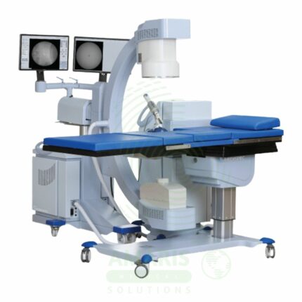
ESWL Lithotripter Systems
A ESWL Lithotripter Systems is a non-invasive surgical device that uses focused, high-energy acoustic shock waves generated outside the body to fragment kidney and upper ureteral stones. The patient lies on a table while integrated imaging (fluoroscopy/ultrasound) is used to target the stone precisely at the shock wave focal point. Treatment success depends on stone size, location, and composition. While avoiding surgical incisions, the procedure requires meticulous safety protocols to manage risks such as renal injury, bleeding, and radiation exposure. It is a capital-intensive, fixed equipment system that defines the modern, non-invasive approach to urolithiasis.
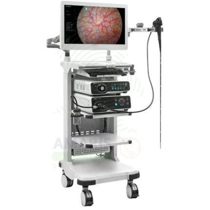
High-Definition Endoscope
A High-Definition Endoscope is an advanced endoscopic system providing superior image resolution, color accuracy, and contrast for diagnostic and therapeutic procedures. Incorporating HD or 4K imaging with advanced visualization technologies such as narrow band imaging, blue light imaging, and autofluorescence, it enables detection of subtle mucosal abnormalities and early neoplasia that may be missed with standard definition systems. Essential for early cancer detection, precise therapeutic intervention, and documentation, it represents the standard of care for advanced endoscopy.
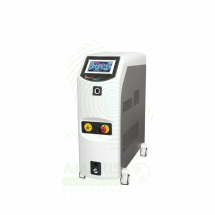
Holmium Laser System
A Holmium Laser System is a versatile, high-power Class IV surgical laser that emits at a 2120 nm wavelength, which is highly absorbed by water and biological tissue. It is the gold-standard tool in urology for laser lithotripsy (stone dusting/fragmentation) and advanced soft tissue procedures like prostate enucleation (HoLEP) and tumor ablation. Its energy is delivered via flexible silica fibers through endoscopes, enabling precise cutting, ablation, and coagulation with excellent hemostasis in a fluid environment. Strict laser safety protocols—including mandatory 2120 nm eyewear, fiber inspection, and controlled activation—are non-negotiable for its safe operation in the OR.
Lithotriptic Scope
A Lithotriptic Scope is a specialized endoscope used for endoscopic fragmentation of urinary tract stones in the kidney, ureter, and bladder. Available as semi-rigid ureteroscopes, flexible ureteroscopes, and nephroscopes, these instruments provide direct visualization for laser, pneumatic, or ultrasonic lithotripsy. Essential in endourology, they enable minimally invasive stone treatment with high stone-free rates, rapid recovery, and preservation of renal function.
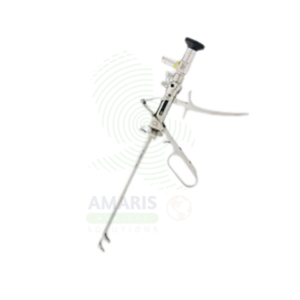
Resectoscope
A Resectoscope is a specialized endoscopic instrument used for transurethral resection of the prostate, bladder tumors, and endometrium. Combining a telescope, working element, and resecting loop, it enables precise cutting and coagulation of tissue under direct visualization. Essential in urology for benign prostatic hyperplasia and bladder cancer treatment, and in gynecology for endometrial resection, it provides minimally invasive treatment with rapid recovery and preservation of organ function.
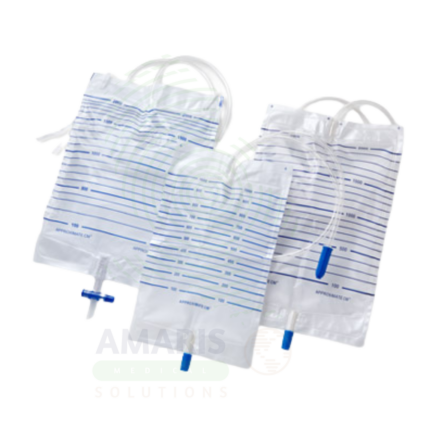
Urine Bags
Urine Bags are sterile, single-use, closed-system medical devices designed for gravity drainage, collection, containment, and measurement of urine from indwelling urinary catheters. Available in two primary configurations: large-capacity drainage bags (2000-4000 mL) for bedridden patients and overnight use, and smaller leg bags (350-750 mL) for ambulatory, community-dwelling patients. Core components include a transparent graduated collection bag, flexible drainage tubing, anti-reflux valve, needleless sampling port, and secure outlet valve. Manufactured from medical-grade PVC (DEHP-free alternatives available) or non-PVC materials, all components are latex-free and terminally sterilized. Critical safety requirements include maintaining the bag below bladder level at all times to prevent retrograde flow, preserving closed system integrity, and adhering to strict single-patient-use protocols. An indispensable component of CAUTI prevention bundles and urinary drainage management across acute care, long-term care, and home healthcare settings.