Dermatoscope and Magnifiers
 Diagnostic Kits
 Vital Signs Monitors
 Stethoscopes and Accessories
 Otoscopes, Ophthalmoscopes, and Retinoscopes
 Reflex Hammers and Neurological Tools
 Scales and Measuring Devices
 Spirometers and Pulmonary Function Tests
 Electrosurgical Units and Accessories
 Cutting Instruments
 Grasping and Holding Instruments
 Hemostatic Instruments
 Specialized Surgical Sets
 Single-Use Procedure Trays and Packs
 Surgical Drapes, Gowns, and Covers
 Tissue Unifying Instruments
 Radiation Protection
 X-Ray Machines and Accessories
 Ultrasound Systems and Probes
 MRI and CT Scanners
 Radiology Consumables
 Bone Densitometers
 Fluoroscopy Equipment
 Imaging Tables and Positioning Aids
 Microscopes and Accessories
 Centrifuges and Separators
 Analyzers
 Incubators and Ovens
 Pipettes, Dispensers, and Lab Glassware
 Refrigerators, Freezers, and Storage Units
 Lab Consumables
 Sterilizers and Autoclaves for Lab Use
 Multi-Parameter Monitors
 Ventilators and Respiratory Support Devices
 Defibrillators and AEDs
 Infusion Pumps and IV Systems
 Patient Warmers and Cooling Devices
 Central Monitoring Stations
 Accessories
 Anesthesia Machines and Workstations
 Oxygen Concentrators and Delivery Systems
 Nebulizers and Inhalers
 CPAP/BiPAP Machines
 Airway Management
 Anesthesia Masks, Circuits, and Bags
 Humidifiers and Heaters
 Respiratory Therapy Accessories
 First Aid Kits and Cabinets
 Emergency Resuscitation Equipment
 Trauma Supplies
 Emergency Carts and Crash Carts
 Burn Care Products
 Bleeding Control
 Automated External Defibrillators (AEDs)
 Transport and Evacuation
 Wheelchairs and Accessories
 Walkers, Crutches, and Canes
 Prosthetics and Orthotics
 Physical Therapy Equipment
 Transfer Devices
 Bathroom Safety
 Orthopedic Traction and Tables
 Hot/Cold Therapy Packs and Units
 Beds and Mattresses
 Chairs and Stools
 Tables
 Cabinets and Storage
 Privacy Screens & Curtains
 Stands and Racks
 Linens and Textiles
 Lighting
 Autoclaves and Sterilizers
 Ultrasonic Cleaners
 Disinfectant Solutions and Wipes
 Sterilization Pouches, Wraps, and Indicators
 Instrument Trays and Containers
 UV and Ozone Disinfection Devices
 Washer Disinfectors
 Wound Care
 Gloves
 Masks and Respirators
 Catheters and Tubing
 Swabs, Applicators, and Sponges
 Incontinence Products
 Personal Protective Equipment (PPE)
 Dental Chairs and Units
 Handpieces and Burs
 Instruments
 Consumables
 Sterilization for Dental Use
 Orthodontic Supplies
 Endodontic Tools
 Slit Lamps and Tonometers
 Lensometers and Phoropters
 Ophthalmic Surgical Instruments
 Eyewear Frames and Lenses
 Contact Lens Supplies
 Vision Testing Charts and Devices
 Eye Care Consumables
 Laser Systems for Eye Care
 ENT Exam Chairs and Tables
 Endoscopes
 Audiometers and Hearing Tests
 ENT Instruments
 Nasal and Throat Packs
 Hearing Aids and Accessories
 Otology Supplies
 Fetal Dopplers and Monitors
 Delivery Beds and Tables
 Gynecological Instruments
 Neonatal Incubators and Warmers
 Breast Pumps and Accessories
 Contraceptive Devices
 Maternity Supports and Pads
 Neonatal Consumables
 Cystoscopes and Urethroscopes
 Dialysis Machines and Supplies
 Urological Catheters and Bags
 Lithotripters
 Prostate Treatment Devices
 Urinary Incontinence Products
 Kidney Stone Management Tools
 Consumables & Disposables
 EEG and EMG Machines
 Neurosurgical Instruments
 Nerve Stimulators
 Headrests and Positioning Aids
 Lumbar Puncture Kits
 Seizure Monitoring Devices
 Consumables
 Rehabilitation for Neurological Conditions
 ECG Machines and Accessories
 Holter Monitors
 Stress Test Systems
 Pacemakers and Defibrillator Accessories
 Vascular Access Devices
 Cardiac Catheters and Guidewires
 Blood Flow Meters
 Consumables
 Orthopedic Instruments
 Casts, Splints, and Padding
 Joint Replacement Supplies
 Prosthetic Limbs and Components
 Bone Grafts and Substitutes
 Traction Devices
 Orthopedic Braces and Supports
 Rehabilitation Aids for Orthopedics
 Home Oxygen Therapy
 Hospital Beds for Home Use
 Mobility Aids
 Bathroom and Daily Living Aids
 Wound Care for Home
 Monitoring Devices
 Enteral Feeding Pumps and Tubes
 Hand Sanitizers and Dispensers
 Face Shields and Goggles
 Isolation Gowns and Suits
 Biohazard Waste Containers
 Air Purifiers and HEPA Filters
 Surface Disinfectants
 Sharps Containers
 Protective Barriers
 Cardiovascular & Endurance Training
 Strength Training & Weightlifting
 Functional Training & Core Conditioning
 Physical Therapy & Rehabilitation
 Sports & Outdoor Recreation
 Gym Flooring & Facility Equipment
 Fitness Monitoring & Accessories
 Kids & Novelties

Body surface conduit fixing device – Type A
WhatsApp Order
The Body Surface Conduit Fixing Device – Type A is a sterile, single‑use adhesive securement device designed to replace sutures for holding percutaneous catheters, drains, and tubes in place. It consists of a breathable non‑woven fabric base coated with a medical‑grade pressure‑sensitive adhesive and an integrated hook‑and‑loop (Velcro) fastener. The device is applied to intact skin adjacent to the catheter exit site, and the fastener is wrapped around the catheter to secure it without occluding the lumen.
By eliminating sutures, the Type A fixator reduces needle‑stick injuries, eliminates suture‑related pain and tension, and minimises micro‑motion of the catheter. This micro‑motion is a known risk factor for catheter‑related bloodstream infections and exit‑site infections, as it disrupts the healing tissue and can wick bacteria along the catheter surface. The broad adhesive base distributes the weight of the drain evenly, preventing pressure damage and accidental pull‑out. The low‑profile, soft fabric design conforms to body contours, making the device comfortable for patients during sleeping, walking, and bathing.
Clinically, the Type A device is indicated for securing chest tubes, abdominal drains, nasogastric tubes, epidural catheters, central venous catheters, peritoneal dialysis catheters, and surgical wound drains. It is suitable for use in surgical wards, intensive care units, interventional radiology, and home care settings. For the healthcare provider, the device simplifies drain management, reduces the need for frequent dressing changes, and lowers the risk of infection. For the patient, it offers greater comfort, reduced pain, and confidence that the drain will remain securely in place during recovery.
Description
Body Surface Conduit Fixing Device – Type A
PRIMARY CLINICAL & DIAGNOSTIC USES
1. Post‑Surgical Securing of Drains and Tubes
Primary Use: Provides a sterile, single‑use, adhesive‑backed fixation device designed to secure a percutaneous catheter, tube, or drain (e.g., chest tube, abdominal drain, nasogastric tube) to a patient’s skin surface without suturing. Type A consists of a body surface fixation base made of a non‑woven fabric coated with medical‑grade pressure‑sensitive adhesive, a hook‑and‑loop fastener (Velcro) section, and a release liner.
How it helps: For the surgeon and the surgical nurse, the Type A fixator replaces traditional sutures for securing drains. Sutures can cause additional trauma, increase the risk of needle‑stick injuries, and may lead to painful tension on the skin. The adhesive base provides a secure, broad surface attachment that distributes the weight of the drain more evenly, reducing the risk of accidental pull‑out (extravasation) and minimising local pressure damage. The hook‑and‑loop closure allows the device to be wrapped around any size of drain up to approximately 36 Fr in diameter. For the patient, this means a more comfortable postoperative recovery with less drain‑site pain and a lower risk of accidental dislodgement during routine activities such as turning in bed or walking.
2. Prevention of Catheter‑Related Infections
Primary Use: By holding the catheter firmly in place, the fixation device eliminates the need for sutures, which can act as wicks for bacteria, and prevents the piston‑like movement of the catheter in and out of the insertion site. This micro‑motion is known to disrupt the formation of the skin tract and can introduce microorganisms into the deeper tissues.
How it helps: For the infection control nurse and the surgeon, the primary benefit is a demonstrable reduction in the risk of catheter‑related bloodstream infections (CRBSI) and exit‑site infections. By minimising movement of the catheter, the device helps maintain the integrity of the healing tissue around the insertion site, creating a more effective barrier against invading pathogens. This also reduces the need for frequent, traumatic dressing changes that can disturb the wound bed. For the patient, a lower infection risk means shorter hospital stays, fewer antibiotic courses, and a safer overall recovery.
3. Improved Patient Comfort and Safety
Primary Use: Designed for patient comfort, the non‑woven base is made from a breathable, soft material that conforms to the body’s contours. The adhesive is medical‑grade and hypoallergenic to minimise skin reactions. The low‑profile design prevents the device from snagging on bedding or clothing.
How it helps: For the patient, shifting from a sutured drain to an adhesive fixation device dramatically reduces the sensation of tugging, pulling, or pinching at the drain site. This translates to less drain‑site pain, greater freedom of movement, and a lower risk of accidental dislodgement during routine activities such as turning over in bed, walking, or bathing. For the nursing staff, the secure fixation means fewer emergency call‑outs for dislodged drains and less time spent reapplying loose tape. The result is a less stressful recovery and an overall improvement in quality of life during the post‑operative period.
SECONDARY & SUPPORTIVE USES
1. Securing Epidural Catheters: As an alternative to sutures or tape, the Type A device can be used to secure an epidural catheter for post‑operative pain management. For the anaesthesiologist and the pain management nurse, the adhesive base holds the catheter in place on the patient’s back, while the hook‑and‑loop closure securely grips the catheter without occluding the lumen, helping to maintain correct placement. For the patient, a securely fixed epidural catheter means more reliable post‑operative pain relief and a lower risk of the catheter being accidentally pulled out during movement or repositioning in bed.
2. Fixation of Nasogastric (NG) and Nasoenteric (NE) Tubes: The device is used to affix NG or NE tubes to the nasal bridge or cheek, replacing the traditional method of taping the tube to the face. For the clinical nutrition nurse and the gastroenterologist, the adhesive is gentle on facial skin, and the secure hold prevents the tube from pulling out or migrating back into the stomach. For the patient, this means less facial skin irritation, reduced risk of tube dislodgement during coughing or vomiting, and improved comfort when the tube must remain in place for extended periods.
3. Securing Central Venous Catheters (CVCs): For temporary or short‑term central lines, the Type A fixator can be used to secure the catheter hub to the skin of the chest or neck. For the interventional radiologist and the critical care nurse, this is particularly useful for dialysis catheters or introducer sheaths where a strong, reliable fixation is necessary to maintain vascular access. For the patient, a properly secured CVC reduces the risk of catheter migration, accidental removal, and the associated bleeding or air embolism, while also minimising the discomfort caused by loose tape or sutures.
4. Securing Peritoneal Dialysis Catheters: The device can be used to fix the external segment of a peritoneal dialysis catheter to the abdominal wall. For the nephrologist and the dialysis nurse, this provides a clean, secure alternative to traditional dressings, reducing the risk of catheter torsion and exit‑site infection. For the patient receiving peritoneal dialysis at home, the Type A fixator offers an easy‑to‑use, reliable method for keeping the catheter in place during daily activities and sleep, improving confidence in self‑care.
5. Fixation of Wound Drains (Jackson‑Pratt, Blake, Hemovac): The device is well suited for securing the tubing of surgical wound drains to the skin near the incision site. For the general surgeon and the wound care nurse, this prevents the drain from pulling on the suture at the insertion point, reducing local tissue damage and patient pain. For the patient, a securely fixed drain is less likely to be accidentally dislodged when changing dressings or moving, which helps ensure that the drain remains functional for the prescribed duration.
6. Securing Chest Tubes Post‑Thoracotomy: For patients after lung surgery or with a pneumothorax, the Type A fixator can be used to secure the chest tube to the thoracic wall. For the thoracic surgeon and the respiratory nurse, the broad adhesive base provides stable fixation even in the presence of chest hair or moisture, while the hook‑and‑loop fastener accommodates the larger diameter of chest tubes. For the patient, this means less pulling at the chest tube insertion site during breathing and movement, reducing pain and the anxiety associated with having a chest tube in place.
KEY PRODUCT FEATURES
1. BASIC IDENTIFICATION ATTRIBUTES
-
Device Type: A sterile, single‑use, adhesive‑backed fixation device for securing percutaneous catheters, tubes, and drains to the body surface.
-
Designation: Body Surface Conduit Fixing Device – Type A; catheter fixation device; drain securement device; tube holder.
-
Key Components:
-
Non‑woven fabric base coated with medical‑grade pressure‑sensitive adhesive (hypoallergenic, latex‑free).
-
Hook‑and‑loop (Velcro) fastener integrated onto the base.
-
Release liner (protective film) covering the adhesive before use.
-
Optional components (depending on manufacturer): documentation label, anti‑rotation tab.
-
2. TECHNICAL & PERFORMANCE PROPERTIES
-
Adhesive type: Medical‑grade acrylic or silicone pressure‑sensitive adhesive (skin‑friendly, residue‑free removal).
-
Base material: Breathable non‑woven fabric (e.g., polyurethane or polyester) that conforms to body contours.
-
Fastener type: Hook‑and‑loop (Velcro) closure, allowing secure wrapping around catheters up to approximately 36 Fr (12 mm) diameter.
-
Sterility: Sterile, single‑patient use.
-
Latex‑free: Yes.
-
Dimensions: Typically 8 cm × 10 cm (varies by manufacturer); the adhesive base is sized to provide stable anchorage without excessive bulk.
-
Adhesion performance: Designed to maintain secure attachment for up to 7–14 days, depending on skin condition and moisture exposure.
-
Removal: Adhesive allows for gentle removal with minimal skin trauma; adhesive remover wipes may be used.
-
Packaging: Individually sealed sterile pouch; typically supplied in boxes of 10, 20, or 50 units.
-
Storage: Store in a cool, dry place below 25 °C (77 °F), away from direct sunlight and moisture.
-
Shelf life: Typically 3 years from date of manufacture.
-
Standards compliance: CE marked (Medical Device Regulation), FDA registered (Class I medical device, product code FRO – Catheter Securement Device).
3. PHYSICAL & OPERATIONAL PROPERTIES
-
Configuration: The device is supplied as a flat adhesive patch with an integrated hook‑and‑loop strap. The release liner is removed before application.
-
Application: The adhesive base is placed on intact skin adjacent to the catheter exit site. The catheter is laid over the base, and the hook‑and‑loop fastener is wrapped around the catheter and secured to itself.
-
Skin compatibility: The non‑woven fabric is breathable, reducing moisture buildup and the risk of skin maceration. The adhesive is hypoallergenic and designed for extended wear.
-
Flexibility: The soft fabric base conforms to body curves (e.g., chest wall, abdomen, back), ensuring comfortable wear even on mobile areas.
-
Visibility: The device does not obscure the catheter exit site, allowing regular inspection of the insertion point without removal.
-
Customisability: The hook‑and‑loop closure can accommodate catheters of varying diameters, from small‑bore feeding tubes to large‑bore chest drains, without the need for different size devices.
-
Removal technique: Adhesive is removed by gently lifting an edge and slowly peeling back parallel to the skin surface. Adhesive remover wipes may be used to ease removal and reduce skin irritation.
4. SAFETY & COMPLIANCE ATTRIBUTES
-
Regulatory Status: Class I medical device (FDA product code FRO – Catheter Securement Device). CE marked under the Medical Device Regulation (EU) 2017/745. Manufactured under ISO 13485 quality management systems.
-
Biocompatibility: The materials are biocompatible per ISO 10993 standards (non‑cytotoxic, non‑irritating, non‑sensitising).
-
Latex‑free: Contains no natural rubber latex, suitable for latex‑allergic patients.
-
Adhesive safety: The pressure‑sensitive adhesive is formulated to provide secure attachment without causing skin stripping or allergic contact dermatitis when used as directed.
-
Radiolucency: The device is typically radiolucent, allowing X‑ray imaging without removal (check manufacturer specifications).
-
MRI compatibility: Non‑ferromagnetic components; safe for MRI up to 3 Tesla (check manufacturer label).
-
Quality Assurance: Each lot is tested for adhesive peel strength and microbial barrier properties.
5. STORAGE & HANDLING ATTRIBUTES
-
Storage: Store in a cool, dry place at room temperature (15–30 °C). Avoid exposure to heat, humidity, and direct sunlight, which can degrade the adhesive.
-
Handling: Always wash and dry hands before handling the device. Use aseptic technique when applying to patients with fresh surgical incisions or indwelling catheters.
-
Application steps: Clean the skin around the catheter exit site with an appropriate skin prep (alcohol or chlorhexidine). Allow the skin to dry completely. Remove the release liner from the adhesive base. Apply the base to the skin, ensuring it lies flat without wrinkles. Place the catheter over the centre of the base. Wrap the hook‑and‑loop fastener around the catheter and secure it to the opposite side of the base. Do not over‑tighten; allow a finger's breadth of space to avoid occluding the lumen.
-
Re‑application: If the device becomes loose or soiled, remove it gently (use adhesive remover if needed), clean the skin, and apply a new sterile device. Do not re‑use a device once removed.
-
Removal: Lift one corner of the adhesive base and slowly peel it back at a 180‑degree angle, supporting the skin with the other hand. Use adhesive remover wipes to help dissolve any residue. Inspect the skin for redness, breakdown, or allergic reaction.
-
Disposal: Discard the used device in clinical waste (non‑sharps) according to facility protocol.
6. LABORATORY & CLINICAL APPLICATIONS
-
Primary Application: Securing various percutaneous conduits (chest tubes, abdominal drains, nasogastric tubes, epidural catheters, central venous catheters, peritoneal dialysis catheters) to the skin surface, replacing or supplementing sutures and tape.
-
Clinical Role: An essential device in surgical wards, intensive care units, interventional radiology, gastroenterology, anaesthesiology, and home care settings. It is indicated for any situation where a catheter or drain must be securely anchored to the body surface to prevent dislodgement, reduce infection risk, and improve patient comfort.
SAFETY HANDLING PRECAUTIONS
1. SAFETY PRECAUTIONS
-
Skin Integrity Assessment: Before application, inspect the skin for any signs of irritation, excoriation, rash, or infection. Do not apply the device to open wounds, actively bleeding areas, or skin with known allergy to medical adhesives.
-
Do Not Over‑Tighten: When securing the hook‑and‑loop fastener around the catheter, leave enough slack to allow a finger to slide easily between the strap and the catheter. Over‑tightening can occlude the catheter lumen, impede drainage, and cause pressure necrosis of the underlying tissue.
-
Avoid Application Over Joints: For catheters near joints (e.g., antecubital fossa), ensure the device is positioned on a relatively flat, non‑mobile area of skin to prevent the adhesive from being repeatedly stressed, which can lead to premature loosening.
-
Monitor for Allergic Reaction: Although the adhesive is hypoallergenic, some patients may develop contact dermatitis. If redness, itching, or blistering develops at the adhesive site, remove the device immediately and consult a clinician.
-
Do Not Re‑use: The device is intended for single use only. Re‑use compromises sterility, reduces adhesive performance, and increases the risk of infection.
-
Radiological and Surgical Procedures: For patients undergoing radiation therapy or repeat imaging, ensure the device does not interfere with the treatment field. If the device must be removed for a procedure, apply a fresh sterile device afterwards.
-
Prevent Moisture Accumulation: In patients with excessive sweating or incontinence, the adhesive may loosen prematurely. Consider using a skin barrier wipe or additional anchoring tape around the edges of the device.
-
Paediatric Use: For small children, ensure the adhesive base does not cover an excessive proportion of the skin surface. Monitor the child for signs of skin irritation more frequently.
-
Training Requirement: Healthcare professionals applying the device should be trained in aseptic technique, proper application, and removal of adhesive‑based securement devices. Patients and caregivers receiving a Type A fixator for home use must receive clear instructions and a demonstration before self‑application.
2. FIRST AID MEASURES
-
Skin Tear upon Removal: If the adhesive tears the skin during removal, clean the area with sterile saline or water, apply an antibiotic ointment and a sterile gauze dressing. If bleeding is profuse or the wound is large, seek medical attention.
-
Allergic Reaction (Contact Dermatitis): If the skin becomes red, itchy or develops a rash under the adhesive, remove the device immediately. Gently cleanse the area with mild soap and water and apply a thin layer of hydrocortisone cream (if not contraindicated). Monitor for improvement; if symptoms persist or worsen, consult a clinician.
-
Infection at Catheter Exit Site: If the exit site becomes red, swollen, tender, or purulent, remove the fixation device, clean the area with chlorhexidine or povidone‑iodine, and apply a new sterile dressing. Notify the medical team, as the catheter may need to be cultured and antibiotics considered.
-
Accidental Catheter Dislodgement: If the catheter is pulled out despite the fixation device, apply direct pressure to the exit site with a sterile gauze pad to control any bleeding. Do not attempt to re‑insert the catheter. Cover the site with an occlusive dressing and immediately notify the responsible clinician.
3. FIRE FIGHTING MEASURES
-
Flammability: The non‑woven fabric and the hook‑and‑loop fastener are combustible. Keep away from open flames, cigarettes, and electrosurgical units when near the patient. The device presents no special fire hazard beyond typical medical textiles.
-
Extinguishing Media: Use water, foam, CO₂, or dry chemical extinguishers for fires involving combustible materials. The device is not a significant contributor to fire spread.
Related products
Centrifuge Tubes
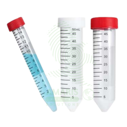
Centrifuge Tubes are specialized disposable containers (polypropylene) designed to withstand high centrifugal forces (up to 30,000 x g) for separating biological fluids based on density. Available in microcentrifuge sizes (0.2-2.0 mL), conical tubes (15 mL, 50 mL), and larger volumes (100-500 mL). Features include leak-proof screw caps with O-rings (conical tubes), snap caps (microcentrifuge tubes), graduated volume markings, writing areas, and sterile/non-sterile options. Certified RNase/DNase-free, pyrogen-free, and autoclavable. Primary clinical applications include blood component separation (serum/plasma), urine sediment preparation, microbiology sample concentration, molecular biology (DNA/RNA extraction), cell culture processing, protein/enzyme analysis, and viral load testing. Critical safety precautions include never exceeding maximum RCF rating, proper balancing by weight, secure cap tightening, inspection for cracks, and disposal as biohazardous waste. Essential consumable in every clinical, research, and industrial laboratory.
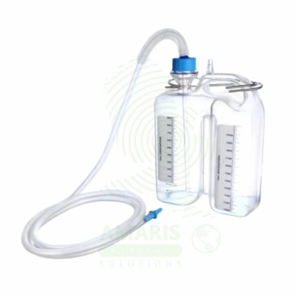
Chest Bottle
A Chest Bottle (Underwater Seal Drainage System) is a three-chamber disposable device used to drain air and fluid from the pleural space via a chest tube. Its critical component is a water seal chamber that acts as a one-way valve, preventing air from re-entering the chest while allowing drainage. It is used to treat pneumothorax, hemothorax, and pleural effusions, and is mandatory post-thoracic surgery. The system must always be kept upright and below the patient's chest level. It provides essential clinical information through tidaling in the water seal (indicating tube patency and lung expansion) and bubbling (indicating an air leak), making it both a therapeutic and diagnostic tool.
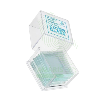
Cover Glass
Cover Glass (coverslips) are thin, transparent glass or plastic squares (e.g., 18×18 mm, 22×22 mm) or rectangles (22×30 mm, 24×50 mm) used to cover specimens on microscope slides for microscopic examination. Available in thickness No. 1 (0.13-0.16 mm) and No. 1.5 (0.16-0.19 mm) for optimal microscopy. Made from optical glass with high light transmission (>90%), refractive index 1.52 matching immersion oil. Packaged in boxes of 100-1000, interleaved with paper to prevent scratching. Primary clinical applications include covering blood films for manual differential counts and malaria diagnosis, wet mount preparations of urine sediment and vaginal secretions, histopathology and cytology slides, Gram-stained bacterial smears, semen analysis, KOH preparations for fungal elements, and India ink preparations for Cryptococcus. Critical safety precautions include handling with forceps (sharp edges), disposing of broken coverslips in sharps containers, avoiding fingerprints on optical surface, and proper disposal of biohazard-contaminated coverslips. Essential consumable in every clinical laboratory performing microscopy.
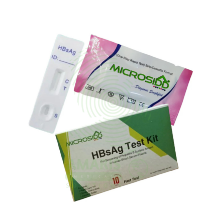
Hepatitis B Test Kit
The Hepatitis B Test Kit is an in vitro diagnostic device for the detection of Hepatitis B virus markers in human blood samples, available in rapid, ELISA, and molecular formats. It detects specific antigens (HBsAg) and antibodies (anti-HBc, anti-HBs) to diagnose acute and chronic infection, monitor treatment response, screen blood donations, and determine immunity status. With high sensitivity (>98%) and specificity (>99%), it provides results in 15 minutes to 4 hours depending on format. Essential applications include prenatal screening to prevent mother-to-child transmission, evaluation of acute hepatitis, pre-immunosuppression screening, and occupational health testing. All patient samples must be handled with universal precautions, and testing must include appropriate quality controls.
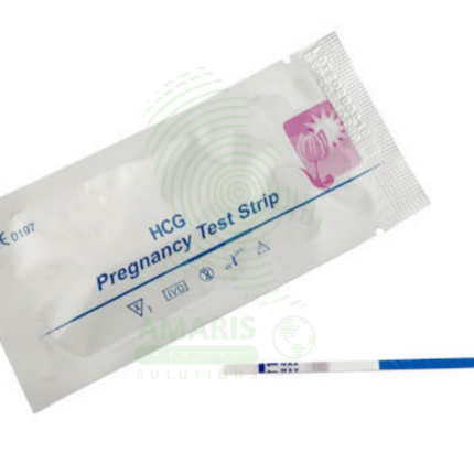
Pregnancy Test Strips
Pregnancy Test Strips are simple, dip-style lateral flow devices designed for the rapid, qualitative detection of human chorionic gonadotropin (hCG) in urine to indicate pregnancy. As the most fundamental format of home pregnancy tests, they offer high sensitivity (often from 10 mIU/mL), affordability, and a quick result within 3-5 minutes. Their ease of use makes them a widely accessible first step in pregnancy detection for personal use and clinical screening. A visible test line, even if faint, is a positive result, though confirmation with a healthcare provider is always recommended.
Thermal Printing Paper Roll
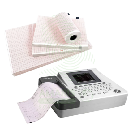
Thermal Printing Paper Roll is a heat-sensitive recording medium used with medical monitoring and diagnostic equipment including ECG machines, fetal monitors, ultrasound printers, and telemetry systems. The paper produces high-contrast, permanent images of physiological waveforms and data when passed over the printer's thermal head. Available in various widths, lengths, and grid patterns to match specific device requirements, it is an essential consumable for creating hard copy records for patient charts, consultation, and legal documentation. Proper storage away from light, heat, and moisture is essential to preserve image quality. BPA-free and archival-grade options are available for specific applications.
Ultrasound Gel
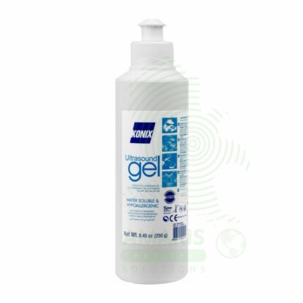
Ultrasound Gel is a water-based, hypoallergenic, acoustically conductive coupling medium essential for diagnostic and therapeutic ultrasound procedures. It eliminates air pockets between the transducer and skin, creating an efficient acoustic pathway for sound wave transmission and reception. Formulated to match the acoustic impedance of human tissue, it ensures high-quality imaging while providing lubrication for smooth probe movement and patient comfort. Available in standard, sterile, and hypoallergenic formulations, it is a single-use consumable critical for all ultrasound examinations. Proper storage, handling, and selection of appropriate sterile versus non-sterile formulations are essential for patient safety and image quality.