Dermatoscope and Magnifiers
 Diagnostic Kits
 Vital Signs Monitors
 Stethoscopes and Accessories
 Otoscopes, Ophthalmoscopes, and Retinoscopes
 Reflex Hammers and Neurological Tools
 Scales and Measuring Devices
 Spirometers and Pulmonary Function Tests
 Electrosurgical Units and Accessories
 Cutting Instruments
 Grasping and Holding Instruments
 Hemostatic Instruments
 Specialized Surgical Sets
 Single-Use Procedure Trays and Packs
 Surgical Drapes, Gowns, and Covers
 Tissue Unifying Instruments
 Radiation Protection
 X-Ray Machines and Accessories
 Ultrasound Systems and Probes
 MRI and CT Scanners
 Radiology Consumables
 Bone Densitometers
 Fluoroscopy Equipment
 Imaging Tables and Positioning Aids
 Microscopes and Accessories
 Centrifuges and Separators
 Analyzers
 Incubators and Ovens
 Pipettes, Dispensers, and Lab Glassware
 Refrigerators, Freezers, and Storage Units
 Lab Consumables
 Sterilizers and Autoclaves for Lab Use
 Multi-Parameter Monitors
 Ventilators and Respiratory Support Devices
 Defibrillators and AEDs
 Infusion Pumps and IV Systems
 Patient Warmers and Cooling Devices
 Central Monitoring Stations
 Accessories
 Anesthesia Machines and Workstations
 Oxygen Concentrators and Delivery Systems
 Nebulizers and Inhalers
 CPAP/BiPAP Machines
 Airway Management
 Anesthesia Masks, Circuits, and Bags
 Humidifiers and Heaters
 Respiratory Therapy Accessories
 First Aid Kits and Cabinets
 Emergency Resuscitation Equipment
 Trauma Supplies
 Emergency Carts and Crash Carts
 Burn Care Products
 Bleeding Control
 Automated External Defibrillators (AEDs)
 Transport and Evacuation
 Wheelchairs and Accessories
 Walkers, Crutches, and Canes
 Prosthetics and Orthotics
 Physical Therapy Equipment
 Transfer Devices
 Bathroom Safety
 Orthopedic Traction and Tables
 Hot/Cold Therapy Packs and Units
 Beds and Mattresses
 Chairs and Stools
 Tables
 Cabinets and Storage
 Privacy Screens & Curtains
 Stands and Racks
 Linens and Textiles
 Lighting
 Autoclaves and Sterilizers
 Ultrasonic Cleaners
 Disinfectant Solutions and Wipes
 Sterilization Pouches, Wraps, and Indicators
 Instrument Trays and Containers
 UV and Ozone Disinfection Devices
 Washer Disinfectors
 Wound Care
 Gloves
 Masks and Respirators
 Catheters and Tubing
 Swabs, Applicators, and Sponges
 Incontinence Products
 Personal Protective Equipment (PPE)
 Dental Chairs and Units
 Handpieces and Burs
 Instruments
 Consumables
 Sterilization for Dental Use
 Orthodontic Supplies
 Endodontic Tools
 Slit Lamps and Tonometers
 Lensometers and Phoropters
 Ophthalmic Surgical Instruments
 Eyewear Frames and Lenses
 Contact Lens Supplies
 Vision Testing Charts and Devices
 Eye Care Consumables
 Laser Systems for Eye Care
 ENT Exam Chairs and Tables
 Endoscopes
 Audiometers and Hearing Tests
 ENT Instruments
 Nasal and Throat Packs
 Hearing Aids and Accessories
 Otology Supplies
 Fetal Dopplers and Monitors
 Delivery Beds and Tables
 Gynecological Instruments
 Neonatal Incubators and Warmers
 Breast Pumps and Accessories
 Contraceptive Devices
 Maternity Supports and Pads
 Neonatal Consumables
 Cystoscopes and Urethroscopes
 Dialysis Machines and Supplies
 Urological Catheters and Bags
 Lithotripters
 Prostate Treatment Devices
 Urinary Incontinence Products
 Kidney Stone Management Tools
 Consumables & Disposables
 EEG and EMG Machines
 Neurosurgical Instruments
 Nerve Stimulators
 Headrests and Positioning Aids
 Lumbar Puncture Kits
 Seizure Monitoring Devices
 Consumables
 Rehabilitation for Neurological Conditions
 ECG Machines and Accessories
 Holter Monitors
 Stress Test Systems
 Pacemakers and Defibrillator Accessories
 Vascular Access Devices
 Cardiac Catheters and Guidewires
 Blood Flow Meters
 Consumables
 Orthopedic Instruments
 Casts, Splints, and Padding
 Joint Replacement Supplies
 Prosthetic Limbs and Components
 Bone Grafts and Substitutes
 Traction Devices
 Orthopedic Braces and Supports
 Rehabilitation Aids for Orthopedics
 Home Oxygen Therapy
 Hospital Beds for Home Use
 Mobility Aids
 Bathroom and Daily Living Aids
 Wound Care for Home
 Monitoring Devices
 Enteral Feeding Pumps and Tubes
 Hand Sanitizers and Dispensers
 Face Shields and Goggles
 Isolation Gowns and Suits
 Biohazard Waste Containers
 Air Purifiers and HEPA Filters
 Surface Disinfectants
 Sharps Containers
 Protective Barriers
 Cardiovascular & Endurance Training
 Strength Training & Weightlifting
 Functional Training & Core Conditioning
 Physical Therapy & Rehabilitation
 Sports & Outdoor Recreation
 Gym Flooring & Facility Equipment
 Fitness Monitoring & Accessories
 Kids & Novelties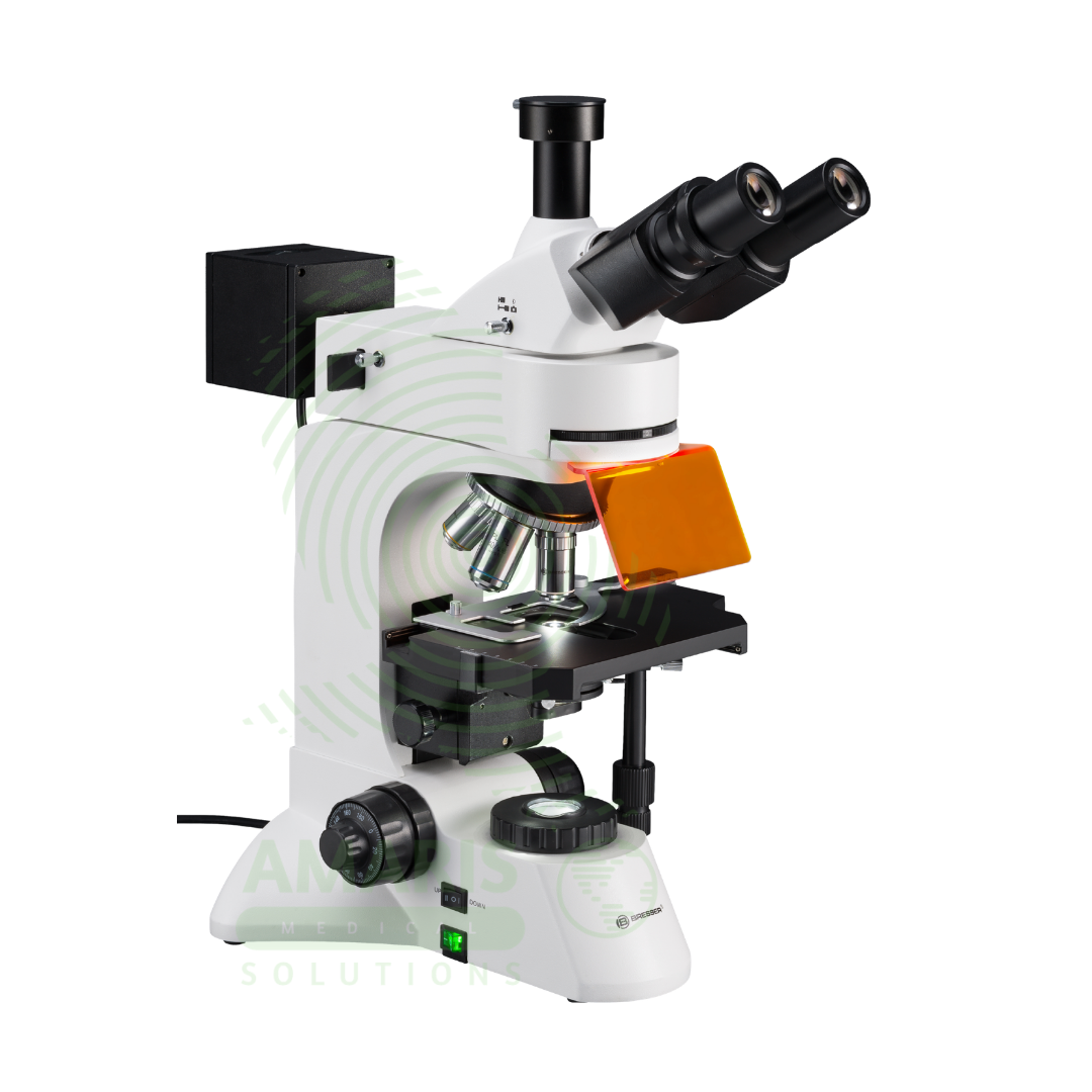
Fluorescent Microscope
$1.00
WhatsApp Order
A Fluorescent Microscope is a specialized Class I medical device designed for visualizing specimens labeled with fluorescent dyes (fluorochromes) that emit light at specific wavelengths when excited by high-intensity illumination. Equipped with high-pressure mercury (HBO), xenon (XBO), metal halide, or LED light sources, interchangeable filter cubes for specific fluorochromes (DAPI, FITC, TRITC, Cy3, Cy5), and high numerical aperture objectives for maximum light collection. Features include epifluorescence design, precision filter turret (4-6 positions), UV-blocking safety shields, and trinocular head for digital imaging. Primary clinical applications include immunofluorescence for autoimmune disease diagnosis (ANA, ANCA, anti-dsDNA, skin/kidney biopsies), fluorescence in situ hybridization (FISH) for genetic abnormalities (aneuploidy, deletions, translocations, HER2 amplification), auramine-rhodamine staining for tuberculosis screening, direct fluorescent antibody (DFA) testing for Legionella, Pneumocystis, Cryptosporidium, Giardia, and respiratory viruses, and FTA-ABS confirmatory testing for syphilis. Essential equipment in immunology, microbiology, pathology, cytogenetics, and molecular diagnostics laboratories requiring sensitive and specific detection of fluorescent signals for diagnosis of infectious, autoimmune, genetic, and neoplastic diseases.
Description
Fluorescent Microscope
PRIMARY CLINICAL & DIAGNOSTIC USES
1. Immunofluorescence for Autoimmune Disease Diagnosis:
-
Primary Use: Fluorescent microscopes are essential for detecting antinuclear antibodies (ANA), anti-dsDNA, and other autoantibodies in autoimmune diseases including systemic lupus erythematosus, rheumatoid arthritis, and Sjögren’s syndrome using HEp-2 cell substrates and fluorescent conjugates.
-
How it helps: Reveals the telltale glowing patterns of autoantibodies attacking a patient’s own tissues, giving rheumatologists the evidence they need to diagnose complex autoimmune conditions and start treatment before irreversible damage occurs.
2. Fluorescence In Situ Hybridization (FISH) for Genetic Abnormalities:
-
Primary Use: Used to visualize fluorescently labeled DNA probes hybridized to chromosomes for detecting genetic abnormalities including aneuploidies, deletions, translocations, and amplifications in prenatal diagnosis, cancer cytogenetics, and genetic disorders.
-
How it helps: Makes chromosomes glow in specific colors, allowing geneticists to see missing pieces, extra copies, or swapped segments that cause conditions from Down syndrome to certain leukemias.
3. Acid-Fast Bacilli (AFB) Detection for Tuberculosis:
-
Primary Use: Fluorescent microscopy with auramine-rhodamine staining enables rapid screening for Mycobacterium tuberculosis in sputum specimens, providing higher sensitivity than conventional Ziehl-Neelsen staining and faster screening of multiple specimens.
-
How it helps: Makes TB bacteria glow like tiny yellow rods against a dark background, allowing laboratory technicians to scan slides quickly and find the organisms causing a persistent, contagious cough.
4. Immunohistochemistry and Immunofluorescence in Pathology:
-
Primary Use: Used to visualize fluorescently labeled antibodies binding to specific antigens in tissue sections for diagnosis and classification of tumors, infectious agents, and immune-mediated diseases.
-
How it helps: Lights up specific proteins on cancer cells, helping pathologists determine exactly what type of tumor a patient has and whether it will respond to targeted therapies.
5. Microbiology and Parasitology Identification:
-
Primary Use: Employed for direct fluorescent antibody (DFA) testing for rapid identification of pathogens including Legionella pneumophila, Pneumocystis jirovecii, Cryptosporidium, Giardia, and respiratory viruses (influenza, RSV, adenovirus).
-
How it helps: Transforms invisible microorganisms into bright, glowing shapes, allowing rapid diagnosis of infections from pneumonia to parasitic diarrhea in minutes rather than days.
6. Fluorescent Treponemal Antibody (FTA-ABS) Test for Syphilis:
-
Primary Use: Used to detect antibodies against Treponema pallidum in confirmatory testing for syphilis, particularly useful in cases where non-treponemal tests (VDRL, RPR) are reactive.
-
How it helps: Confirms whether a positive screening test truly means a patient has syphilis, preventing both unnecessary treatment and missed diagnosis of this serious infection.
7. Cytogenetics and Molecular Diagnostics:
-
Primary Use: Essential for visualizing fluorescent signals in various molecular diagnostic techniques including chromosome painting, telomere length assessment, and detection of microdeletions and microduplications.
-
How it helps: Allows geneticists to see the subtle chromosomal changes that cause developmental delays, birth defects, and inherited conditions, providing answers for families seeking to understand their child’s condition.
SECONDARY & SUPPORTIVE USES
1. Research and Developmental Biology: Used extensively in research for visualizing fluorescent proteins (GFP, RFP), cellular structures, and dynamic processes in living and fixed cells, advancing our understanding of how life works at the cellular level.
2. Neuroscience and Neuropathology: Employed for visualizing neuronal pathways, protein aggregates (amyloid plaques, Lewy bodies), and neurodegenerative disease markers, helping researchers understand diseases like Alzheimer’s and Parkinson’s.
3. Cell Biology and Organelle Studies: Used to visualize specific organelles (mitochondria, endoplasmic reticulum, nucleus) using fluorescent dyes and probes, revealing the inner workings of cells.
4. Drug Development and Pharmacology: Employed in high-content screening and drug discovery studies using fluorescent assays, accelerating the development of new medications.
5. Environmental Microbiology: Used for detecting fluorescently labeled microorganisms in water, soil, and environmental samples, monitoring environmental safety.
6. Food Safety and Quality Control: Employed for rapid detection of pathogens in food products using fluorescent antibody techniques, helping prevent foodborne illness outbreaks.
7. Forensic Science: Used for visualizing fluorescently labeled evidence in forensic investigations, helping solve crimes and bring justice to victims.
KEY PRODUCT FEATURES
1. BASIC IDENTIFICATION ATTRIBUTES
-
Product Type: Specialized microscope designed for visualizing specimens labeled with fluorescent dyes (fluorochromes) that emit light at specific wavelengths when excited by high-intensity illumination.
-
Common Names: Fluorescence Microscope, Fluorescent Microscope, Epifluorescence Microscope, UV Microscope, Immunofluorescence Microscope, FISH Microscope.
-
Optical Configuration: Binocular or trinocular observation tubes with fluorescence-specific optics.
-
Fluorescence Illumination: High-intensity light sources including mercury vapor (HBO), xenon (XBO), metal halide, or LED with specific excitation/emission wavelengths.
-
Filter Cubes: Interchangeable filter cubes containing excitation filter, dichroic mirror, and emission barrier filter for specific fluorochromes.
-
Common Fluorochromes: DAPI (blue), FITC (green), TRITC/Cy3 (red), Cy5 (far-red), Auramine (yellow), Rhodamine (red-orange).
-
Objectives: High numerical aperture (NA) objectives specifically designed for fluorescence to maximize light collection.
-
Magnification Range: 40× to 1000× (oil immersion) depending on applications.
-
Condenser: Specialized fluorescence condenser or brightfield condenser with fluorescence capability.
-
Stage: Mechanical stage with precise X-Y controls for scanning specimens.
-
Focusing: Coaxial coarse and fine focus with high precision for fluorescence work.
-
Camera Port: Trinocular head for digital camera attachment and image capture.
2. TECHNICAL & PERFORMANCE PROPERTIES
-
Fluorescence Illumination Sources:
-
Mercury Vapor (HBO): 50W or 100W; intense line spectrum at specific wavelengths; requires warm-up (10-15 min) and cool-down; bulb life 200-300 hours.
-
Xenon (XBO): 75W or 150W; continuous spectrum; good for quantitative work; bulb life 400-500 hours.
-
Metal Halide: 120W; longer life (2,000+ hours); rapid start; stable output.
-
LED: Long life (25,000+ hours); instant on/off; specific wavelengths; minimal heat; increasingly common.
-
-
Filter Cubes: Precision-machined with:
-
Excitation Filter: Selects specific wavelengths to excite fluorochrome.
-
Dichroic Mirror: Reflects excitation light to specimen; transmits emitted fluorescence.
-
Emission (Barrier) Filter: Blocks residual excitation light; transmits specific fluorescence wavelength.
-
-
Objective Numerical Aperture (NA): Higher NA (1.3-1.4 for oil) critical for fluorescence; light collection efficiency proportional to NA⁴.
-
Optical System: Infinity-corrected plan apochromatic or plan fluorite objectives for maximum light transmission and chromatic correction.
-
Epifluorescence Design: Excitation and emission light paths through the same objective; standard for most applications.
-
Darkfield Condenser: Optional for specific applications requiring darkfield fluorescence.
-
Neutral Density Filters: For reducing excitation intensity to prevent photobleaching.
-
Heat Protection: Heat absorption filters to protect specimens from thermal damage.
-
Shutter: Automatic or manual shutter to control excitation exposure and minimize photobleaching.
3. PHYSICAL & OPERATIONAL PROPERTIES
-
Dimensions: 30-40 cm W × 50-70 cm D × 50-60 cm H (plus lamp housing).
-
Weight: 15-30 kg depending on configuration and light source.
-
Construction: Heavy-duty cast metal base and stand for vibration-free operation; light-tight lamp housing.
-
Viewing Heads: Binocular or trinocular with adjustable inclination.
-
Nosepiece: Revolving quintuple or sextuple nosepiece with fluorescence and brightfield objectives.
-
Filter Turret: Rotating turret holding 4-6 filter cubes for different fluorochromes.
-
Stage: Mechanical stage with low-position coaxial controls; some models with heated stage for live cell work.
-
Focus Mechanism: Coaxial coarse and fine focus with high precision; fine focus graduation 1-2 microns.
-
Light Source: External lamp housing with collector lenses and alignment mechanisms.
-
Lamp Alignment: Centering and focusing controls for optimal illumination.
-
Heat Management: Cooling fans and heat filters to protect specimens.
-
UV Shield: Protective shield to block UV light from the operator.
-
Camera Port: Standard C-mount or specific camera attachment for digital imaging.
4. SAFETY & COMPLIANCE ATTRIBUTES
-
Regulatory Status: Class I medical device (FDA, CE marked for IVD use).
-
UV Safety: UV-blocking shields and eyepieces; safety interlocks on lamp housing.
-
Electrical Safety: Compliant with IEC 61010-1; high-voltage components in lamp power supply.
-
Lamp Safety: Mercury and xenon lamps contain hazardous materials; operate at high pressure and temperature.
-
Heat Management: Adequate ventilation required; heat filters and cooling fans.
-
Chemical Safety: Specimens may contain infectious agents; follow universal precautions.
-
Cleaning: Surfaces designed for cleaning with mild detergents and disinfectants.
-
Quality Management: Manufactured under ISO 13485 or ISO 9001 certified processes.
-
UV Exposure: Operators should avoid direct exposure to UV light; use protective shields.
-
Lamp Disposal: Mercury lamps require special disposal as hazardous waste.
5. STORAGE & HANDLING ATTRIBUTES
-
Storage: Store in a clean, dry environment when not in use; use dust cover; protect from vibration.
-
Installation: Professional installation recommended; place on rigid, vibration-free table; ensure adequate ventilation for lamp housing.
-
Lamp Operation: Allow warm-up (10-15 min) before use; minimize on/off cycles; record lamp hours; allow cool-down before moving.
-
Filter Cubes: Store in dust-free container; handle by edges; clean with lens paper if needed.
-
Cleaning: Clean lenses with lens paper and approved optical cleaner. Clean stage and frame with mild detergent and soft cloth.
-
Objective Care: Keep objectives clean; use immersion oil only with oil objectives; clean immediately after use.
-
Köhler Illumination: Align brightfield Köhler first; align fluorescence illumination per manufacturer.
-
Lamp Alignment: Check and adjust lamp alignment periodically; misalignment reduces intensity.
-
Annual Maintenance: Professional cleaning, alignment, and calibration recommended.
-
Lamp Replacement: Follow manufacturer procedure; allow to cool; use protective gloves; dispose properly.
6. LABORATORY & CLINICAL APPLICATIONS
-
Primary Application: Detection and visualization of fluorescently labeled specimens for diagnostic immunofluorescence, FISH, and fluorescent staining techniques.
-
Immunofluorescence Applications:
-
ANA Testing: HEp-2 cell patterns (homogeneous, speckled, nucleolar, centromere) at 400×.
-
ANCA Testing: Cytoplasmic (c-ANCA) and perinuclear (p-ANCA) patterns at 400×.
-
Anti-dsDNA: Crithidia luciliae kinetoplast staining at 400×.
-
Skin Immunofluorescence: Linear vs. granular IgG deposits in pemphigus/pemphigoid.
-
Kidney Immunofluorescence: Immune complex deposition patterns in glomerulonephritis.
-
-
FISH Applications:
-
Prenatal Aneuploidy Detection: Trisomy 13, 18, 21; sex chromosome abnormalities at 1000× oil.
-
HER2/neu Amplification: Breast cancer prognosis and treatment selection at 1000× oil.
-
BCR-ABL Fusion: Chronic myeloid leukemia diagnosis and monitoring.
-
Deletion Syndromes: 22q11.2 (DiGeorge), 15q11.2 (Angelman/Prader-Willi).
-
Translocation Detection: Lymphoma and leukemia-associated translocations.
-
-
Microbiology Applications:
-
AFB Smears: Auramine-rhodamine staining for TB screening at 400×.
-
DFA for Legionella: Direct fluorescent antibody for Legionella pneumophila at 400×.
-
Pneumocystis jirovecii: DFA or calcofluor white at 400×.
-
Cryptosporidium and Giardia: DFA in stool specimens at 400×.
-
Respiratory Viruses: DFA for influenza, RSV, adenovirus, parainfluenza at 400×.
-
-
Pathology Applications:
-
Immunohistochemistry: Fluorescent detection of tumor markers, prognostic factors.
-
Amyloid Detection: Thioflavin T or Congo red with fluorescence.
-
Neuropathology: Detection of amyloid plaques, neurofibrillary tangles.
-
-
Cytogenetics Applications:
-
Chromosome Painting: Whole chromosome probes for karyotype analysis.
-
Telomere Length Assessment: Quantitative FISH for telomere studies.
-
Spectral Karyotyping (SKY): Simultaneous visualization of all chromosomes.
-
-
Syphilis Testing:
-
FTA-ABS: Confirmatory testing for syphilis using T. pallidum antigen.
-
SAFETY HANDLING PRECAUTIONS
1. SAFETY PRECAUTIONS
-
UV Exposure: Never look directly at UV light source; ensure UV shields and blocking eyepieces are in place. Operators should wear UV-blocking safety glasses when aligning lamps.
-
Lamp Safety: Mercury and xenon lamps operate at high pressure and temperature; risk of explosion if mishandled. Allow to cool before handling; use protective gloves; follow manufacturer procedures.
-
Electrical Safety: Lamp power supplies use high voltage; disconnect power before servicing; ensure proper grounding.
-
Photobleaching: Minimize exposure of fluorochromes to excitation light; use neutral density filters; acquire images quickly.
-
Heat Management: Ensure adequate ventilation; do not block cooling fans; allow warm-up and cool-down periods.
-
Chemical Safety: Specimens may contain infectious agents; follow universal precautions; clean spills immediately.
-
Lens Care: Never touch lenses with fingers; use only lens paper and approved cleaners.
-
Oil Immersion: Use only with 100× objectives; clean immediately after use.
-
Filter Cube Handling: Handle by edges; avoid touching optical surfaces; store in dust-free containers.
-
Training: Operators must be trained on fluorescence microscope operation, safety procedures, and lamp handling.
2. FIRST AID MEASURES
-
UV Exposure (Eye): If eyes are exposed to UV, rest eyes; seek medical attention if symptoms (pain, redness, tearing) develop.
-
Lamp Breakage (Mercury): Evacuate area; ventilate; follow hazardous material spill protocol; mercury is toxic. Use specialized cleanup; dispose of hazardous waste.
-
Chemical Spill (Stains, Reagents): Follow chemical spill protocol; use appropriate PPE; clean affected areas.
-
Specimen Spill on Microscope: Disconnect power; carefully clean with appropriate disinfectant; dry thoroughly.
-
Electrical Shock: Disconnect power; seek medical attention if needed.
-
Burn from Hot Lamp: Cool area with water; seek medical attention for severe burns.
3. FIRE FIGHTING MEASURES
-
Flammability: Plastic components and immersion oil are combustible; mercury vapor lamps may release toxic gases if involved in fire.
-
Extinguishing Media: For electrical fire, use CO₂ or dry chemical (Class C) extinguisher.
-
Power Off: Disconnect power if safe to do so.
-
Mercury Lamp Fire: May release toxic mercury vapor; use SCBA in enclosed spaces.
-
Evacuation: Evacuate area; follow facility fire protocols.
Related products
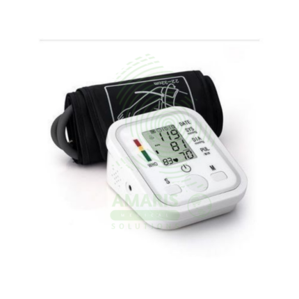
Digital Blood Pressure Monitor
A Digital Blood Pressure Monitor is an automatic electronic device that measures and displays systolic pressure, diastolic pressure, and pulse rate using the oscillometric method. Designed primarily for home and clinical use, it offers a user-friendly alternative to manual aneroid devices, requiring minimal training. Key features often include irregular heartbeat detection, multi-user memory, and connectivity for data tracking. Its accuracy, when clinically validated and used with correct technique, makes it an indispensable tool for the diagnosis and management of hypertension, empowering patients and supporting healthcare providers with reliable ambulatory data.
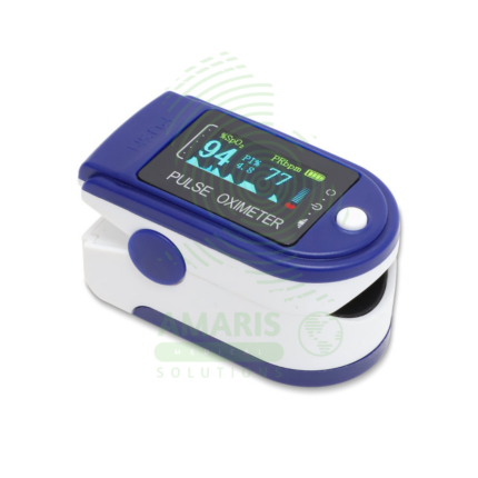
Fingertip Pulse Oximeter
A Fingertip Pulse Oximeter is a compact, non-invasive medical device that clips onto a finger to measure peripheral arterial oxygen saturation (SpO2) and pulse rate in seconds. Utilizing light-based technology, it provides critical, real-time data on respiratory and circulatory status. Its portability and ease of use make it indispensable in hospitals (from ORs to general wards), for patients managing chronic lung conditions at home, and for situational monitoring in sports or high-altitude environments. While an extremely valuable screening tool, readings must be interpreted in context, considering factors like patient movement, perfusion, and clinical symptoms.
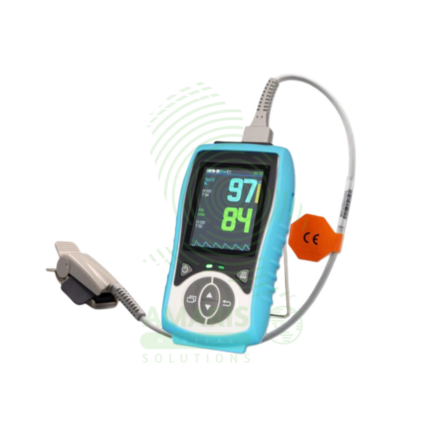
Handheld Pulse Oximeter
A Handheld Pulse Oximeter is a professional-grade, portable medical device used by healthcare providers for accurate and reliable monitoring of blood oxygen saturation (SpO2) and pulse rate. It consists of a durable, feature-rich monitor unit and separate, interchangeable sensors for use on fingers, toes, or ears. Superior to basic fingertip models, it offers advanced functions like configurable alarms, detailed plethysmograph display, trend memory, and data connectivity. Its robust design, clinical accuracy under challenging conditions (motion, low perfusion), and versatility make it an essential tool in hospitals, clinics, ambulances, and home health for patient assessment and monitoring.
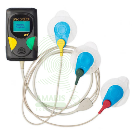
Holter ECG Monitor
A Holter ECG Monitor is a portable, wearable medical device used for continuous, long-term recording of a patient's heart's electrical activity over 24 to 48 hours or more. It is the cornerstone diagnostic tool for detecting intermittent cardiac arrhythmias, evaluating unexplained symptoms like syncope or palpitations, and monitoring the efficacy of antiarrhythmic treatments or implanted cardiac device function. Consisting of a small digital recorder connected to chest electrodes (or an all-in-one adhesive patch), it allows patients to maintain their normal daily routine while capturing a comprehensive electrocardiographic record.
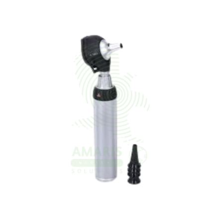
LED Otoscope
An LED Otoscope is a handheld medical device used for illuminating and magnifying the external ear canal and tympanic membrane. Featuring a bright, cool, and long-lasting LED light source, it provides superior visualization for diagnosing common conditions like ear infections, wax impaction, and perforations. Its design, incorporating a reusable handle/head and disposable specula, ensures hygiene and patient safety. As an essential tool in family medicine, pediatrics, and ENT, it enables clinicians to perform accurate otoscopic examinations and, when equipped with an insufflator, crucial pneumatic otoscopy to assess middle ear function.
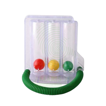
Spirometer
A Spirometer is an essential diagnostic medical device used to measure how much and how quickly air can be inhaled and exhaled by the lungs. By performing forced breathing maneuvers, it generates key parameters—primarily Forced Vital Capacity (FVC) and Forced Expiratory Volume in 1 second (FEV1)—that are fundamental for diagnosing and monitoring obstructive and restrictive lung diseases like Asthma and COPD. Requiring daily calibration and strict adherence to international standards (ATS/ERS), it provides the objective, reproducible data needed for clinical decision-making in pulmonology, primary care, occupational health, and pre-operative assessment.
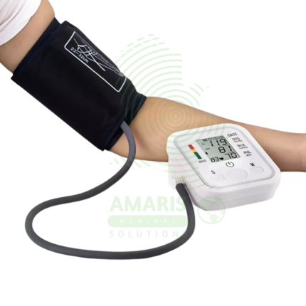
Upper Arm Blood Pressure Monitor
The Upper Arm Blood Pressure Monitor is a clinically validated, automatic digital device designed for accurate and comfortable blood pressure and pulse measurement at home or in professional settings. Known for its user-friendly features like Intellisense inflation technology, a large clear display, and comprehensive functions including irregular heartbeat detection, multi-user memory, and a WHO-based hypertension indicator, it provides reliable data for effective hypertension management. Its robust construction, adherence to international safety standards (CE, FDA), and focus on patient comfort make it a trusted choice for individuals and healthcare providers seeking a dependable tool for cardiovascular health monitoring.