Dermatoscope and Magnifiers
 Diagnostic Kits
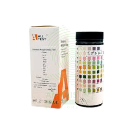
 Vital Signs Monitors
 Stethoscopes and Accessories
 Otoscopes, Ophthalmoscopes, and Retinoscopes
 Reflex Hammers and Neurological Tools
 Scales and Measuring Devices
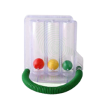
 Spirometers and Pulmonary Function Tests
 Electrosurgical Units and Accessories
 Cutting Instruments
 Grasping and Holding Instruments
 Hemostatic Instruments
 Specialized Surgical Sets
 Single-Use Procedure Trays and Packs
 Surgical Drapes, Gowns, and Covers
 Tissue Unifying Instruments
 Radiation Protection
 X-Ray Machines and Accessories
 Ultrasound Systems and Probes
 MRI and CT Scanners
 Radiology Consumables
 Bone Densitometers
 Fluoroscopy Equipment
 Imaging Tables and Positioning Aids
 Microscopes and Accessories
 Centrifuges and Separators
 Analyzers
 Incubators and Ovens
 Pipettes, Dispensers, and Lab Glassware
 Refrigerators, Freezers, and Storage Units
 Lab Consumables
 Sterilizers and Autoclaves for Lab Use
 Multi-Parameter Monitors
 Ventilators and Respiratory Support Devices
 Defibrillators and AEDs
 Infusion Pumps and IV Systems
 Patient Warmers and Cooling Devices
 Central Monitoring Stations
 Accessories
 Anesthesia Machines and Workstations
 Oxygen Concentrators and Delivery Systems
 Nebulizers and Inhalers
 CPAP/BiPAP Machines
 Airway Management
 Anesthesia Masks, Circuits, and Bags
 Humidifiers and Heaters
 Respiratory Therapy Accessories
 First Aid Kits and Cabinets
 Emergency Resuscitation Equipment
 Trauma Supplies
 Emergency Carts and Crash Carts
 Burn Care Products
 Bleeding Control
 Automated External Defibrillators (AEDs)
 Transport and Evacuation
 Wheelchairs and Accessories
 Walkers, Crutches, and Canes
 Prosthetics and Orthotics
 Physical Therapy Equipment
 Transfer Devices
 Bathroom Safety
 Orthopedic Traction and Tables
 Hot/Cold Therapy Packs and Units
 Beds and Mattresses
 Chairs and Stools
 Tables
 Cabinets and Storage
 Privacy Screens & Curtains
 Stands and Racks
 Linens and Textiles
 Lighting
 Autoclaves and Sterilizers
 Ultrasonic Cleaners
 Disinfectant Solutions and Wipes
 Sterilization Pouches, Wraps, and Indicators
 Instrument Trays and Containers
 UV and Ozone Disinfection Devices
 Washer Disinfectors
 Wound Care
 Gloves
 Masks and Respirators
 Catheters and Tubing
 Swabs, Applicators, and Sponges
 Incontinence Products
 Personal Protective Equipment (PPE)
 Dental Chairs and Units
 Handpieces and Burs
 Instruments
 Consumables
 Sterilization for Dental Use
 Orthodontic Supplies
 Endodontic Tools
 Slit Lamps and Tonometers
 Lensometers and Phoropters
 Ophthalmic Surgical Instruments
 Eyewear Frames and Lenses
 Contact Lens Supplies
 Vision Testing Charts and Devices
 Eye Care Consumables
 Laser Systems for Eye Care
 ENT Exam Chairs and Tables
 Endoscopes
 Audiometers and Hearing Tests
 ENT Instruments
 Nasal and Throat Packs
 Hearing Aids and Accessories
 Otology Supplies
 Fetal Dopplers and Monitors
 Delivery Beds and Tables
 Gynecological Instruments
 Neonatal Incubators and Warmers
 Breast Pumps and Accessories
 Contraceptive Devices
 Maternity Supports and Pads
 Neonatal Consumables
 Cystoscopes and Urethroscopes
 Dialysis Machines and Supplies
 Urological Catheters and Bags
 Lithotripters
 Prostate Treatment Devices
 Urinary Incontinence Products
 Kidney Stone Management Tools
 Consumables & Disposables
 EEG and EMG Machines
 Neurosurgical Instruments
 Nerve Stimulators
 Headrests and Positioning Aids
 Lumbar Puncture Kits
 Seizure Monitoring Devices
 Consumables
 Rehabilitation for Neurological Conditions
 ECG Machines and Accessories
 Holter Monitors
 Stress Test Systems
 Pacemakers and Defibrillator Accessories
 Vascular Access Devices
 Cardiac Catheters and Guidewires
 Blood Flow Meters
 Consumables
 Orthopedic Instruments
 Casts, Splints, and Padding
 Joint Replacement Supplies
 Prosthetic Limbs and Components
 Bone Grafts and Substitutes
 Traction Devices
 Orthopedic Braces and Supports
 Rehabilitation Aids for Orthopedics
 Home Oxygen Therapy
 Hospital Beds for Home Use
 Mobility Aids
 Bathroom and Daily Living Aids
 Wound Care for Home
 Monitoring Devices
 Enteral Feeding Pumps and Tubes
 Hand Sanitizers and Dispensers
 Face Shields and Goggles
 Isolation Gowns and Suits
 Biohazard Waste Containers
 Air Purifiers and HEPA Filters
 Surface Disinfectants
 Sharps Containers
 Protective Barriers
 Cardiovascular & Endurance Training
 Strength Training & Weightlifting
 Functional Training & Core Conditioning
 Physical Therapy & Rehabilitation
 Sports & Outdoor Recreation
 Gym Flooring & Facility Equipment
 Fitness Monitoring & Accessories
 Kids & Novelties
 Amaris Medical")
Binocular Microscope
WhatsApp Order
A Binocular Microscope is a high-precision, compound optical instrument fundamental to clinical diagnostics. Featuring dual eyepieces for comfortable viewing and a suite of parfocal objective lenses (4x, 10x, 40x, 100x oil), it provides magnifications from 40x to 1000x. Its integrated LED illumination, mechanical stage, and Abbe condenser enable the detailed examination of stained blood films, tissue sections, microbiological specimens, and cytological preparations. As the primary tool for pathologists and laboratory scientists, it is indispensable for definitive diagnoses in hematology, histopathology, microbiology, and urinalysis, demanding skilled operation and meticulous maintenance for optimal performance.
Description
Binocular Microscope
PRIMARY CLINICAL & DIAGNOSTIC USES
1. Hematological Analysis:
-
Primary Use: The cornerstone tool for performing manual blood film examination (peripheral blood smear). Used to identify and differentiate blood cells, diagnose anemias, detect malarial parasites, and identify abnormalities in white blood cell morphology indicative of leukemia or infection.
-
How it helps: Gives hematologists and laboratory professionals a direct window into the cellular makeup of blood, revealing the subtle changes in cell appearance that signal everything from vitamin deficiencies to life-threatening cancers.
2. Histopathological Diagnosis:
-
Primary Use: Essential in pathology laboratories for examining tissue biopsies. Pathologists use it to diagnose cancer, determine tumor grade and margins, identify infectious agents in tissues, and assess inflammatory conditions.
-
How it helps: Transforms tiny tissue samples into definitive diagnoses, allowing pathologists to tell patients and their surgeons whether a growth is cancerous, how aggressive it appears, and whether all of it has been removed.
3. Microbiological Identification:
-
Primary Use: Used to directly visualize microorganisms from clinical samples (sputum, urine, CSF, stool). Enables Gram staining for bacterial classification, acid-fast staining for Tuberculosis, wet mount preparation for parasites, and fungal morphology assessment.
-
How it helps: Brings invisible pathogens into clear view, allowing microbiologists to identify the specific bacteria, fungi, or parasites causing an infection and guide targeted antibiotic therapy.
4. Cytological Examination:
-
Primary Use: Used in cytopathology for examining cells from body fluids (pleural, peritoneal), cerebrospinal fluid (CSF) analysis, and Pap smears for cervical cancer screening.
-
How it helps: Detects cancer at its earliest stages by revealing abnormal cells shed into body fluids, catching malignancies before they form tumors large enough to be felt or seen on imaging.
5. Urinalysis:
-
Primary Use: Critical for the microscopic examination of urine sediment to detect cells (RBCs, WBCs), casts, crystals, bacteria, and parasites, aiding in the diagnosis of renal and urinary tract diseases.
-
How it helps: Reveals the hidden story in a urine sample, showing doctors whether kidney damage, urinary tract infections, or metabolic disorders are affecting a patient’s health.
SECONDARY & SUPPORTIVE USES
1. Fertility & Andrology: Used to analyze sperm count, motility, and morphology in semen analysis, helping couples understand and address fertility challenges.
2. Parasitology: The definitive method for identifying ova and parasites (O&P) in stool samples, diagnosing intestinal infections that cause chronic diarrhea and malnutrition.
3. Quality Control in Clinical Labs: Used to verify automated analyzer flags and perform manual differential counts, ensuring laboratory results are accurate before they reach patients.
4. Forensic Science: Used to examine trace evidence like hair, fibers, and biological fluids, helping solve crimes and bring justice to victims.
5. Educational & Training: A fundamental teaching tool in medical, nursing, and laboratory science education, training the next generation of healthcare professionals to recognize the cellular signatures of disease.
KEY PRODUCT FEATURES
1. BASIC IDENTIFICATION ATTRIBUTES
-
Type: Compound Light Microscope.
-
Eyepiece Configuration: Binocular head with two eyepieces (typically 10x magnification each), reducing eye strain during prolonged use compared to monocular models.
-
Illumination: Uses a built-in LED or halogen light source for Köhler illumination, providing bright, even, and adjustable lighting. Superior to mirror-based systems.
-
Mechanical Stage: An integrated, mechanical stage with two coaxial knobs for precise and smooth X-Y movement of the slide, essential for systematic sample scanning.
2. TECHNICAL & PERFORMANCE PROPERTIES
-
Magnification System: Total magnification is the product of the eyepiece and the objective lens.
-
Eyepieces (Oculars): Standard 10x Widefield (WF10x).
-
Objective Lenses: A rotating nosepiece typically holds four parfocal objectives:
-
4x (Scanning): For locating areas of interest.
-
10x (Low Power): For general overview and counting.
-
40x (High Dry): For detailed cell morphology (used with fine focus).
-
100x (Oil Immersion): For examining bacteria, fine blood cell details, and microorganisms. Requires immersion oil between the lens and slide.
-
-
-
Numerical Aperture (NA) & Resolution: The 100x oil immersion objective has a high NA (~1.25), which, combined with proper illumination, provides the maximum resolving power (~0.2 µm) to see fine details.
-
Focusing System: Coarse and fine focus knobs for precise adjustment. Fine focus is critical at high magnifications (40x and 100x).
3. PHYSICAL & OPERATIONAL PROPERTIES
-
Construction: Robust, heavy base to minimize vibration. Arm and stage made of durable metal (often aluminum alloy).
-
Condenser & Diaphragm: An Abbe condenser (often NA 1.25) with an iris diaphragm is mounted below the stage. It concentrates and controls the angle of light entering the objective, which is crucial for resolution, contrast, and depth of field. Proper adjustment (Köhler illumination) is key to image quality.
-
Ergonomics: The binocular head is often adjustable for interpupillary distance. Eyepieces may be adjustable for diopter difference.
4. SAFETY & COMPLIANCE ATTRIBUTES
-
Electrical Safety: Complies with IEC 61010-1 (Safety requirements for electrical equipment for measurement, control, and laboratory use).
-
Optical Standards: Lenses are corrected for optical aberrations (achromatic or plan objectives). Plan objectives provide a flat field of view, essential for pathology.
-
Eye Safety: The light source intensity is within safe limits to prevent eye damage.
5. STORAGE & HANDLING ATTRIBUTES
-
Storage: Cover with a dust cover when not in use. Store in a clean, dry, and stable environment. Avoid humid areas to prevent lens fungus.
-
Transportation: Always carry with two hands—one on the arm, one under the base. Never tilt or swing.
-
Lens Care: Clean lenses only with lens paper and appropriate lens cleaner (ether-alcohol mixture for oil). Never use regular paper, cloth, or fingers, as this will scratch the coatings. Remove immersion oil from the 100x objective immediately after use.
-
General Cleaning: Wipe the body with a soft, slightly damp cloth. Avoid solvents.
6. LABORATORY & CLINICAL APPLICATIONS
-
Primary Application: The central instrument for microscopic analysis in clinical hematology, microbiology, histopathology, and cytology laboratories.
-
User-Dependent Tool: The quality of the diagnosis depends heavily on the skill and training of the microscopist (e.g., Clinical Laboratory Scientist, Pathologist).
SAFETY HANDLING PRECAUTIONS
1. SAFETY PRECAUTIONS
-
Electrical Safety: Unplug before cleaning. Ensure proper grounding.
-
Eye Strain: Adjust interpupillary distance and diopter rings for comfortable viewing. Take regular breaks during prolonged use.
-
Chemical & Biohazard: Slides may contain hazardous stains (e.g., xylene-based) or infectious agents. Handle with care, wear gloves, and dispose of slides and coverslips in appropriate sharps/biohazard containers.
-
Immersion Oil: Use only dedicated microscopy immersion oil. Wipe off excess oil from the objective and slide immediately after use to prevent damage and build-up.
2. FIRST AID MEASURES
-
Electrical Shock: Unplug the microscope. If someone is injured, provide first aid and seek medical attention.
-
Chemical Exposure: If immersion oil or lens cleaner gets in eyes, rinse immediately with plenty of water for 15 minutes and seek medical advice.
-
Broken Glass: Handle broken slides or coverslips with care. Dispose in a sharps container.
3. FIRE FIGHTING MEASURES
-
Flammability: Plastic components, wiring, and immersion oil are combustible.
-
Extinguishing Media: For electrical fires, use a CO₂ or dry chemical extinguisher.
Related products
Anti-Streptol Olysin O Titer (ASOT)
Anti-Streptol Olysin O Titer (ASOT) is a quantitative or semi-quantitative serological test (latex agglutination, turbidimetry, nephelometry, or ELISA) for detecting antibodies against streptolysin O, an exotoxin produced by Group A Streptococcus. Elevated or rising titers indicate recent streptococcal infection and are essential for diagnosing post-streptococcal sequelae including acute rheumatic fever (Jones criteria) and post-streptococcal glomerulonephritis. The test requires serum samples; acute and convalescent (2-4 weeks apart) with fourfold rise confirms recent infection. Reference range typically <200-250 Todd units/mL (adults), varies by age and population. Primary clinical applications include diagnosis of Group A streptococcal infections, acute rheumatic fever evaluation, post-streptococcal glomerulonephritis diagnosis, differentiation of acute vs. past infection, evaluation of unexplained arthritis or carditis, pediatric inflammatory conditions (PANDAS), and monitoring disease activity in rheumatic fever. Critical safety precautions include proper timing of acute and convalescent samples, awareness of false negatives/positives, clinical correlation for diagnosis, and standard biohazard precautions. Essential test for rheumatology, nephrology, cardiology, and infectious disease practice.
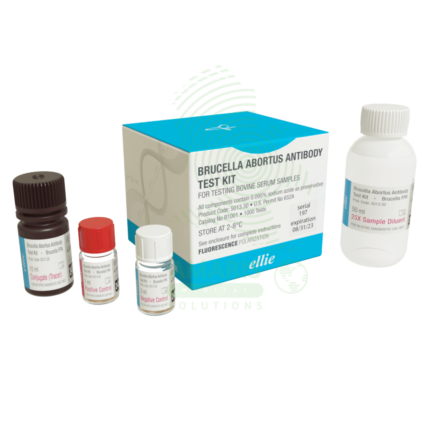
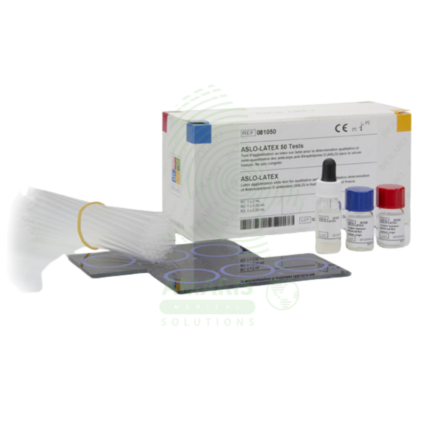
Brucella Set
A Brucella Set is a comprehensive serological test system (Class II medical device, CE-marked/FDA-cleared) for qualitative and quantitative detection of antibodies against Brucella species (B. abortus, B. melitensis, B. suis, B. canis) using multiple methodologies including tube agglutination, slide agglutination, and 2-mercaptoethanol (2-ME) testing. The set includes killed, stained Brucella antigens, positive and negative controls, dilution tubes, reaction cards, and 2-ME reagent for IgM/IgG differentiation. Diagnostic titer ≥1:160 or fourfold rise confirms brucellosis. Primary clinical applications include diagnosis of brucellosis in endemic regions (undulant fever, night sweats, arthralgia), fever of unknown origin investigation, occupational health screening in high-risk workers (veterinarians, farmers, slaughterhouse workers), diagnosis of focal complications (sacroiliitis, spondylitis, meningitis, endocarditis), treatment monitoring (fourfold decline indicates response), outbreak investigation, and public health surveillance. Critical safety precautions include handling all samples in biosafety cabinet (Brucella is highly infectious via aerosol), 2-ME reagent toxicity (use fume hood), awareness of prozone phenomenon (serial dilutions essential), cross-reactions with other Gram-negative bacteria, and mandatory reporting to public health authorities. Essential test for brucellosis diagnosis in endemic regions and occupational health settings.
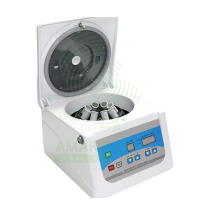
Electric Centrifuge
An Electric Centrifuge is a Class I medical device that uses rapid rotation to separate fluids of different densities, essential in clinical laboratories for processing blood, urine, and other biological specimens. Available in benchtop, refrigerated, microcentrifuge, hematocrit, and high-speed models with fixed-angle, swinging-bucket, or vertical rotors. Speed range 1,000-30,000+ RPM with RCF up to 65,000+ x g. Features include brushless induction motor, digital controls, imbalance detection, lid lock safety, and programmable protocols. Primary clinical applications include separation of serum/plasma for chemistry and hematology testing, urine sediment analysis for microscopy, platelet-rich plasma (PRP) preparation for regenerative medicine, concentration of bacteria for microbiology, cytology fluid analysis, blood banking, and molecular biology sample processing. Critical safety precautions include proper balancing of loads (weight matching), use of sealed rotors for biohazardous samples, regular rotor inspection for corrosion or cracks, never exceeding maximum rated speed, and never opening lid during operation. Essential equipment in clinical, research, and teaching laboratories.
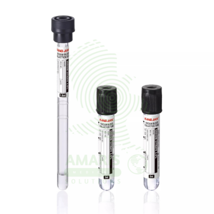
ESR Tubes Glass
ESR Tubes Glass are specialized glass tubes (300 mm length, 2.55 mm bore diameter, graduated 0-200 mm in 1 mm intervals) meeting ICSH and CLSI Westergren specifications for measuring erythrocyte sedimentation rate, a non-specific marker of inflammation. Used with citrated or diluted EDTA blood, filled to exactly 200 mm, placed vertically in Westergren stand, and read at 60 minutes. Primary clinical applications include determination of ESR for diagnosing and monitoring inflammatory conditions (infections, autoimmune disorders, malignancies), monitoring disease activity in rheumatoid arthritis, assessment of temporal arteritis and polymyalgia rheumatica, evaluation of infection and inflammatory response, screening for occult inflammatory conditions, and monitoring chronic inflammatory diseases (SLE, IBD, vasculitis). Critical safety precautions include handling glass tubes carefully to avoid breakage (sharps hazard), proper disposal in sharps containers as biohazardous waste, ensuring vertical alignment and constant temperature (18-25°C), reading at exactly 60 minutes, and avoiding air bubbles and vibration. Essential consumable for reference Westergren ESR testing in clinical laboratories.
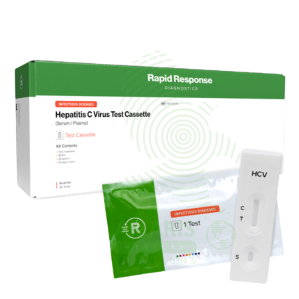
Hepatitis C Test Kit
The Hepatitis C Test Kit is an in vitro diagnostic device for the detection of Hepatitis C virus antibodies or RNA in human blood, serum, or plasma samples. Available in rapid, laboratory immunoassay, and molecular formats, it screens for HCV infection, confirms active infection with RNA testing, and monitors treatment response. With high sensitivity and specificity exceeding 99%, it is essential for identifying infected individuals, linking them to curative direct-acting antiviral therapy, and confirming cure. Essential applications include risk-based screening of high-risk populations, birth cohort screening of baby boomers, evaluation of acute hepatitis, management of occupational exposures, and monitoring of antiviral therapy that can cure over 95% of infections. All patient samples must be handled with universal precautions, and positive antibody results require confirmatory testing.
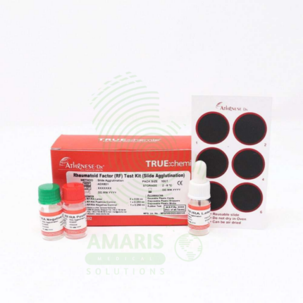
Rheumatoid factor
Rheumatoid Factor (RF) testing is a quantitative or semi-quantitative serological assay (nephelometry, turbidimetry, latex agglutination, or ELISA) for detecting autoantibodies against the Fc portion of immunoglobulin G, primarily of the IgM isotype. RF is a key marker in the ACR/EULAR classification criteria for rheumatoid arthritis, with sensitivity 60-80% and specificity 70-90%. Test requires serum samples; results reported in IU/mL (reference <15-20 IU/mL) or titer. Primary clinical applications include diagnosis and classification of rheumatoid arthritis, prognostic assessment (high-titer RF correlates with severe, erosive disease), differential diagnosis of inflammatory arthritis, monitoring disease activity, diagnosis of Sjögren's syndrome, evaluation of mixed cryoglobulinemia (hepatitis C-associated), and assessment of other autoimmune and chronic inflammatory conditions. Critical safety precautions include awareness of false positives (other autoimmune diseases, chronic infections, healthy elderly), false negatives (seronegative RA 20-30%), need for clinical correlation, and anti-CCP antibody testing to improve diagnostic accuracy. Essential test for rheumatology practice, autoimmune disease evaluation, and differential diagnosis of inflammatory arthritis.
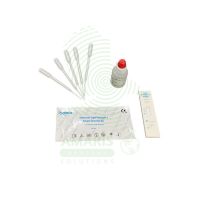
Salmonella Typhi Test Peratop
The Salmonella typhi test peratop is a CE-marked rapid immunochromatographic assay for qualitative detection of IgG and IgM antibodies to Salmonella enterica serotype Typhi in human serum, plasma, or whole blood, providing results in 15-20 minutes. The test uses recombinant S. typhi antigens immobilized on a membrane to capture specific antibodies, with separate lines for IgG and IgM interpretation. The kit includes individually foil-wrapped test cassettes, sample diluent, disposable pipettes, and package insert. Primary clinical applications include rapid diagnosis of typhoid fever in endemic regions (Indian subcontinent, Southeast Asia, Africa, South America), differentiation of acute (IgM positive) from past infection (IgG only), outbreak investigation and control, screening in febrile patients, travel medicine assessment, antibiotic stewardship, and maternal/child health in endemic areas. Critical safety precautions include proper specimen handling (universal precautions), quality control verification, awareness of window period (repeat testing if early infection suspected), and clinical correlation of results. Essential rapid test for typhoid fever diagnosis in resource-limited and endemic settings.