Dermatoscope and Magnifiers
 Diagnostic Kits
 Vital Signs Monitors
 Stethoscopes and Accessories
 Otoscopes, Ophthalmoscopes, and Retinoscopes
 Reflex Hammers and Neurological Tools
 Scales and Measuring Devices
 Spirometers and Pulmonary Function Tests
 Electrosurgical Units and Accessories
 Cutting Instruments
 Grasping and Holding Instruments
 Hemostatic Instruments
 Specialized Surgical Sets
 Single-Use Procedure Trays and Packs
 Surgical Drapes, Gowns, and Covers
 Tissue Unifying Instruments
 Radiation Protection
 X-Ray Machines and Accessories
 Ultrasound Systems and Probes
 MRI and CT Scanners
 Radiology Consumables
 Bone Densitometers
 Fluoroscopy Equipment
 Imaging Tables and Positioning Aids
 Microscopes and Accessories
 Centrifuges and Separators
 Analyzers
 Incubators and Ovens
 Pipettes, Dispensers, and Lab Glassware
 Refrigerators, Freezers, and Storage Units
 Lab Consumables
 Sterilizers and Autoclaves for Lab Use
 Multi-Parameter Monitors
 Ventilators and Respiratory Support Devices
 Defibrillators and AEDs
 Infusion Pumps and IV Systems
 Patient Warmers and Cooling Devices
 Central Monitoring Stations
 Accessories
 Anesthesia Machines and Workstations
 Oxygen Concentrators and Delivery Systems
 Nebulizers and Inhalers
 CPAP/BiPAP Machines
 Airway Management
 Anesthesia Masks, Circuits, and Bags
 Humidifiers and Heaters
 Respiratory Therapy Accessories
 First Aid Kits and Cabinets
 Emergency Resuscitation Equipment
 Trauma Supplies
 Emergency Carts and Crash Carts
 Burn Care Products
 Bleeding Control
 Automated External Defibrillators (AEDs)
 Transport and Evacuation
 Wheelchairs and Accessories
 Walkers, Crutches, and Canes
 Prosthetics and Orthotics
 Physical Therapy Equipment
 Transfer Devices
 Bathroom Safety
 Orthopedic Traction and Tables
 Hot/Cold Therapy Packs and Units
 Beds and Mattresses
 Chairs and Stools
 Tables
 Cabinets and Storage
 Privacy Screens & Curtains
 Stands and Racks
 Linens and Textiles
 Lighting
 Autoclaves and Sterilizers
 Ultrasonic Cleaners
 Disinfectant Solutions and Wipes
 Sterilization Pouches, Wraps, and Indicators
 Instrument Trays and Containers
 UV and Ozone Disinfection Devices
 Washer Disinfectors
 Wound Care
 Gloves
 Masks and Respirators
 Catheters and Tubing
 Swabs, Applicators, and Sponges
 Incontinence Products
 Personal Protective Equipment (PPE)
 Dental Chairs and Units
 Handpieces and Burs
 Instruments
 Consumables
 Sterilization for Dental Use
 Orthodontic Supplies
 Endodontic Tools
 Slit Lamps and Tonometers
 Lensometers and Phoropters
 Ophthalmic Surgical Instruments
 Eyewear Frames and Lenses
 Contact Lens Supplies
 Vision Testing Charts and Devices
 Eye Care Consumables
 Laser Systems for Eye Care
 ENT Exam Chairs and Tables
 Endoscopes
 Audiometers and Hearing Tests
 ENT Instruments
 Nasal and Throat Packs
 Hearing Aids and Accessories
 Otology Supplies
 Fetal Dopplers and Monitors
 Delivery Beds and Tables
 Gynecological Instruments
 Neonatal Incubators and Warmers
 Breast Pumps and Accessories
 Contraceptive Devices
 Maternity Supports and Pads
 Neonatal Consumables
 Cystoscopes and Urethroscopes
 Dialysis Machines and Supplies
 Urological Catheters and Bags
 Lithotripters
 Prostate Treatment Devices
 Urinary Incontinence Products
 Kidney Stone Management Tools
 Consumables & Disposables
 EEG and EMG Machines
 Neurosurgical Instruments
 Nerve Stimulators
 Headrests and Positioning Aids
 Lumbar Puncture Kits
 Seizure Monitoring Devices
 Consumables
 Rehabilitation for Neurological Conditions
 ECG Machines and Accessories
 Holter Monitors
 Stress Test Systems
 Pacemakers and Defibrillator Accessories
 Vascular Access Devices
 Cardiac Catheters and Guidewires
 Blood Flow Meters
 Consumables
 Orthopedic Instruments
 Casts, Splints, and Padding
 Joint Replacement Supplies
 Prosthetic Limbs and Components
 Bone Grafts and Substitutes
 Traction Devices
 Orthopedic Braces and Supports
 Rehabilitation Aids for Orthopedics
 Home Oxygen Therapy
 Hospital Beds for Home Use
 Mobility Aids
 Bathroom and Daily Living Aids
 Wound Care for Home
 Monitoring Devices
 Enteral Feeding Pumps and Tubes
 Hand Sanitizers and Dispensers
 Face Shields and Goggles
 Isolation Gowns and Suits
 Biohazard Waste Containers
 Air Purifiers and HEPA Filters
 Surface Disinfectants
 Sharps Containers
 Protective Barriers
 Cardiovascular & Endurance Training
 Strength Training & Weightlifting
 Functional Training & Core Conditioning
 Physical Therapy & Rehabilitation
 Sports & Outdoor Recreation
 Gym Flooring & Facility Equipment
 Fitness Monitoring & Accessories
 Kids & Novelties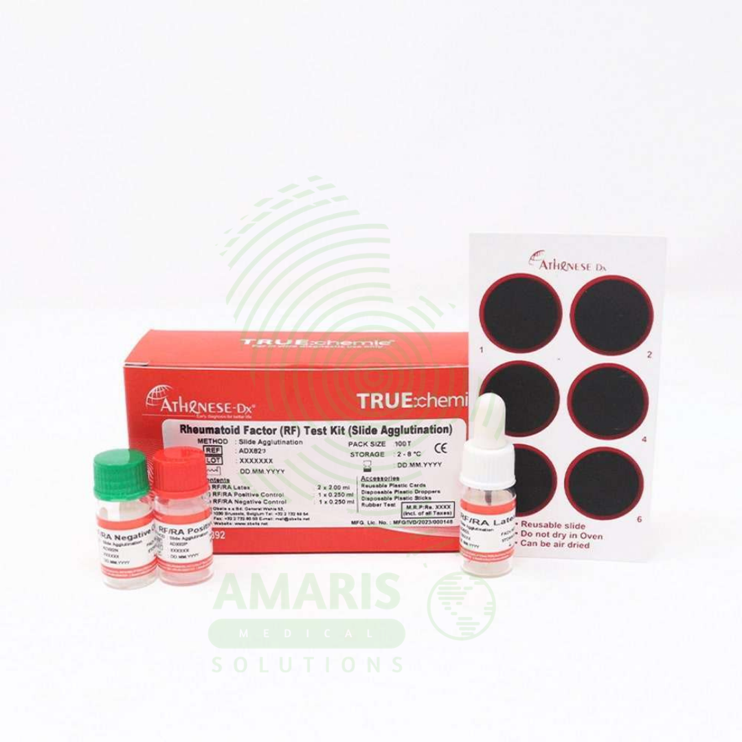
Rheumatoid factor
$1.00
WhatsApp Order
Rheumatoid Factor (RF) testing is a quantitative or semi-quantitative serological assay (nephelometry, turbidimetry, latex agglutination, or ELISA) for detecting autoantibodies against the Fc portion of immunoglobulin G, primarily of the IgM isotype. RF is a key marker in the ACR/EULAR classification criteria for rheumatoid arthritis, with sensitivity 60-80% and specificity 70-90%. Test requires serum samples; results reported in IU/mL (reference <15-20 IU/mL) or titer. Primary clinical applications include diagnosis and classification of rheumatoid arthritis, prognostic assessment (high-titer RF correlates with severe, erosive disease), differential diagnosis of inflammatory arthritis, monitoring disease activity, diagnosis of Sjögren’s syndrome, evaluation of mixed cryoglobulinemia (hepatitis C-associated), and assessment of other autoimmune and chronic inflammatory conditions. Critical safety precautions include awareness of false positives (other autoimmune diseases, chronic infections, healthy elderly), false negatives (seronegative RA 20-30%), need for clinical correlation, and anti-CCP antibody testing to improve diagnostic accuracy. Essential test for rheumatology practice, autoimmune disease evaluation, and differential diagnosis of inflammatory arthritis.
Description
Rheumatoid factor
PRIMARY CLINICAL & DIAGNOSTIC USES
1. Diagnosis and Classification of Rheumatoid Arthritis:
-
Primary Use: Rheumatoid factor (RF) testing is a key serological marker used in the classification criteria for rheumatoid arthritis (RA) established by the American College of Rheumatology (ACR) and European League Against Rheumatism (EULAR). Positive RF, particularly in combination with anti-CCP antibodies, supports the diagnosis of RA in patients presenting with inflammatory arthritis, morning stiffness, and symmetric joint involvement.
-
How it helps: Provides the objective laboratory evidence that helps rheumatologists distinguish rheumatoid arthritis from other causes of joint pain, ensuring patients receive the correct diagnosis and appropriate treatment from the start.
2. Prognostic Assessment in Rheumatoid Arthritis:
-
Primary Use: The presence and titer of RF correlate with more severe, erosive disease, extra-articular manifestations (rheumatoid nodules, vasculitis, interstitial lung disease), and poorer functional outcomes. High-titer RF helps identify patients who may benefit from more aggressive early intervention and biologic therapy.
-
How it helps: Gives rheumatologists valuable insight into how aggressive a patient’s disease is likely to be, helping them decide who needs early, intensive treatment to prevent joint damage and disability.
3. Differential Diagnosis of Inflammatory Arthritis:
-
Primary Use: RF testing helps distinguish rheumatoid arthritis from other forms of inflammatory arthritis such as psoriatic arthritis, reactive arthritis, ankylosing spondylitis, and osteoarthritis, which are typically RF-negative.
-
How it helps: Helps solve the diagnostic puzzle of inflammatory arthritis, with RF status providing a crucial clue that points toward rheumatoid arthritis versus other conditions with different treatments and prognoses.
4. Monitoring Disease Activity and Treatment Response:
-
Primary Use: Serial RF measurements can be used to monitor disease activity in some patients, though anti-CCP antibodies and acute phase reactants (ESR, CRP) are more commonly used for this purpose. Decreasing RF titers may indicate response to therapy in some cases.
-
How it helps: Offers an additional tool for tracking how well treatment is working, with falling RF levels providing objective evidence that therapy is controlling the autoimmune process.
5. Diagnosis of Sjögren’s Syndrome:
-
Primary Use: RF is frequently positive in primary Sjögren’s syndrome, an autoimmune disorder characterized by dry eyes (keratoconjunctivitis sicca) and dry mouth (xerostomia). RF testing is part of the diagnostic workup for suspected Sjögren’s syndrome.
-
How it helps: Contributes to the diagnosis of this often-overlooked condition, providing laboratory evidence that supports the diagnosis in patients suffering from debilitating dryness and fatigue.
6. Evaluation of Mixed Cryoglobulinemia:
-
Primary Use: RF is often present in mixed cryoglobulinemia, a condition associated with hepatitis C infection, where immune complexes deposit in small vessels causing vasculitis, purpura, and glomerulonephritis.
-
How it helps: Helps diagnose this serious complication of hepatitis C, identifying patients whose immune complexes are causing blood vessel inflammation that can damage skin, nerves, and kidneys.
7. Assessment of Other Autoimmune and Chronic Inflammatory Conditions:
-
Primary Use: RF may be elevated in systemic lupus erythematosus (SLE), systemic sclerosis, mixed connective tissue disease, sarcoidosis, and chronic infections such as subacute bacterial endocarditis, tuberculosis, and leprosy, though with lower specificity.
-
How it helps: Provides a clue that an autoimmune or inflammatory process is at work, even when the diagnosis is not immediately clear, guiding further diagnostic investigation.
SECONDARY & SUPPORTIVE USES
1. Screening in Unexplained Polyarthritis: Used in the initial evaluation of patients presenting with unexplained polyarthritis, arthralgias, or morning stiffness to assess for possible autoimmune etiology, helping direct the diagnostic workup.
2. Preoperative Assessment in Joint Replacement Surgery: May be ordered in patients undergoing elective joint replacement to evaluate for underlying inflammatory arthritis that could affect surgical planning and postoperative management.
3. Evaluation of Sicca Symptoms: Used in patients presenting with dry eyes and dry mouth to support the diagnosis of Sjögren’s syndrome, providing objective evidence for a condition often diagnosed based on symptoms alone.
4. Assessment of Unexplained Vasculitis or Neuropathy: RF testing may be part of the workup for unexplained vasculitis, peripheral neuropathy, or mononeuritis multiplex where autoimmune etiology is suspected, uncovering hidden autoimmune causes.
5. Research and Epidemiological Studies: Used in studies investigating prevalence, risk factors, and outcomes of rheumatoid arthritis and other autoimmune diseases, contributing to scientific understanding and better treatments.
6. Monitoring of Hepatitis C Patients: Serial RF testing may be used in hepatitis C patients to monitor for development of mixed cryoglobulinemia, catching this complication early when it is most treatable.
7. Differential Diagnosis of Interstitial Lung Disease: RF testing may be included in the evaluation of unexplained interstitial lung disease to assess for underlying connective tissue disease, identifying autoimmune causes of lung scarring.
KEY PRODUCT FEATURES
1. BASIC IDENTIFICATION ATTRIBUTES
-
Test Type: Quantitative or semi-quantitative serological test for detection of autoantibodies against the Fc portion of immunoglobulin G (IgG).
-
Common Names: Rheumatoid Factor, RF, RF Test, Rheumatoid Factor Antibodies.
-
Antibody Isotypes Detected: Primarily IgM RF; some assays detect IgA and IgG RF as well.
-
Sample Type: Serum (venous blood).
-
Methodologies:
-
Nephelometry/Turbidimetry: Automated quantitative immunoassay (most common in modern laboratories).
-
Latex Agglutination: Semi-quantitative, rapid screening test.
-
ELISA: Enzyme-linked immunosorbent assay for quantitative measurement and isotype-specific detection.
-
Waaler-Rose Test: Traditional sheep cell agglutination assay (historical).
-
-
Reporting Units: IU/mL (international units per milliliter) or titer (e.g., 1:40, 1:80, 1:160).
-
Reference Range: Varies by laboratory and methodology; typically <15-20 IU/mL is considered negative.
2. TECHNICAL & PERFORMANCE PROPERTIES
-
Sensitivity for Rheumatoid Arthritis: 60-80% (higher in established disease, lower in early RA).
-
Specificity for Rheumatoid Arthritis: 70-90% (false positives occur in other autoimmune diseases, chronic infections, and healthy elderly individuals).
-
Positive Predictive Value: Higher with high-titer RF and when combined with anti-CCP antibodies.
-
Negative Predictive Value: A negative RF does not rule out RA (seronegative RA comprises 20-30% of cases).
-
Detection Limit: Varies by method; nephelometry typically detects 5-10 IU/mL.
-
Cross-Reactivity: May occur with other autoantibodies; false positives in liver disease, chronic infections, and inflammatory conditions.
-
Prozone Effect: Very high RF concentrations may cause false-negative results in agglutination assays (rare).
3. PHYSICAL & OPERATIONAL PROPERTIES
-
Kit Components (Latex Agglutination Method):
-
Latex Reagent: Polystyrene latex particles coated with human IgG.
-
Positive Control: Serum with known RF titer.
-
Negative Control: Non-reactive serum.
-
Dilution Buffer: For sample preparation.
-
Reaction Cards: Black glass or plastic cards for agglutination reading.
-
Mixing Sticks: Disposable sticks for sample-reagent mixing.
-
-
Kit Components (Nephelometry/Turbidimetry):
-
Reagent 1: Buffer/diluent.
-
Reagent 2: Latex particles coated with human IgG.
-
Calibrators: Set of known RF concentrations.
-
Controls: Commercial controls at various levels.
-
-
Kit Components (ELISA):
-
Microplate: Coated with human IgG.
-
Conjugate: Enzyme-labeled anti-human IgM (or other isotype).
-
Substrate: Chromogenic substrate for color development.
-
Standards and Controls: For quantitation and quality control.
-
-
Packaging Sizes: 50-200 tests per kit depending on methodology.
-
Storage Requirements: 2-8°C; do not freeze.
-
Shelf Life: 12-24 months depending on manufacturer.
4. SAFETY & COMPLIANCE ATTRIBUTES
-
Regulatory Status: Class II medical device requiring FDA 510(k) clearance; CE marked for IVD use.
-
Quality Control: Must run positive and negative controls with each batch; calibration curves for quantitative methods.
-
Biohazard Precautions: All patient samples potentially infectious for HIV, hepatitis B/C, and other bloodborne pathogens.
-
Chemical Safety: Reagents contain preservatives (sodium azide); avoid contact with eyes and skin.
-
Disposal: Used test materials disposed as biohazardous waste.
-
Reporting: Results reported to the ordering physician; positive results interpreted in clinical context.
5. STORAGE & HANDLING ATTRIBUTES
-
Storage Temperature: 2-8°C; do not freeze; protect from light.
-
Sample Handling: Serum stable 7 days at 2-8°C; longer storage requires freezing at -20°C (avoid repeated freeze-thaw).
-
Reagent Preparation: Bring reagents to room temperature before use; mix gently; avoid foam.
-
Expiration: Never use reagents beyond expiration date; performance compromised.
-
Quality Control: Run controls with each batch; document results per laboratory protocol.
-
Disposal: Dispose of contaminated materials in biohazard containers.
6. LABORATORY & CLINICAL APPLICATIONS
-
Primary Application: Detection and quantitation of rheumatoid factor autoantibodies for diagnosis, classification, and prognostic assessment of rheumatoid arthritis and other autoimmune conditions.
-
Testing Indications:
-
Suspected rheumatoid arthritis (inflammatory arthritis, morning stiffness, joint swelling).
-
Classification per ACR/EULAR criteria (requires RF and/or anti-CCP testing).
-
Unexplained polyarthritis or arthralgias.
-
Suspected Sjögren's syndrome (with sicca symptoms).
-
Evaluation of mixed cryoglobulinemia (especially with hepatitis C).
-
Unexplained vasculitis, neuropathy, or interstitial lung disease.
-
-
Interpretation:
-
Negative (<15-20 IU/mL): Does not rule out RA; consider anti-CCP testing.
-
Low-Positive (20-80 IU/mL): May be seen in RA, other autoimmune diseases, infections, or elderly.
-
High-Positive (>80 IU/mL): Strongly suggestive of RA, especially with compatible clinical presentation.
-
False Positives: Chronic infections (endocarditis, TB, hepatitis C), other autoimmune diseases (SLE, Sjögren's, sarcoidosis), aging.
-
-
Limitations: Not specific for RA; 3-5% of healthy individuals (especially elderly) may be positive; 20-30% of RA patients are seronegative.
SAFETY HANDLING PRECAUTIONS
1. SAFETY PRECAUTIONS
-
Biological Hazard: All serum samples potentially infectious; wear gloves and appropriate PPE.
-
Hand Hygiene: Wash hands thoroughly after handling specimens and reagents.
-
Quality Control: Always run controls with each batch; invalid if controls fail.
-
Clinical Correlation: RF alone is not diagnostic; must be interpreted with clinical presentation, anti-CCP antibodies, and other laboratory findings.
-
Age-Related Positivity: Be aware that low-titer RF can occur in healthy elderly individuals without autoimmune disease.
-
False Negatives: Seronegative RA occurs in 20-30% of cases; anti-CCP testing improves diagnostic yield.
-
Isotype-Specific Testing: IgM RF is most common; IgA and IgG RF may provide additional information in some cases.
2. FIRST AID MEASURES
-
Needlestick/Sharps Injury: If injury occurs during blood collection, wash area vigorously with soap and water; report immediately; follow institutional post-exposure prophylaxis protocol.
-
Blood Spill: Contain with absorbent material; disinfect area with 10% bleach solution; dispose as biohazard waste.
-
Reagent Contact (Eyes): Flush eyes with copious water for 15 minutes; seek medical attention.
-
Reagent Ingestion: Contact poison control center; do not induce vomiting unless instructed.
3. FIRE FIGHTING MEASURES
-
Flammability: Liquid reagents contain preservatives; plastic components combustible.
-
Extinguishing Media: Use water, foam, CO₂, or dry chemical as appropriate.
-
Sodium Azide Hazard: Reagents may contain sodium azide; may react with lead/copper plumbing to form explosive azides; flush with copious water when disposing.
Related products
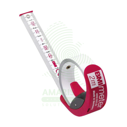
BMI Tape
A BMI Tape is a rapid screening tool that estimates an individual's Body Mass Index (BMI) category by measuring body circumference, most commonly the waist. It features a color-coded scale that directly displays BMI ranges (Underweight, Normal, Overweight, Obese), eliminating the need for separate height and weight measurements and manual calculation. While highly useful for community health screenings, wellness programs, and patient education due to its portability and simplicity, it is an approximate method. Its accuracy is limited compared to BMI calculated from precise clinical measurements, and it should be used as an initial screening indicator rather than a definitive diagnostic tool.
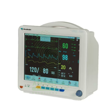
ECG Machine
An ECG Machine is a Class II medical device that records and displays the electrical activity of the heart through surface electrodes, producing an electrocardiogram for diagnosis of cardiac conditions. Standard diagnostic machines record 12 simultaneous leads (3 limb, 6 precordial, 1 ground) with frequency response 0.05-150 Hz, sampling rate 500-1,000 Hz, and high-resolution (5-10 µV) signal acquisition. Features include color touchscreen display, thermal array printer, computerized interpretation algorithms, internal memory (50-500+ ECGs), and network connectivity for EMR integration. Lead wires (AHA or IEC color coding) connect to disposable adhesive electrodes. Portable models (5-15 kg) with rechargeable batteries enable bedside and mobile use; cart-mounted units provide full diagnostic capability. Primary clinical applications include diagnosis of arrhythmias (AF, VT, bradycardia), detection of myocardial ischemia/infarction (STEMI, NSTEMI), evaluation of chest pain, preoperative cardiac risk assessment, monitoring electrolyte imbalances, assessment of chamber enlargement, and drug effect/toxicity monitoring. Essential diagnostic equipment in emergency departments, cardiology clinics, ICUs, operating rooms, and primary care settings worldwide.
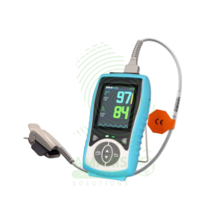
Handheld Pulse Oximeter
A Handheld Pulse Oximeter is a professional-grade, portable medical device used by healthcare providers for accurate and reliable monitoring of blood oxygen saturation (SpO2) and pulse rate. It consists of a durable, feature-rich monitor unit and separate, interchangeable sensors for use on fingers, toes, or ears. Superior to basic fingertip models, it offers advanced functions like configurable alarms, detailed plethysmograph display, trend memory, and data connectivity. Its robust design, clinical accuracy under challenging conditions (motion, low perfusion), and versatility make it an essential tool in hospitals, clinics, ambulances, and home health for patient assessment and monitoring.
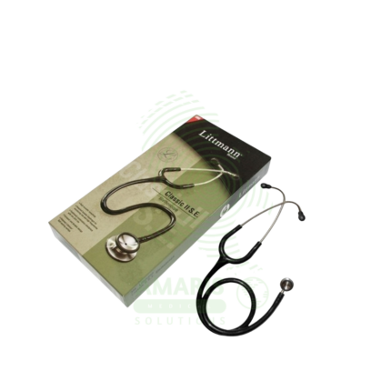
Littmann Stethoscope (Classic II)
A Littmann Stethoscope (Classic II) is a premium, acoustic dual-head stethoscope renowned for its diagnostic clarity, durability, and innovative tunable diaphragm technology. It allows healthcare professionals to hear both high and low-frequency sounds by simply adjusting pressure on the single-sided chestpiece, eliminating the need to rotate it. With its superior acoustics, comfortable fit, and wide range of colors, it is the instrument of choice for medical students, nurses, and physicians across all specialties who require reliable performance for physical assessment, auscultation of heart and lung sounds, and manual blood pressure measurement.
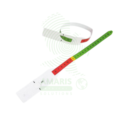
MUAC Tape
A MUAC Tape (Mid-Upper Arm Circumference) is a specialized, color-coded measuring tape used globally as the primary field tool for screening children aged 6-59 months for acute malnutrition. Its simple design—featuring red, yellow, and green zones corresponding to Severe Acute Malnutrition, Moderate Acute Malnutrition, and normal nutritional status—allows for instant, non-invasive assessment without the need for scales, calculation, or literacy. Durable, portable, and inexpensive, it is the cornerstone of community-based nutrition programs, emergency relief efforts, and public health surveillance, enabling early detection and life-saving intervention for malnourished children in even the most resource-constrained settings.
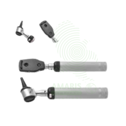
Ophthalmoscope
An Ophthalmoscope is a handheld diagnostic instrument used to examine the interior of the eye, specifically the fundus (retina, optic disc, blood vessels, and macula). By providing illuminated magnification, it serves as a vital screening tool for detecting ocular diseases like glaucoma and retinal disorders, and more importantly, for identifying signs of systemic conditions such as diabetes, hypertension, and increased intracranial pressure. Its operation involves selecting appropriate apertures and compensating lenses to obtain a clear view. While offering a detailed but narrow field of vision, mastery of the direct ophthalmoscope remains a cornerstone skill in general medicine, neurology, and ophthalmology for assessing vascular and neurological health.
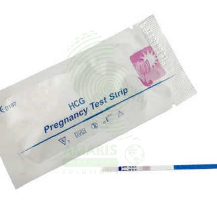
Pregnancy Test Strips
Pregnancy Test Strips are simple, dip-style lateral flow devices designed for the rapid, qualitative detection of human chorionic gonadotropin (hCG) in urine to indicate pregnancy. As the most fundamental format of home pregnancy tests, they offer high sensitivity (often from 10 mIU/mL), affordability, and a quick result within 3-5 minutes. Their ease of use makes them a widely accessible first step in pregnancy detection for personal use and clinical screening. A visible test line, even if faint, is a positive result, though confirmation with a healthcare provider is always recommended.