Dermatoscope and Magnifiers
 Diagnostic Kits
 Vital Signs Monitors
 Stethoscopes and Accessories
 Otoscopes, Ophthalmoscopes, and Retinoscopes
 Reflex Hammers and Neurological Tools
 Scales and Measuring Devices
 Spirometers and Pulmonary Function Tests
 Electrosurgical Units and Accessories
 Cutting Instruments
 Grasping and Holding Instruments
 Hemostatic Instruments
 Specialized Surgical Sets
 Single-Use Procedure Trays and Packs
 Surgical Drapes, Gowns, and Covers
 Tissue Unifying Instruments
 Radiation Protection
 X-Ray Machines and Accessories
 Ultrasound Systems and Probes
 MRI and CT Scanners
 Radiology Consumables
 Bone Densitometers
 Fluoroscopy Equipment
 Imaging Tables and Positioning Aids
 Microscopes and Accessories
 Centrifuges and Separators
 Analyzers
 Incubators and Ovens
 Pipettes, Dispensers, and Lab Glassware
 Refrigerators, Freezers, and Storage Units
 Lab Consumables
 Sterilizers and Autoclaves for Lab Use
 Multi-Parameter Monitors
 Ventilators and Respiratory Support Devices
 Defibrillators and AEDs
 Infusion Pumps and IV Systems
 Patient Warmers and Cooling Devices
 Central Monitoring Stations
 Accessories
 Anesthesia Machines and Workstations
 Oxygen Concentrators and Delivery Systems
 Nebulizers and Inhalers
 CPAP/BiPAP Machines
 Airway Management
 Anesthesia Masks, Circuits, and Bags
 Humidifiers and Heaters
 Respiratory Therapy Accessories
 First Aid Kits and Cabinets
 Emergency Resuscitation Equipment
 Trauma Supplies
 Emergency Carts and Crash Carts
 Burn Care Products
 Bleeding Control
 Automated External Defibrillators (AEDs)
 Transport and Evacuation
 Wheelchairs and Accessories
 Walkers, Crutches, and Canes
 Prosthetics and Orthotics
 Physical Therapy Equipment
 Transfer Devices
 Bathroom Safety
 Orthopedic Traction and Tables
 Hot/Cold Therapy Packs and Units
 Beds and Mattresses
 Chairs and Stools
 Tables
 Cabinets and Storage
 Privacy Screens & Curtains
 Stands and Racks
 Linens and Textiles
 Lighting
 Autoclaves and Sterilizers
 Ultrasonic Cleaners
 Disinfectant Solutions and Wipes
 Sterilization Pouches, Wraps, and Indicators
 Instrument Trays and Containers
 UV and Ozone Disinfection Devices
 Washer Disinfectors
 Wound Care
 Gloves
 Masks and Respirators
 Catheters and Tubing
 Swabs, Applicators, and Sponges
 Incontinence Products
 Personal Protective Equipment (PPE)
 Dental Chairs and Units
 Handpieces and Burs
 Instruments
 Consumables
 Sterilization for Dental Use
 Orthodontic Supplies
 Endodontic Tools
 Slit Lamps and Tonometers
 Lensometers and Phoropters
 Ophthalmic Surgical Instruments
 Eyewear Frames and Lenses
 Contact Lens Supplies
 Vision Testing Charts and Devices
 Eye Care Consumables
 Laser Systems for Eye Care
 ENT Exam Chairs and Tables
 Endoscopes
 Audiometers and Hearing Tests
 ENT Instruments
 Nasal and Throat Packs
 Hearing Aids and Accessories
 Otology Supplies
 Fetal Dopplers and Monitors
 Delivery Beds and Tables
 Gynecological Instruments
 Neonatal Incubators and Warmers
 Breast Pumps and Accessories
 Contraceptive Devices
 Maternity Supports and Pads
 Neonatal Consumables
 Cystoscopes and Urethroscopes
 Dialysis Machines and Supplies
 Urological Catheters and Bags
 Lithotripters
 Prostate Treatment Devices
 Urinary Incontinence Products
 Kidney Stone Management Tools
 Consumables & Disposables
 EEG and EMG Machines
 Neurosurgical Instruments
 Nerve Stimulators
 Headrests and Positioning Aids
 Lumbar Puncture Kits
 Seizure Monitoring Devices
 Consumables
 Rehabilitation for Neurological Conditions
 ECG Machines and Accessories
 Holter Monitors
 Stress Test Systems
 Pacemakers and Defibrillator Accessories
 Vascular Access Devices
 Cardiac Catheters and Guidewires
 Blood Flow Meters
 Consumables
 Orthopedic Instruments
 Casts, Splints, and Padding
 Joint Replacement Supplies
 Prosthetic Limbs and Components
 Bone Grafts and Substitutes
 Traction Devices
 Orthopedic Braces and Supports
 Rehabilitation Aids for Orthopedics
 Home Oxygen Therapy
 Hospital Beds for Home Use
 Mobility Aids
 Bathroom and Daily Living Aids
 Wound Care for Home
 Monitoring Devices
 Enteral Feeding Pumps and Tubes
 Hand Sanitizers and Dispensers
 Face Shields and Goggles
 Isolation Gowns and Suits
 Biohazard Waste Containers
 Air Purifiers and HEPA Filters
 Surface Disinfectants
 Sharps Containers
 Protective Barriers
 Cardiovascular & Endurance Training
 Strength Training & Weightlifting
 Functional Training & Core Conditioning
 Physical Therapy & Rehabilitation
 Sports & Outdoor Recreation
 Gym Flooring & Facility Equipment
 Fitness Monitoring & Accessories
 Kids & Novelties
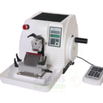
Cryostat Microtome
WhatsApp Order
A Cryostat Microtome is a refrigerated instrument used to rapidly freeze and section tissue specimens for intraoperative frozen section diagnosis, immunofluorescence studies, and research applications. Essential in pathology departments, it enables rapid microscopic examination of tissue during surgery, allowing surgeons to determine margin status and guide resection. The cryostat preserves tissue antigenicity for immunohistochemistry and molecular testing, supporting comprehensive diagnostic and research capabilities.
Description
Cryostat Microtome
PRIMARY CLINICAL & DIAGNOSTIC USES
1. Frozen Section Preparation for Intraoperative Pathology
-
Primary Use: A cryostat is a refrigerated microtome used to rapidly freeze and section tissue specimens for immediate microscopic examination during surgical procedures. Frozen sections provide rapid pathological diagnosis while the patient remains in the operating room, guiding surgical decisions such as margin assessment, lymph node evaluation, and tumor identification.
-
How it helps: For the pathologist and surgeon, the cryostat enables rapid intraoperative diagnosis—allowing the surgical team to determine if margins are clear, if lymph nodes contain metastatic disease, or if a lesion is malignant while the patient is still on the operating table. For the patient, this means definitive surgical treatment can be completed in a single procedure, reducing the need for second surgeries and improving outcomes.
2. Rapid Tissue Sectioning for Histological Analysis
-
Primary Use: Produces thin sections of frozen tissue (typically 4-10 microns) that are mounted on slides, stained, and examined under a microscope. The cryostat allows for rapid processing without the time-consuming fixation, embedding, and paraffin processing required for permanent sections.
-
How it helps: For the histotechnologist and pathologist, the cryostat enables rapid tissue sectioning that preserves antigenicity for immunohistochemistry and molecular testing—expanding the diagnostic capabilities beyond routine histology. For the patient, rapid sectioning means faster diagnosis and quicker initiation of treatment.
3. Frozen Section for Margin Assessment
-
Primary Use: Used extensively in oncologic surgery to assess surgical margins during tumor resection. The pathologist examines frozen sections of the resected specimen’s margins to determine if tumor cells extend to the edge of the resected tissue, guiding the surgeon to remove additional tissue if needed.
-
How it helps: For the surgical oncologist, intraoperative margin assessment ensures complete tumor resection—reducing the need for re-excision surgeries and improving local control rates. For the patient, this means a single surgery with clear margins offers the best chance for cure without the need for additional procedures.
4. Immunofluorescence and Enzyme Histochemistry
-
Primary Use: Cryostat sections preserve antigenicity, making them ideal for immunofluorescence studies in autoimmune kidney disease, skin diseases, and other conditions requiring antibody-based detection of proteins. Fresh frozen tissue sections are used for direct and indirect immunofluorescence.
-
How it helps: For the pathologist and immunopathologist, cryostat sections enable immunofluorescence testing for autoimmune diseases such as lupus nephritis, pemphigus, and Goodpasture syndrome—providing definitive diagnosis and guiding immunosuppressive therapy. For the patient, accurate immunofluorescence diagnosis enables targeted treatment and improved outcomes.
5. Research Applications in Neuroscience and Pharmacology
-
Primary Use: Cryostats are used in research laboratories to section fresh frozen brain tissue, tumors, and other specimens for histological, immunohistochemical, and molecular studies. The ability to section frozen tissue preserves RNA and protein integrity for advanced molecular analysis.
-
How it helps: For the research scientist, the cryostat enables high-quality sectioning of fresh frozen tissue for neuroscience research, cancer biology, and drug development—preserving delicate structures and molecular integrity for downstream applications. For future patients, research using cryostat sections contributes to understanding disease mechanisms and developing new therapies.
SECONDARY & SUPPORTIVE USES
1. Enzyme Histochemistry: Sectioning for enzyme localization studies in tissue.
2. In Situ Hybridization: Sectioning for RNA and DNA detection in tissue.
3. Lipid and Fat Staining: Frozen sections preserve lipid content for fat staining studies.
4. Neuropathology: Sectioning of brain tissue for rapid diagnosis of neurodegenerative diseases.
5. Pediatric Pathology: Frozen section diagnosis in pediatric surgical cases.
6. Veterinary Pathology: Cryostat use in veterinary diagnostic and research settings.
KEY PRODUCT FEATURES
1. BASIC IDENTIFICATION ATTRIBUTES
-
Device Type: A refrigerated microtome for rapid freezing and sectioning of tissue specimens.
-
Designation: Cryostat Microtome, Cryostat, Frozen Section Microtome, Cryotome.
-
Key Components:
-
Refrigerated Chamber: Maintains temperature between -20°C and -40°C.
-
Microtome: Precision cutting mechanism with adjustable thickness.
-
Specimen Holder (Chuck): Clamp for mounting frozen tissue blocks.
-
Anti-Roll Guide: Device to prevent section curling during cutting.
-
Control Panel: Temperature, section thickness, and cutting speed controls.
-
Specimen Cooling: Rapid freeze station for tissue embedding.
-
Vacuum System: For section collection and cleanup.
-
2. TECHNICAL & PERFORMANCE PROPERTIES
-
Temperature Range: -20°C to -40°C (chamber); -50°C for specimen cooling.
-
Section Thickness: 1-100 microns adjustable.
-
Specimen Size: Up to 40 x 50 mm typical.
-
Cutting Speed: Variable; 1-10 mm/s.
-
Defrost Cycle: Automatic or manual defrost.
-
Refrigerant: Environmentally compatible refrigerants.
-
Temperature Stability: ±0.5°C typical.
3. PHYSICAL & OPERATIONAL PROPERTIES
-
Construction: Stainless steel chamber; durable exterior.
-
Dimensions: Benchtop or freestanding units.
-
Controls: Digital interface with temperature monitoring.
-
Portability: Stationary installation.
-
Cleaning: Manual cleaning; UV disinfection options.
4. SAFETY & COMPLIANCE ATTRIBUTES
-
Regulatory Status: Class I or Class II medical device.
-
Electrical Safety: Compliant with electrical safety standards.
-
Refrigerant Safety: Compliant with environmental regulations.
-
Sharp Instrument: Microtome blade requires careful handling.
5. STORAGE & HANDLING ATTRIBUTES
-
Storage: Stored in laboratory or pathology suite.
-
Cleaning: Regular cleaning of chamber; decontamination protocols.
-
Maintenance: Regular blade replacement; temperature calibration.
-
Defrosting: Regular defrost cycles to prevent ice buildup.
6. LABORATORY & CLINICAL APPLICATIONS
-
Primary Application: Frozen section preparation for intraoperative pathology, immunofluorescence, and research.
-
Clinical Role: Essential equipment in pathology departments and research laboratories.
SAFETY HANDLING PRECAUTIONS
1. SAFETY PRECAUTIONS
-
Blade Handling: Use extreme caution when handling microtome blades; use blade remover; dispose in sharps container.
-
Cold Temperature: Use appropriate gloves when handling specimens and chamber components.
-
Electrical Safety: Ensure proper grounding; avoid water near electrical components.
-
Cleaning: Decontaminate after processing infectious specimens.
2. FIRST AID MEASURES
-
Blade Cut: If cut occurs, clean wound; apply pressure; seek medical attention if needed.
-
Cold Injury: If cold injury occurs, warm area gradually; seek medical attention if tissue damage.
-
Chemical Exposure: If cryogen or fixative contacts skin or eyes, rinse thoroughly; seek medical attention if irritation persists.
3. FIRE FIGHTING MEASURES
-
Flammability: Electrical components and refrigerants may pose fire risk.
-
Extinguishing Media: For electrical fire, use CO₂ or dry chemical extinguisher.
Related products
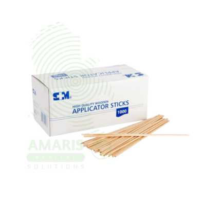
Applicator Sticks
Applicator Sticks are single-use medical devices with absorbent tips (cotton, rayon, flocked polyester) on wood, plastic, or paper shafts for collecting clinical specimens (throat, nasal, wound, cervical, urethral, rectal) for microbiological culture, rapid testing, and molecular diagnostics. Sterile individually wrapped or non-sterile bulk formats. Flocked swabs provide >90% specimen release for PCR testing. Primary applications include specimen collection for infection diagnosis, wound care and antiseptic application, gynecological sampling (Pap smears, STI testing), eye/ear specimen collection, laboratory slide preparation, and forensic evidence collection. Critical precautions include single-use only, sterile technique, proper collection method, and biohazard disposal. Essential consumable in all clinical settings.
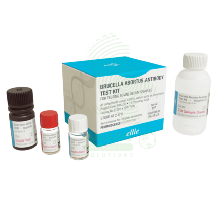
Brucella Set
A Brucella Set is a comprehensive serological test system (Class II medical device, CE-marked/FDA-cleared) for qualitative and quantitative detection of antibodies against Brucella species (B. abortus, B. melitensis, B. suis, B. canis) using multiple methodologies including tube agglutination, slide agglutination, and 2-mercaptoethanol (2-ME) testing. The set includes killed, stained Brucella antigens, positive and negative controls, dilution tubes, reaction cards, and 2-ME reagent for IgM/IgG differentiation. Diagnostic titer ≥1:160 or fourfold rise confirms brucellosis. Primary clinical applications include diagnosis of brucellosis in endemic regions (undulant fever, night sweats, arthralgia), fever of unknown origin investigation, occupational health screening in high-risk workers (veterinarians, farmers, slaughterhouse workers), diagnosis of focal complications (sacroiliitis, spondylitis, meningitis, endocarditis), treatment monitoring (fourfold decline indicates response), outbreak investigation, and public health surveillance. Critical safety precautions include handling all samples in biosafety cabinet (Brucella is highly infectious via aerosol), 2-ME reagent toxicity (use fume hood), awareness of prozone phenomenon (serial dilutions essential), cross-reactions with other Gram-negative bacteria, and mandatory reporting to public health authorities. Essential test for brucellosis diagnosis in endemic regions and occupational health settings.
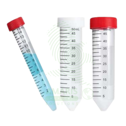
Centrifuge Tubes
Centrifuge Tubes are specialized disposable containers (polypropylene) designed to withstand high centrifugal forces (up to 30,000 x g) for separating biological fluids based on density. Available in microcentrifuge sizes (0.2-2.0 mL), conical tubes (15 mL, 50 mL), and larger volumes (100-500 mL). Features include leak-proof screw caps with O-rings (conical tubes), snap caps (microcentrifuge tubes), graduated volume markings, writing areas, and sterile/non-sterile options. Certified RNase/DNase-free, pyrogen-free, and autoclavable. Primary clinical applications include blood component separation (serum/plasma), urine sediment preparation, microbiology sample concentration, molecular biology (DNA/RNA extraction), cell culture processing, protein/enzyme analysis, and viral load testing. Critical safety precautions include never exceeding maximum RCF rating, proper balancing by weight, secure cap tightening, inspection for cracks, and disposal as biohazardous waste. Essential consumable in every clinical, research, and industrial laboratory.
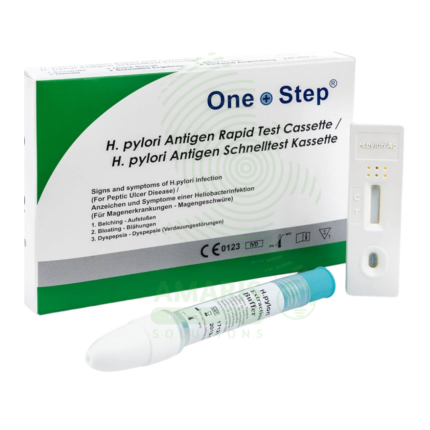
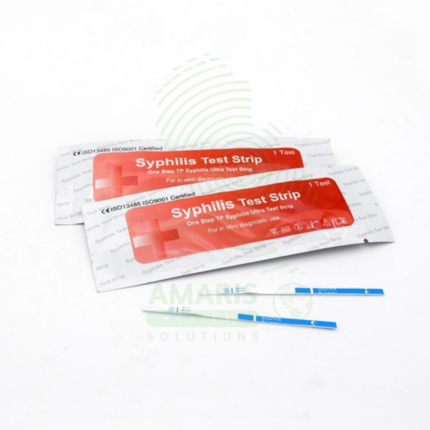
H.pylori test kits (Antigen)
H.pylori test kits (Antigen) are Class II medical devices (FDA-cleared/CE-marked) for rapid immunochromatographic or enzyme immunoassay detection of Helicobacter pylori antigens in stool specimens, enabling non-invasive diagnosis of active infection and post-treatment eradication confirmation. Test principles use monoclonal or polyclonal antibodies specific to H. pylori antigens, producing visible results in 10-20 minutes (rapid tests) or 1-2 hours (EIA). Sensitivity 90-98%, specificity 92-99% compared to gold standard methods. Primary clinical applications include rapid diagnosis of H. pylori infection in patients with dyspepsia, initial screening in uncomplicated dyspepsia (test-and-treat strategy), confirmation of active infection before therapy, post-treatment eradication confirmation (4-6 weeks after therapy), pediatric testing (preferred non-invasive method), screening in high-risk populations (family history of gastric cancer), and monitoring in patients with peptic ulcer disease or MALT lymphoma. Critical safety precautions include mandatory washout period (4 weeks off antibiotics, 2 weeks off PPIs) to avoid false negatives, proper specimen handling (universal precautions), refrigerated storage of kits (2-30°C), quality control verification, and disposal as biohazardous waste. Essential non-invasive tool for H. pylori management in gastroenterology and primary care.
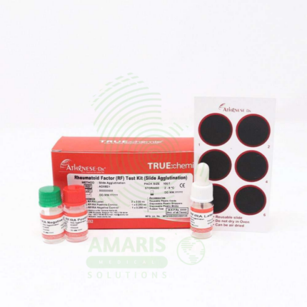
Rheumatoid factor
Rheumatoid Factor (RF) testing is a quantitative or semi-quantitative serological assay (nephelometry, turbidimetry, latex agglutination, or ELISA) for detecting autoantibodies against the Fc portion of immunoglobulin G, primarily of the IgM isotype. RF is a key marker in the ACR/EULAR classification criteria for rheumatoid arthritis, with sensitivity 60-80% and specificity 70-90%. Test requires serum samples; results reported in IU/mL (reference <15-20 IU/mL) or titer. Primary clinical applications include diagnosis and classification of rheumatoid arthritis, prognostic assessment (high-titer RF correlates with severe, erosive disease), differential diagnosis of inflammatory arthritis, monitoring disease activity, diagnosis of Sjögren's syndrome, evaluation of mixed cryoglobulinemia (hepatitis C-associated), and assessment of other autoimmune and chronic inflammatory conditions. Critical safety precautions include awareness of false positives (other autoimmune diseases, chronic infections, healthy elderly), false negatives (seronegative RA 20-30%), need for clinical correlation, and anti-CCP antibody testing to improve diagnostic accuracy. Essential test for rheumatology practice, autoimmune disease evaluation, and differential diagnosis of inflammatory arthritis.
Rota virus
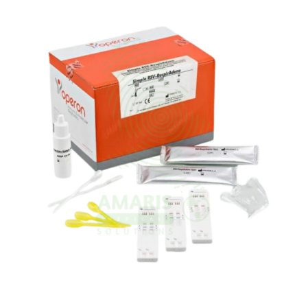
Rota virus rapid tests are immunochromatographic assays (CE-marked, FDA-cleared for some brands) for qualitative detection of rotavirus group A antigens in stool specimens, providing results in 10-20 minutes. The test principle uses monoclonal or polyclonal antibodies against rotavirus antigens (typically VP6 protein) immobilized on membranes, capturing antigens that form a visible line. Sensitivity 90-98%, specificity 95-99% compared to ELISA or PCR. The kit includes individually foil-wrapped test devices, sample diluent, and collection applicators. Primary clinical applications include rapid diagnosis of acute gastroenteritis in children (rotavirus is the leading cause of severe diarrhea in under-5 children), outbreak investigation in daycare centers and schools, hospital infection control and isolation precautions, differential diagnosis of acute diarrhea (viral vs. bacterial vs. parasitic), vaccine effectiveness monitoring and surveillance, assessment of dehydration risk, and evaluation of immunocompromised patients. Critical safety precautions include proper specimen collection (clean container, avoid contamination), biological hazard precautions (all stool potentially infectious), quality control verification, and awareness that a negative test does not rule out other causes of gastroenteritis. Essential tool for pediatric gastroenteritis management, infection control, and public health surveillance.
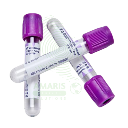
Vacutainer (EDTA/PLAIN)
Vacutainer (EDTA/PLAIN) is a sterile, evacuated blood collection tube (lavender/purple top) containing EDTA anticoagulant (K2EDTA or K3EDTA, 1.5-2.0 mg/mL blood) for hematology, blood banking, flow cytometry, and molecular diagnostics. Available in draw volumes 2-10 mL, glass or PET plastic, with color-coded stopper and sterile interior. EDTA chelates calcium, preventing coagulation while preserving cellular morphology. Primary clinical applications include complete blood count (CBC) and differential, blood typing and antibody screening (ABO/Rh, Coombs), hemoglobin A1c (HbA1c) testing, flow cytometry and immunophenotyping (CD4 counts), molecular diagnostics and PCR testing, sickle cell screening and hemoglobin electrophoresis, and parasitology (malaria smears). Critical safety precautions include filling to completion (correct blood-to-additive ratio), gentle inversion 8-10 times immediately after collection, never using for chemistry or coagulation testing, and proper disposal as biohazardous waste. Essential collection tube for hematology and immunohematology.