Dermatoscope and Magnifiers
 Diagnostic Kits
 Vital Signs Monitors
 Stethoscopes and Accessories
 Otoscopes, Ophthalmoscopes, and Retinoscopes
 Reflex Hammers and Neurological Tools
 Scales and Measuring Devices
 Spirometers and Pulmonary Function Tests
 Electrosurgical Units and Accessories
 Cutting Instruments
 Grasping and Holding Instruments
 Hemostatic Instruments
 Specialized Surgical Sets
 Single-Use Procedure Trays and Packs
 Surgical Drapes, Gowns, and Covers
 Tissue Unifying Instruments
 Radiation Protection
 X-Ray Machines and Accessories
 Ultrasound Systems and Probes
 MRI and CT Scanners
 Radiology Consumables
 Bone Densitometers
 Fluoroscopy Equipment
 Imaging Tables and Positioning Aids
 Microscopes and Accessories
 Centrifuges and Separators
 Analyzers
 Incubators and Ovens
 Pipettes, Dispensers, and Lab Glassware
 Refrigerators, Freezers, and Storage Units
 Lab Consumables
 Sterilizers and Autoclaves for Lab Use
 Multi-Parameter Monitors
 Ventilators and Respiratory Support Devices
 Defibrillators and AEDs
 Infusion Pumps and IV Systems
 Patient Warmers and Cooling Devices
 Central Monitoring Stations
 Accessories
 Anesthesia Machines and Workstations
 Oxygen Concentrators and Delivery Systems
 Nebulizers and Inhalers
 CPAP/BiPAP Machines
 Airway Management
 Anesthesia Masks, Circuits, and Bags
 Humidifiers and Heaters
 Respiratory Therapy Accessories
 First Aid Kits and Cabinets
 Emergency Resuscitation Equipment
 Trauma Supplies
 Emergency Carts and Crash Carts
 Burn Care Products
 Bleeding Control
 Automated External Defibrillators (AEDs)
 Transport and Evacuation
 Wheelchairs and Accessories
 Walkers, Crutches, and Canes
 Prosthetics and Orthotics
 Physical Therapy Equipment
 Transfer Devices
 Bathroom Safety
 Orthopedic Traction and Tables
 Hot/Cold Therapy Packs and Units
 Beds and Mattresses
 Chairs and Stools
 Tables
 Cabinets and Storage
 Privacy Screens & Curtains
 Stands and Racks
 Linens and Textiles
 Lighting
 Autoclaves and Sterilizers
 Ultrasonic Cleaners
 Disinfectant Solutions and Wipes
 Sterilization Pouches, Wraps, and Indicators
 Instrument Trays and Containers
 UV and Ozone Disinfection Devices
 Washer Disinfectors
 Wound Care
 Gloves
 Masks and Respirators
 Catheters and Tubing
 Swabs, Applicators, and Sponges
 Incontinence Products
 Personal Protective Equipment (PPE)
 Dental Chairs and Units
 Handpieces and Burs
 Instruments
 Consumables
 Sterilization for Dental Use
 Orthodontic Supplies
 Endodontic Tools
 Slit Lamps and Tonometers
 Lensometers and Phoropters
 Ophthalmic Surgical Instruments
 Eyewear Frames and Lenses
 Contact Lens Supplies
 Vision Testing Charts and Devices
 Eye Care Consumables
 Laser Systems for Eye Care
 ENT Exam Chairs and Tables
 Endoscopes
 Audiometers and Hearing Tests
 ENT Instruments
 Nasal and Throat Packs
 Hearing Aids and Accessories
 Otology Supplies
 Fetal Dopplers and Monitors
 Delivery Beds and Tables
 Gynecological Instruments
 Neonatal Incubators and Warmers
 Breast Pumps and Accessories
 Contraceptive Devices
 Maternity Supports and Pads
 Neonatal Consumables
 Cystoscopes and Urethroscopes
 Dialysis Machines and Supplies
 Urological Catheters and Bags
 Lithotripters
 Prostate Treatment Devices
 Urinary Incontinence Products
 Kidney Stone Management Tools
 Consumables & Disposables
 EEG and EMG Machines
 Neurosurgical Instruments
 Nerve Stimulators
 Headrests and Positioning Aids
 Lumbar Puncture Kits
 Seizure Monitoring Devices
 Consumables
 Rehabilitation for Neurological Conditions
 ECG Machines and Accessories
 Holter Monitors
 Stress Test Systems
 Pacemakers and Defibrillator Accessories
 Vascular Access Devices
 Cardiac Catheters and Guidewires
 Blood Flow Meters
 Consumables
 Orthopedic Instruments
 Casts, Splints, and Padding
 Joint Replacement Supplies
 Prosthetic Limbs and Components
 Bone Grafts and Substitutes
 Traction Devices
 Orthopedic Braces and Supports
 Rehabilitation Aids for Orthopedics
 Home Oxygen Therapy
 Hospital Beds for Home Use
 Mobility Aids
 Bathroom and Daily Living Aids
 Wound Care for Home
 Monitoring Devices
 Enteral Feeding Pumps and Tubes
 Hand Sanitizers and Dispensers
 Face Shields and Goggles
 Isolation Gowns and Suits
 Biohazard Waste Containers
 Air Purifiers and HEPA Filters
 Surface Disinfectants
 Sharps Containers
 Protective Barriers
 Cardiovascular & Endurance Training
 Strength Training & Weightlifting
 Functional Training & Core Conditioning
 Physical Therapy & Rehabilitation
 Sports & Outdoor Recreation
 Gym Flooring & Facility Equipment
 Fitness Monitoring & Accessories
 Kids & Novelties
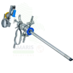
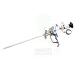
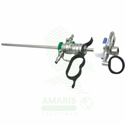
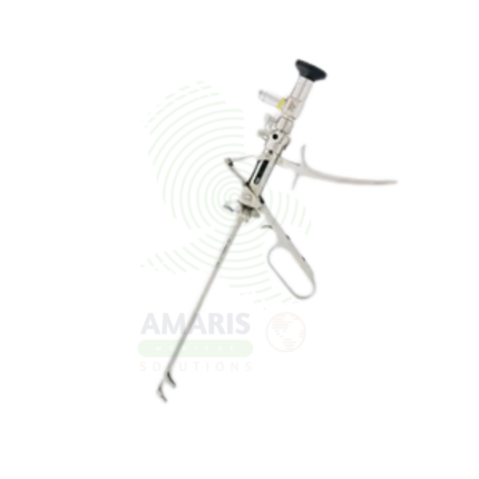
Cystoscope
WhatsApp Order
A Cystoscope is a specialized endoscope used for visualization of the urethra and bladder for diagnostic and therapeutic urologic procedures. Rigid cystoscopes provide superior optics for operative procedures, while flexible cystoscopes offer enhanced patient comfort for diagnostic examinations. Essential for evaluation of hematuria, diagnosis and surveillance of bladder cancer, stent management, and treatment of bladder stones and urethral strictures, cystoscopy is the cornerstone of urologic practice.
Description
Cystoscope
PRIMARY CLINICAL & DIAGNOSTIC USES
1. Direct Visualization of the Bladder and Urethra
-
Primary Use: Provides direct visualization of the urethra, bladder neck, and bladder mucosa for diagnostic evaluation of hematuria, recurrent urinary tract infections, lower urinary tract symptoms, urinary incontinence, and suspicion of bladder tumors or stones. The cystoscope is inserted through the urethra to examine the entire bladder interior.
-
How it helps: For the urologist, the cystoscope transforms the bladder from a hidden, inaccessible organ into a clearly visualized cavity—revealing tumors that bleed, stones that obstruct, inflamed mucosa that causes symptoms, and anatomical abnormalities that explain incontinence. For the patient with blood in their urine, recurrent infections, or bothersome voiding symptoms, cystoscopy provides answers that imaging alone cannot, guiding diagnosis and treatment with certainty.
2. Diagnosis of Bladder Tumors
-
Primary Use: Essential for the diagnosis, staging, and surveillance of bladder cancer. Cystoscopy allows direct visualization of papillary tumors, carcinoma in situ, and other suspicious lesions. Biopsy forceps passed through the cystoscope obtain tissue samples for histopathological confirmation.
-
How it helps: For the urologic oncologist, cystoscopy provides the definitive diagnostic tool for bladder cancer—revealing the number, size, location, and appearance of tumors, and enabling directed biopsy for pathological diagnosis. For the patient with suspected bladder cancer, cystoscopy provides the critical information needed to determine the stage, grade, and treatment plan.
3. Evaluation of Hematuria
-
Primary Use: The gold standard for evaluating painless gross hematuria and microscopic hematuria. Cystoscopy identifies the source of bleeding including tumors, stones, inflammation, and vascular abnormalities that may not be visible on imaging.
-
How it helps: For the urologist, cystoscopy provides definitive evaluation of hematuria—identifying the source of bleeding and ruling out malignancy. For the patient with blood in their urine, a normal cystoscopy provides reassurance, while abnormal findings guide timely treatment.
4. Ureteral Stent Placement and Removal
-
Primary Use: Used for placement and removal of ureteral stents under direct visualization. The cystoscope provides access to the ureteral orifices for passage of guidewires and stent delivery systems.
-
How it helps: For the urologist, cystoscopic stent placement provides reliable drainage for obstructed ureters—relieving pain, preserving renal function, and protecting ureteral repairs. For the patient with ureteral obstruction, stent placement offers relief and protects kidney function.
5. Removal of Bladder Stones and Foreign Bodies
-
Primary Use: Used to visualize and remove bladder stones, retained stents, and other foreign bodies. Stone crushing instruments, graspers, and retrieval devices are passed through the cystoscope under direct visualization.
-
How it helps: For the urologist, cystoscopic stone removal eliminates the source of obstruction and infection without open surgery. For the patient, this means rapid relief of symptoms and avoidance of more invasive procedures.
SECONDARY & SUPPORTIVE USES
1. Retrograde Pyelography: Injection of contrast through ureteral catheters for imaging of the ureters and renal collecting systems.
2. Urethral Stricture Evaluation: Assessment of urethral caliber, location, and severity of strictures.
3. Bladder Biopsy: Directed biopsy of suspicious lesions for pathological diagnosis.
4. Bladder Instillation: Administration of intravesical medications such as BCG or chemotherapy under direct visualization.
5. Post-Treatment Surveillance: Regular cystoscopic surveillance following bladder cancer treatment.
6. Pediatric Cystoscopy: Smaller caliber cystoscopes for pediatric patients.
KEY PRODUCT FEATURES
1. BASIC IDENTIFICATION ATTRIBUTES
-
Device Type: A specialized endoscope designed for visualization of the urethra and bladder.
-
Designation: Cystoscope, Flexible Cystoscope, Rigid Cystoscope, Video Cystoscope.
-
Types:
-
Rigid Cystoscope: Metal sheath with rod lens optics; provides superior optics and larger working channel.
-
Flexible Cystoscope: Fiberoptic or digital flexible shaft; allows easier navigation of the urethra and improved patient comfort.
-
Video Cystoscope: Integrated camera at the distal tip for high-definition visualization.
-
-
Key Components:
-
Telescope: Rod lens system or digital camera.
-
Sheath: Outer tube with irrigation channels.
-
Light Source: Fiberoptic or LED illumination.
-
Working Channel: Passage for instruments, catheters, and irrigation.
-
Bridge: Connects telescope to sheath; allows instrument passage.
-
2. TECHNICAL & PERFORMANCE PROPERTIES
-
Sheath Diameter: 12-24 French for rigid; 14-18 French for flexible.
-
Telescope Diameter: 2.7-4 mm.
-
Viewing Angle: 0°, 12°, 30°, 70° optics.
-
Working Channel: 1-3 mm for instrument passage.
-
Flexible Tip: 120-180° deflection for flexible cystoscopes.
-
Resolution: HD for video cystoscopes.
3. PHYSICAL & OPERATIONAL PROPERTIES
-
Construction: Precision-machined stainless steel (rigid); polymer with fiberoptics (flexible).
-
Ergonomics: Designed for comfortable single-handed operation.
-
Sterilization: Steam autoclave for rigid; low-temperature sterilization for flexible.
-
Portability: Fixed in procedure rooms or portable for bedside use.
4. SAFETY & COMPLIANCE ATTRIBUTES
-
Regulatory Status: Class II medical device regulated by FDA.
-
Biocompatibility: Materials safe for intraurethral and intravesical use.
-
Electrical Safety: Compliant for use with light sources and cameras.
5. STORAGE & HANDLING ATTRIBUTES
-
Storage: Store in protective cases; hang flexible cystoscopes in storage cabinets.
-
Cleaning: Thorough cleaning after each use; ultrasonic cleaning recommended.
-
Sterilization: Steam autoclave for rigid; high-level disinfection for flexible.
-
Inspection: Inspect for lens damage, light transmission, and tip deflection.
6. LABORATORY & CLINICAL APPLICATIONS
-
Primary Application: Diagnosis and treatment of bladder and urethral conditions.
-
Clinical Role: Essential in urology for bladder cancer diagnosis, hematuria evaluation, and stent management.
SAFETY HANDLING PRECAUTIONS
1. SAFETY PRECAUTIONS
-
Sterility: Ensure sterility before intravesical use.
-
Lubrication: Use sterile lubricant for patient comfort.
-
Gentle Insertion: Insert with care to avoid urethral trauma.
-
Infection Prevention: Use sterile technique; consider prophylactic antibiotics in high-risk patients.
2. FIRST AID MEASURES
-
Urethral Injury: If urethral injury occurs, assess for bleeding; consider urethral catheter placement.
-
Bladder Perforation: If perforation occurs, stop procedure; assess for extravasation; manage expectantly or surgically.
3. FIRE FIGHTING MEASURES
-
Flammability: Metal components are non-flammable; plastic parts may burn.
-
Extinguishing Media: Use water, foam, or CO₂ as appropriate for surrounding materials.
Related products
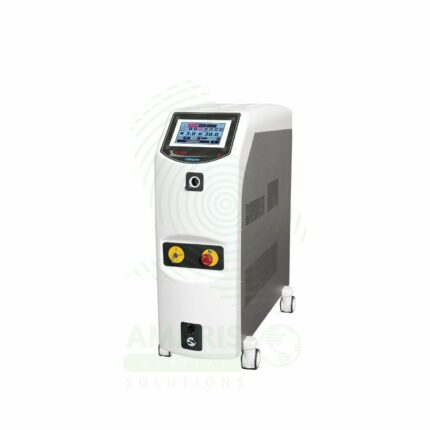
Holmium Laser System
A Holmium Laser System is a versatile, high-power Class IV surgical laser that emits at a 2120 nm wavelength, which is highly absorbed by water and biological tissue. It is the gold-standard tool in urology for laser lithotripsy (stone dusting/fragmentation) and advanced soft tissue procedures like prostate enucleation (HoLEP) and tumor ablation. Its energy is delivered via flexible silica fibers through endoscopes, enabling precise cutting, ablation, and coagulation with excellent hemostasis in a fluid environment. Strict laser safety protocols—including mandatory 2120 nm eyewear, fiber inspection, and controlled activation—are non-negotiable for its safe operation in the OR.
Lithotriptic Scope
A Lithotriptic Scope is a specialized endoscope used for endoscopic fragmentation of urinary tract stones in the kidney, ureter, and bladder. Available as semi-rigid ureteroscopes, flexible ureteroscopes, and nephroscopes, these instruments provide direct visualization for laser, pneumatic, or ultrasonic lithotripsy. Essential in endourology, they enable minimally invasive stone treatment with high stone-free rates, rapid recovery, and preservation of renal function.
Multi-functional Stirrups
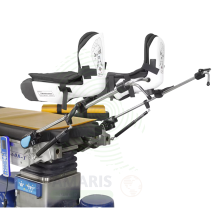
Multi-functional Stirrups are adjustable leg supports used for positioning patients in lithotomy position for gynecologic, urologic, obstetric, and colorectal procedures. Featuring adjustable height, abduction, and rotation, they provide customized positioning for patient comfort and optimal clinician access. Secure locking mechanisms, pressure-reducing padding, and compatibility with standard examination and operating tables make them essential accessories for women's health, urology, and colorectal surgery.
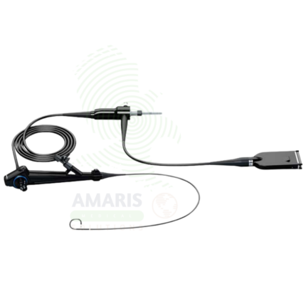
Ureteroscope
A Ureteroscope is a specialized endoscope used for visualization and treatment of the ureter and renal collecting system. Available in semi-rigid and flexible configurations, it enables diagnostic evaluation of ureteral pathology, laser lithotripsy for stone fragmentation, biopsy of upper tract tumors, and treatment of ureteral strictures. Essential in endourology, it offers minimally invasive diagnosis and treatment with preservation of renal function and rapid recovery.

Urethrotome
A Urethrotome is a specialized endoscopic instrument used for internal urethrotomy, the minimally invasive treatment for urethral strictures. Combining a visualizing telescope with a cutting knife or laser fiber, it enables precise incision of strictures under direct vision through the urethra. Essential in urology for managing urethral strictures, it offers a minimally invasive alternative to open urethroplasty with rapid recovery and preservation of erectile function.
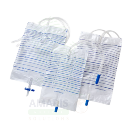
Urine Bags
Urine Bags are sterile, single-use, closed-system medical devices designed for gravity drainage, collection, containment, and measurement of urine from indwelling urinary catheters. Available in two primary configurations: large-capacity drainage bags (2000-4000 mL) for bedridden patients and overnight use, and smaller leg bags (350-750 mL) for ambulatory, community-dwelling patients. Core components include a transparent graduated collection bag, flexible drainage tubing, anti-reflux valve, needleless sampling port, and secure outlet valve. Manufactured from medical-grade PVC (DEHP-free alternatives available) or non-PVC materials, all components are latex-free and terminally sterilized. Critical safety requirements include maintaining the bag below bladder level at all times to prevent retrograde flow, preserving closed system integrity, and adhering to strict single-patient-use protocols. An indispensable component of CAUTI prevention bundles and urinary drainage management across acute care, long-term care, and home healthcare settings.
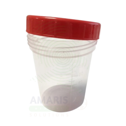
Urine Containers
Urine Containers are sterile or non-sterile, single-use medical devices manufactured from medical-grade polypropylene or polyethylene for the collection, containment, preservation, and transport of urine specimens for diagnostic testing. Available in capacities ranging from 30 mL to 3 liters, they include standard universal containers (plain), preserved containers with boric acid for urine culture, large-volume containers for 24-hour collections, pediatric adhesive collection bags, and DOT-specification drug testing kits with integrated temperature strips and tamper-evident features. Critical performance attributes include leak-proof sealing, chemical inertness, clear volume graduations, and preservative efficacy at specified fill volumes. Strict single-use protocol, adherence to fill line requirements, and handling under universal precautions are essential for specimen integrity and healthcare worker safety. An indispensable consumable in clinical diagnostics, occupational health, and forensic toxicology.