Dermatoscope and Magnifiers
 Diagnostic Kits
 Vital Signs Monitors
 Stethoscopes and Accessories
 Otoscopes, Ophthalmoscopes, and Retinoscopes
 Reflex Hammers and Neurological Tools
 Scales and Measuring Devices
 Spirometers and Pulmonary Function Tests
 Electrosurgical Units and Accessories
 Cutting Instruments
 Grasping and Holding Instruments
 Hemostatic Instruments
 Specialized Surgical Sets
 Single-Use Procedure Trays and Packs
 Surgical Drapes, Gowns, and Covers
 Tissue Unifying Instruments
 Radiation Protection
 X-Ray Machines and Accessories
 Ultrasound Systems and Probes
 MRI and CT Scanners
 Radiology Consumables
 Bone Densitometers
 Fluoroscopy Equipment
 Imaging Tables and Positioning Aids
 Microscopes and Accessories
 Centrifuges and Separators
 Analyzers
 Incubators and Ovens
 Pipettes, Dispensers, and Lab Glassware
 Refrigerators, Freezers, and Storage Units
 Lab Consumables
 Sterilizers and Autoclaves for Lab Use
 Multi-Parameter Monitors
 Ventilators and Respiratory Support Devices
 Defibrillators and AEDs
 Infusion Pumps and IV Systems
 Patient Warmers and Cooling Devices
 Central Monitoring Stations
 Accessories
 Anesthesia Machines and Workstations
 Oxygen Concentrators and Delivery Systems
 Nebulizers and Inhalers
 CPAP/BiPAP Machines
 Airway Management
 Anesthesia Masks, Circuits, and Bags
 Humidifiers and Heaters
 Respiratory Therapy Accessories
 First Aid Kits and Cabinets
 Emergency Resuscitation Equipment
 Trauma Supplies
 Emergency Carts and Crash Carts
 Burn Care Products
 Bleeding Control
 Automated External Defibrillators (AEDs)
 Transport and Evacuation
 Wheelchairs and Accessories
 Walkers, Crutches, and Canes
 Prosthetics and Orthotics
 Physical Therapy Equipment
 Transfer Devices
 Bathroom Safety
 Orthopedic Traction and Tables
 Hot/Cold Therapy Packs and Units
 Beds and Mattresses
 Chairs and Stools
 Tables
 Cabinets and Storage
 Privacy Screens & Curtains
 Stands and Racks
 Linens and Textiles
 Lighting
 Autoclaves and Sterilizers
 Ultrasonic Cleaners
 Disinfectant Solutions and Wipes
 Sterilization Pouches, Wraps, and Indicators
 Instrument Trays and Containers
 UV and Ozone Disinfection Devices
 Washer Disinfectors
 Wound Care
 Gloves
 Masks and Respirators
 Catheters and Tubing
 Swabs, Applicators, and Sponges
 Incontinence Products
 Personal Protective Equipment (PPE)
 Dental Chairs and Units
 Handpieces and Burs
 Instruments
 Consumables
 Sterilization for Dental Use
 Orthodontic Supplies
 Endodontic Tools
 Slit Lamps and Tonometers
 Lensometers and Phoropters
 Ophthalmic Surgical Instruments
 Eyewear Frames and Lenses
 Contact Lens Supplies
 Vision Testing Charts and Devices
 Eye Care Consumables
 Laser Systems for Eye Care
 ENT Exam Chairs and Tables
 Endoscopes
 Audiometers and Hearing Tests
 ENT Instruments
 Nasal and Throat Packs
 Hearing Aids and Accessories
 Otology Supplies
 Fetal Dopplers and Monitors
 Delivery Beds and Tables
 Gynecological Instruments
 Neonatal Incubators and Warmers
 Breast Pumps and Accessories
 Contraceptive Devices
 Maternity Supports and Pads
 Neonatal Consumables
 Cystoscopes and Urethroscopes
 Dialysis Machines and Supplies
 Urological Catheters and Bags
 Lithotripters
 Prostate Treatment Devices
 Urinary Incontinence Products
 Kidney Stone Management Tools
 Consumables & Disposables
 EEG and EMG Machines
 Neurosurgical Instruments
 Nerve Stimulators
 Headrests and Positioning Aids
 Lumbar Puncture Kits
 Seizure Monitoring Devices
 Consumables
 Rehabilitation for Neurological Conditions
 ECG Machines and Accessories
 Holter Monitors
 Stress Test Systems
 Pacemakers and Defibrillator Accessories
 Vascular Access Devices
 Cardiac Catheters and Guidewires
 Blood Flow Meters
 Consumables
 Orthopedic Instruments
 Casts, Splints, and Padding
 Joint Replacement Supplies
 Prosthetic Limbs and Components
 Bone Grafts and Substitutes
 Traction Devices
 Orthopedic Braces and Supports
 Rehabilitation Aids for Orthopedics
 Home Oxygen Therapy
 Hospital Beds for Home Use
 Mobility Aids
 Bathroom and Daily Living Aids
 Wound Care for Home
 Monitoring Devices
 Enteral Feeding Pumps and Tubes
 Hand Sanitizers and Dispensers
 Face Shields and Goggles
 Isolation Gowns and Suits
 Biohazard Waste Containers
 Air Purifiers and HEPA Filters
 Surface Disinfectants
 Sharps Containers
 Protective Barriers
 Cardiovascular & Endurance Training
 Strength Training & Weightlifting
 Functional Training & Core Conditioning
 Physical Therapy & Rehabilitation
 Sports & Outdoor Recreation
 Gym Flooring & Facility Equipment
 Fitness Monitoring & Accessories
 Kids & Novelties
Digital Ceiling X-ray
WhatsApp Order
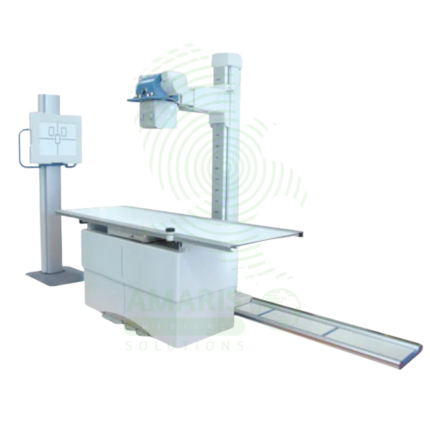
A Digital Ceiling X-ray is a ceiling-mounted digital radiography system for general diagnostic imaging of the skeletal, chest, abdominal, and extremity anatomy. The ceiling-mounted tube assembly provides full room coverage for flexible patient positioning, while digital flat panel detectors produce immediate high-resolution images for rapid diagnosis. Integrated with PACS and RIS, it supports efficient digital workflow from image acquisition to interpretation. Used in radiology departments, emergency rooms, and outpatient imaging centers.
Description
Digital Ceiling X-ray
PRIMARY CLINICAL & DIAGNOSTIC USES
1. Diagnosis of Skeletal Fractures and Dislocations
-
Primary Use: Produces high-resolution digital images of bones to identify breaks, cracks, dislocations, and alignment issues in any part of the body from extremities to the spine. The ceiling-mounted system allows flexible positioning for trauma and orthopedic imaging.
-
How it helps: For the orthopedic surgeon, emergency physician, and radiologist, the digital ceiling X-ray provides the essential view of bony structures needed to diagnose and manage skeletal injuries—revealing the exact location and orientation of fractures, documenting joint dislocations before and after reduction, and guiding decisions about casting, surgery, or conservative management. For the patient with a suspected fracture, the digital X-ray provides definitive answers and directs the course of treatment.
2. Detection of Pulmonary and Chest Conditions
-
Primary Use: Essential for evaluating the lungs and chest cavity to diagnose pneumonia, tuberculosis, lung cancer, pneumothorax, pleural effusion, and assessing heart size and major blood vessels. The ceiling-mounted system allows for upright, supine, and lateral chest imaging.
-
How it helps: For the pulmonologist, cardiologist, emergency physician, and primary care provider, the chest X-ray is a window into the thoracic cavity—revealing infiltrates that indicate pneumonia, the sharp line of a collapsed lung, the enlarged cardiac silhouette of heart failure, or the mass of a lung tumor. For the patient with cough, shortness of breath, or chest pain, a chest X-ray provides rapid, low-cost diagnostic information that guides further evaluation and treatment.
3. Abdominal Imaging
-
Primary Use: Used to detect intestinal obstruction, kidney stones, free air indicating perforation, and calcifications, often serving as the initial test for acute abdominal pain. The ceiling-mounted system allows for flexible positioning for erect and supine abdominal views.
-
How it helps: For the emergency physician and general surgeon evaluating acute abdominal pain, the abdominal X-ray provides rapid screening for life-threatening conditions—revealing the dilated loops of bowel in obstruction, the calcified stones in the renal tract, or the free air under the diaphragm that signals a perforated viscus requiring emergency surgery. For the patient with severe abdominal pain, this quick study can identify the need for immediate intervention.
4. Guiding Orthopedic and Surgical Procedures
-
Primary Use: Provides real-time imaging (fluoroscopy) to guide the placement of hardware during fracture fixation, spinal instrumentation, joint replacements, and other surgical or interventional procedures. The ceiling-mounted system allows for easy positioning around the patient.
-
How it helps: For the orthopedic and trauma surgeon, fluoroscopic guidance during surgery transforms a blind procedure into a visually guided precision operation—confirming that screws are correctly placed within bone, ensuring that fracture fragments are perfectly aligned, and verifying that joint replacement components are positioned optimally. For the patient, this real-time imaging means their surgeon can achieve perfect alignment and hardware placement without guesswork, reducing the need for repeat surgeries.
5. Digital Imaging for Rapid Results
-
Primary Use: Digital detectors produce immediate images that can be viewed, enhanced, and transmitted electronically to PACS (Picture Archiving and Communication System) for interpretation by radiologists and referring physicians, eliminating film processing delays.
-
How it helps: For the radiology team and referring physicians, digital X-ray provides immediate image availability—eliminating the wait for film processing, allowing for image enhancement to improve visualization, and enabling rapid transmission to specialists for interpretation. For the patient, faster results mean quicker diagnosis and treatment decisions.
SECONDARY & SUPPORTIVE USES
1. Dental and Maxillofacial Imaging: Used for imaging the jaw, facial bones, and sinuses to diagnose fractures, infections, and sinusitis.
2. Assessment of Arthritis and Degenerative Joint Disease: Reveals joint space narrowing, bone spurs, and other changes characteristic of osteoarthritis and other arthritic conditions.
3. Monitoring Treatment Progress: Used to track the healing of fractures, resolution of pneumonia, or progression of diseases like scoliosis over time.
4. Foreign Body Localization: Identifies and locates swallowed or embedded foreign objects in soft tissue or the gastrointestinal tract.
5. Preventive and Screening Exams: Such as chest X-rays for pre-employment or immigration physicals.
6. Pediatric Imaging: Ceiling-mounted system allows for easy positioning of pediatric patients with minimal handling.
KEY PRODUCT FEATURES
1. BASIC IDENTIFICATION ATTRIBUTES
-
Device Type: A ceiling-mounted digital X-ray system for general radiography and fluoroscopy.
-
Designation: Digital Ceiling X-ray, Ceiling-Mounted X-ray, Digital Radiography System, DR System.
-
Key Components:
-
Ceiling-Mounted Tube Assembly: X-ray tube suspended from ceiling rails for full room coverage.
-
Digital Detector: Flat panel detector (FPD) for image capture.
-
Generator: High-frequency generator for X-ray production.
-
Control Console: Workstation for image acquisition and processing.
-
X-ray Table: Tilting or fixed table for patient positioning.
-
Wall Stand: Vertical Bucky for upright imaging.
-
2. TECHNICAL & PERFORMANCE PROPERTIES
-
Detector Type: Flat panel digital detector (amorphous silicon or CMOS).
-
Detector Size: Typically 14x17 inches or 17x17 inches.
-
Pixel Matrix: 3-10 megapixels depending on detector.
-
Generator Power: Typically 50-80 kW.
-
Tube Rotation: Full 360-degree rotation for flexible positioning.
-
Room Coverage: Ceiling-mounted system covers full room for multiple imaging positions.
3. PHYSICAL & OPERATIONAL PROPERTIES
-
Mounting: Ceiling-mounted with longitudinal, transverse, and vertical travel.
-
Table: Tilting table (90/90 or 90/15) or fixed table.
-
Wall Stand: Vertical movement for upright imaging.
-
Workflow: Integrated with RIS and PACS for digital workflow.
-
Image Processing: Advanced processing algorithms for image optimization.
4. SAFETY & COMPLIANCE ATTRIBUTES
-
Regulatory Status: Class II medical device regulated by FDA.
-
Radiation Safety: AEC (Automatic Exposure Control) for dose optimization; pediatric protocols available.
-
Collimation: Automatic and manual collimation for beam restriction.
-
Shielding: Built-in filtration for beam hardening.
5. STORAGE & HANDLING ATTRIBUTES
-
Storage: Permanent installation in radiology suite.
-
Room Requirements: Lead-shielded walls, controlled access.
-
Maintenance: Regular quality control testing and calibration.
6. LABORATORY & CLINICAL APPLICATIONS
-
Primary Application: General radiography for skeletal, chest, abdominal, and extremity imaging.
-
Clinical Role: Essential equipment in radiology departments, emergency rooms, and outpatient imaging centers.
SAFETY HANDLING PRECAUTIONS
1. SAFETY PRECAUTIONS
-
Radiation Dose: Follow ALARA principles; use AEC and proper collimation.
-
Pregnancy: Screen for pregnancy; use shielding when appropriate.
-
Pediatric Protocols: Use age-appropriate exposure settings and shielding.
-
Positioning: Ensure proper patient positioning to minimize repeat exposures.
-
Lead Markers: Use lead markers to identify anatomical orientation.
2. FIRST AID MEASURES
-
Patient Fall: If patient falls from table, assess for injury; seek medical attention if needed.
-
Equipment Malfunction: If equipment fails during use, remove the patient; contact the service provider.
3. FIRE FIGHTING MEASURES
-
Flammability: Equipment is non-flammable; fire risk from electrical components.
-
Extinguishing Media: Use CO₂ or dry chemical extinguisher for electrical fires.
Related products
Analogue Fixed X-ray Machine
An Analogue Fixed X-ray Machine is a permanent installation X-ray system using traditional film cassettes for general radiography in radiology departments and imaging centers. Featuring ceiling-mounted tube assemblies, tilting tables, and wall stands, it provides essential diagnostic imaging for skeletal, chest, abdominal, and extremity examinations using film technology. Film cassettes are processed in darkroom facilities, producing permanent physical images for patient records and consultation. Used in facilities without digital radiography, as backup for digital systems, and in resource-limited settings.
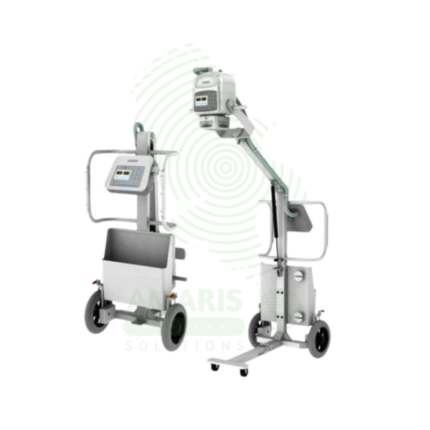
Analogue Mobile X-ray Machine
An Analogue Mobile X-ray Machine is a battery-powered, portable X-ray system using traditional film cassettes for bedside imaging in intensive care units, neonatal intensive care units, emergency departments, and operating rooms. The mobile unit enables chest, abdominal, and extremity imaging at the patient's bedside, eliminating the risks associated with transporting critically ill patients. Film cassettes are processed in darkroom facilities for image development. Used in hospitals without digital radiography, as backup for digital systems, and in resource-limited settings where digital infrastructure is not available.
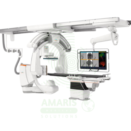
Angiographic Intervention System
An Angiographic Intervention System is a high-end fluoroscopic imaging system designed for guiding minimally invasive vascular and interventional procedures. Essential for cardiac catheterization laboratories, interventional radiology suites, and hybrid operating rooms, it provides real-time, high-resolution visualization of blood vessels, catheters, and devices during coronary interventions, peripheral vascular procedures, neurovascular interventions, and structural heart procedures. Advanced features include rotational angiography for 3D reconstruction, digital subtraction angiography, and dose reduction technologies, enabling precise treatment with minimal radiation exposure.
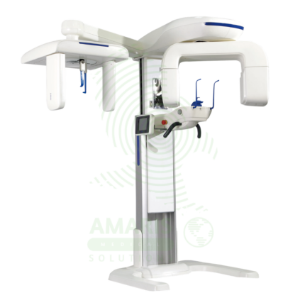
Dental X-ray Machine
A Dental X-ray Machine is a specialized radiographic system designed for imaging teeth, jaws, and facial structures. It encompasses intraoral units for detailed tooth-specific views, panoramic machines for wide screening shots, and advanced Cone Beam CT (CBCT) scanners for 3D surgical planning. Utilizing low-dose radiation and digital imaging technology, it is indispensable for diagnosing cavities, gum disease, infections, and planning treatments like implants, orthodontics, and oral surgery. Its safe operation requires strict adherence to radiation protection protocols, including the use of lead aprons, proper collimation, and operator training to ensure patient and staff safety while obtaining critical diagnostic information.
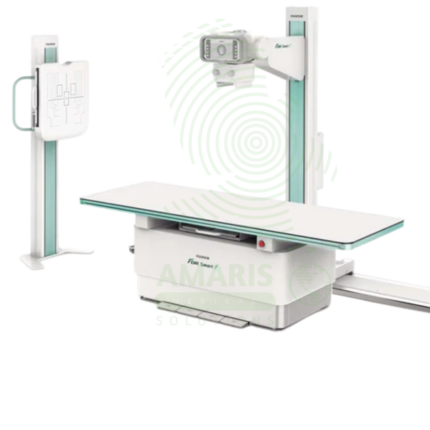
Digital Fixed X-ray
A Digital Fixed X-ray is a permanent installation digital radiography system designed for high-volume general imaging in radiology departments and outpatient imaging centers. Featuring digital flat panel detectors, ceiling-mounted tube assemblies, and tilting tables, it provides high-resolution images for skeletal, chest, abdominal, and extremity examinations. Integrated with PACS and RIS, it supports efficient digital workflow from image acquisition to interpretation, enabling rapid diagnosis and treatment planning.
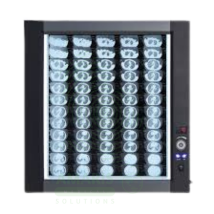
LED Medical Film Viewer
An LED Medical Film Viewer is a light box designed for viewing and interpreting analog X-ray films. Using LED backlight technology, it provides uniform, high-luminance illumination with instant on capability and long life. Available in single, dual, and multi-panel configurations, it supports side-by-side comparison of current and prior studies, pre-operative planning, and group teaching. Essential for radiology departments, orthopedic clinics, emergency departments, and operating rooms where film-based imaging is still used.
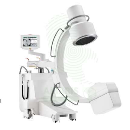
Mobile C-arm Surgical System
A Mobile C-arm Surgical System is a portable fluoroscopic imaging device used for real-time intraoperative guidance during orthopedic, spinal, vascular, and pain management procedures. The C-shaped arm allows flexible positioning around the patient, providing AP, lateral, and oblique views to verify instrument placement, fracture reduction, and device deployment. Essential for minimally invasive surgery, it enables surgeons to achieve precision and accuracy while reducing operative time and improving patient outcomes.