Dermatoscope and Magnifiers
 Diagnostic Kits
 Vital Signs Monitors
 Stethoscopes and Accessories
 Otoscopes, Ophthalmoscopes, and Retinoscopes
 Reflex Hammers and Neurological Tools
 Scales and Measuring Devices
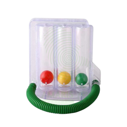
 Spirometers and Pulmonary Function Tests
 Electrosurgical Units and Accessories
 Cutting Instruments
 Grasping and Holding Instruments
 Hemostatic Instruments
 Specialized Surgical Sets
 Single-Use Procedure Trays and Packs
 Surgical Drapes, Gowns, and Covers
 Tissue Unifying Instruments
 Radiation Protection
 X-Ray Machines and Accessories
 Ultrasound Systems and Probes
 MRI and CT Scanners
 Radiology Consumables
 Bone Densitometers
 Fluoroscopy Equipment
 Imaging Tables and Positioning Aids
 Microscopes and Accessories
 Centrifuges and Separators
 Analyzers
 Incubators and Ovens
 Pipettes, Dispensers, and Lab Glassware
 Refrigerators, Freezers, and Storage Units
 Lab Consumables
 Sterilizers and Autoclaves for Lab Use
 Multi-Parameter Monitors
 Ventilators and Respiratory Support Devices
 Defibrillators and AEDs
 Infusion Pumps and IV Systems
 Patient Warmers and Cooling Devices
 Central Monitoring Stations
 Accessories
 Anesthesia Machines and Workstations
 Oxygen Concentrators and Delivery Systems
 Nebulizers and Inhalers
 CPAP/BiPAP Machines
 Airway Management
 Anesthesia Masks, Circuits, and Bags
 Humidifiers and Heaters
 Respiratory Therapy Accessories
 First Aid Kits and Cabinets
 Emergency Resuscitation Equipment
 Trauma Supplies
 Emergency Carts and Crash Carts
 Burn Care Products
 Bleeding Control
 Automated External Defibrillators (AEDs)
 Transport and Evacuation
 Wheelchairs and Accessories
 Walkers, Crutches, and Canes
 Prosthetics and Orthotics
 Physical Therapy Equipment
 Transfer Devices
 Bathroom Safety
 Orthopedic Traction and Tables
 Hot/Cold Therapy Packs and Units
 Beds and Mattresses
 Chairs and Stools
 Tables
 Cabinets and Storage
 Privacy Screens & Curtains
 Stands and Racks
 Linens and Textiles
 Lighting
 Autoclaves and Sterilizers
 Ultrasonic Cleaners
 Disinfectant Solutions and Wipes
 Sterilization Pouches, Wraps, and Indicators
 Instrument Trays and Containers
 UV and Ozone Disinfection Devices
 Washer Disinfectors
 Wound Care
 Gloves
 Masks and Respirators
 Catheters and Tubing
 Swabs, Applicators, and Sponges
 Incontinence Products
 Personal Protective Equipment (PPE)
 Dental Chairs and Units
 Handpieces and Burs
 Instruments
 Consumables
 Sterilization for Dental Use
 Orthodontic Supplies
 Endodontic Tools
 Slit Lamps and Tonometers
 Lensometers and Phoropters
 Ophthalmic Surgical Instruments
 Eyewear Frames and Lenses
 Contact Lens Supplies
 Vision Testing Charts and Devices
 Eye Care Consumables
 Laser Systems for Eye Care
 ENT Exam Chairs and Tables
 Endoscopes
 Audiometers and Hearing Tests
 ENT Instruments
 Nasal and Throat Packs
 Hearing Aids and Accessories
 Otology Supplies
 Fetal Dopplers and Monitors
 Delivery Beds and Tables
 Gynecological Instruments
 Neonatal Incubators and Warmers
 Breast Pumps and Accessories
 Contraceptive Devices
 Maternity Supports and Pads
 Neonatal Consumables
 Cystoscopes and Urethroscopes
 Dialysis Machines and Supplies
 Urological Catheters and Bags
 Lithotripters
 Prostate Treatment Devices
 Urinary Incontinence Products
 Kidney Stone Management Tools
 Consumables & Disposables
 EEG and EMG Machines
 Neurosurgical Instruments
 Nerve Stimulators
 Headrests and Positioning Aids
 Lumbar Puncture Kits
 Seizure Monitoring Devices
 Consumables
 Rehabilitation for Neurological Conditions
 ECG Machines and Accessories
 Holter Monitors
 Stress Test Systems
 Pacemakers and Defibrillator Accessories
 Vascular Access Devices
 Cardiac Catheters and Guidewires
 Blood Flow Meters
 Consumables
 Orthopedic Instruments
 Casts, Splints, and Padding
 Joint Replacement Supplies
 Prosthetic Limbs and Components
 Bone Grafts and Substitutes
 Traction Devices
 Orthopedic Braces and Supports
 Rehabilitation Aids for Orthopedics
 Home Oxygen Therapy
 Hospital Beds for Home Use
 Mobility Aids
 Bathroom and Daily Living Aids
 Wound Care for Home
 Monitoring Devices
 Enteral Feeding Pumps and Tubes
 Hand Sanitizers and Dispensers
 Face Shields and Goggles
 Isolation Gowns and Suits
 Biohazard Waste Containers
 Air Purifiers and HEPA Filters
 Surface Disinfectants
 Sharps Containers
 Protective Barriers
 Cardiovascular & Endurance Training
 Strength Training & Weightlifting
 Functional Training & Core Conditioning
 Physical Therapy & Rehabilitation
 Sports & Outdoor Recreation
 Gym Flooring & Facility Equipment
 Fitness Monitoring & Accessories
 Kids & Novelties
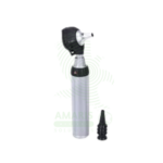
Ophthalmoscope
WhatsApp Order
An Ophthalmoscope is a handheld diagnostic instrument used to examine the interior of the eye, specifically the fundus (retina, optic disc, blood vessels, and macula). By providing illuminated magnification, it serves as a vital screening tool for detecting ocular diseases like glaucoma and retinal disorders, and more importantly, for identifying signs of systemic conditions such as diabetes, hypertension, and increased intracranial pressure. Its operation involves selecting appropriate apertures and compensating lenses to obtain a clear view. While offering a detailed but narrow field of vision, mastery of the direct ophthalmoscope remains a cornerstone skill in general medicine, neurology, and ophthalmology for assessing vascular and neurological health.
Description
Ophthalmoscope
PRIMARY CLINICAL & DIAGNOSTIC USES
1. Direct Ophthalmoscopy for Fundus Examination:
-
Primary Use: The primary use is to perform a direct ophthalmoscopic examination of the internal structures of the eye, known as the fundus. This includes visualizing the optic disc (for signs of glaucoma or papilledema), retinal blood vessels (for signs of hypertension, diabetes, atherosclerosis), macula (for age-related macular degeneration), and the peripheral retina.
-
How it helps: Offers physicians a unique window into the body’s circulatory and nervous systems, allowing direct visualization of blood vessels and nerve tissue without any incision—a view available nowhere else in the body.
2. Screening for Systemic Disease Manifestations:
-
Primary Use: A critical non-ophthalmic tool. It allows clinicians to directly observe pathological changes caused by systemic diseases, making it indispensable for diagnosing and monitoring conditions like Diabetic Retinopathy, Hypertensive Retinopathy, increased Intracranial Pressure (papilledema), and retinal emboli from carotid artery disease.
-
How it helps: Reveals the silent damage that diabetes and high blood pressure inflict on small blood vessels, giving doctors a way to catch complications early and adjust treatment before patients experience vision loss or other serious consequences.
3. Evaluation of Ocular Symptoms and Emergencies:
-
Primary Use: Used to assess patients presenting with visual disturbances (floaters, flashes, vision loss), eye pain, or trauma to detect conditions like retinal detachment, vitreous hemorrhage, optic neuritis, or foreign bodies.
-
How it helps: Provides emergency physicians and eye doctors with the ability to see inside the eye during a crisis, quickly identifying sight-threatening emergencies that require immediate intervention.
4. Routine Physical Examination Component:
-
Primary Use: A standard part of the comprehensive physical exam, especially for patients with systemic risk factors (diabetes, hypertension) or new neurological symptoms (headache, focal deficits).
-
How it helps: Adds a crucial layer to routine check-ups, often revealing the first signs of conditions like high blood pressure or diabetes before patients have any other symptoms.
5. Neurological Assessment:
-
Primary Use: Examination of the optic disc and retina is a key part of the neurological exam, providing clues to diseases of the optic nerve and central nervous system.
-
How it helps: Helps neurologists distinguish between different causes of vision problems and headaches, guiding further testing and treatment for conditions affecting the brain and nerves.
SECONDARY & SUPPORTIVE USES
1. Pediatric Screening: Used to check for the red reflex in newborns and infants, which can reveal congenital cataracts, retinoblastoma (a pediatric eye cancer), or other media opacities, potentially saving an infant’s vision or life.
2. Glaucoma Suspect Evaluation: Assesses the optic nerve head for cupping, a hallmark of glaucomatous damage, helping detect this silent thief of sight before significant vision loss occurs.
3. Teaching and Medical Education: A fundamental instrument for teaching students and residents about the link between systemic disease and ocular findings, training the next generation of physicians to recognize these crucial signs.
KEY PRODUCT FEATURES
1. BASIC IDENTIFICATION ATTRIBUTES
-
Device Type: Handheld, binocular optical instrument for examining the interior of the eye.
-
Common Types:
-
Direct Ophthalmoscope: The most common type used by non-ophthalmologists. Provides a magnified (approx. 15x), upright, virtual image of the fundus, but with a small field of view (about 5-8 degrees). The examiner looks directly into the patient's eye.
-
Indirect Ophthalmoscope: Used primarily by ophthalmologists. Provides a wider, stereoscopic (3D) view of the retina but requires a condensing lens held in front of the patient's eye and more skill to use.
-
-
Light Source: Modern models use LED illumination, offering bright, white, cool light with long battery life, superior to older halogen or incandescent bulbs.
2. TECHNICAL & PERFORMANCE PROPERTIES
-
Optical System: The head contains an illumination system, a viewing aperture, and a dial of lenses (rheostat). This dial allows the examiner to rotate through a range of plus (convex, red numbers) and minus (concave, green or black numbers) diopter lenses (typically ±20 to ±40 D) to compensate for the refractive errors of both the examiner and the patient, bringing the fundus into sharp focus.
-
Aperture Selection: A selector wheel offers different light beam patterns:
-
Large/Small Spot: Standard illumination.
-
Slit Beam: For assessing contour and elevation of lesions.
-
Red-Free (Green) Filter: Enhances the contrast of blood vessels and hemorrhages (appear black).
-
Grid: For approximating the size of lesions.
-
Fixation Target: A small star or circle to assess macular function.
-
Blue Light: For fluorescein staining examination (with cobalt blue filter).
-
-
Magnification and Field: Provides high magnification (~15x) but a very limited field of view, requiring systematic scanning to examine the entire retina.
3. PHYSICAL & OPERATIONAL PROPERTIES
-
Design: Consists of a head containing optics and light, attached to a handle containing the power source (batteries). The head often tilts for comfort.
-
Power Source: Standard alkaline or rechargeable batteries (often C or D cells, or proprietary rechargeable packs).
-
Portability: Handheld and portable, designed for use at the bedside or in the clinic.
4. SAFETY & COMPLIANCE ATTRIBUTES
-
Regulatory Status: Classified as a Class I medical device (low-risk diagnostic instrument). Requires CE Marking and compliance with general safety standards.
-
Light Safety: LED output is within safe limits for ocular exposure during a standard examination.
5. STORAGE & HANDLING ATTRIBUTES
-
Storage: Store in a protective case or holster. Keep in a dry place.
-
Cleaning: Wipe the head, particularly the viewing window and lens contact area, with a soft, lint-free cloth dampened with alcohol after each patient use. Never immerse in liquid. Disposable eye shields are available for some models.
-
Battery Care: Replace or recharge batteries regularly to ensure bright, consistent illumination.
6. LABORATORY & CLINICAL APPLICATIONS
-
Primary Application: A screening and diagnostic extension of the physical and neurological examination, providing a unique "window" to vascular and neurological health.
-
Skill-Intensive Tool: Proficiency requires significant practice to master alignment, focus, and interpretation of findings. It is considered one of the more challenging physical exam skills.
SAFETY HANDLING PRECAUTIONS
1. SAFETY PRECAUTIONS
-
Infection Control: Clean the device between patients. In theory, there is a minimal risk of contact, but good hygiene is essential.
-
Patient Comfort: Darken the room to facilitate pupil dilation. Instruct the patient to fixate on a distant point. Approach from slightly to the side to avoid the "nose-to-nose" discomfort. Use your right eye to examine the patient's right eye, and left for left, to maintain a comfortable working distance.
-
Mydriatics: For a better view, dilating eye drops (mydriatics) may be used by trained personnel, but contraindications (e.g., narrow-angle glaucoma) must be ruled out.
2. FIRST AID MEASURES
-
General: The device is non-invasive. If a patient experiences severe photophobia or discomfort, stop the exam.
3. FIRE FIGHTING MEASURES
-
Flammability: Plastic and electronic components are combustible.
-
Extinguishing Media: Use a CO₂ or dry chemical extinguisher for electrical fires.
Related products
ECG Machine
An ECG Machine is a Class II medical device that records and displays the electrical activity of the heart through surface electrodes, producing an electrocardiogram for diagnosis of cardiac conditions. Standard diagnostic machines record 12 simultaneous leads (3 limb, 6 precordial, 1 ground) with frequency response 0.05-150 Hz, sampling rate 500-1,000 Hz, and high-resolution (5-10 µV) signal acquisition. Features include color touchscreen display, thermal array printer, computerized interpretation algorithms, internal memory (50-500+ ECGs), and network connectivity for EMR integration. Lead wires (AHA or IEC color coding) connect to disposable adhesive electrodes. Portable models (5-15 kg) with rechargeable batteries enable bedside and mobile use; cart-mounted units provide full diagnostic capability. Primary clinical applications include diagnosis of arrhythmias (AF, VT, bradycardia), detection of myocardial ischemia/infarction (STEMI, NSTEMI), evaluation of chest pain, preoperative cardiac risk assessment, monitoring electrolyte imbalances, assessment of chamber enlargement, and drug effect/toxicity monitoring. Essential diagnostic equipment in emergency departments, cardiology clinics, ICUs, operating rooms, and primary care settings worldwide.
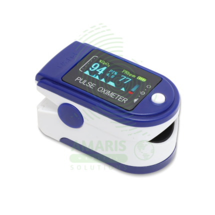
Fingertip Pulse Oximeter
A Fingertip Pulse Oximeter is a compact, non-invasive medical device that clips onto a finger to measure peripheral arterial oxygen saturation (SpO2) and pulse rate in seconds. Utilizing light-based technology, it provides critical, real-time data on respiratory and circulatory status. Its portability and ease of use make it indispensable in hospitals (from ORs to general wards), for patients managing chronic lung conditions at home, and for situational monitoring in sports or high-altitude environments. While an extremely valuable screening tool, readings must be interpreted in context, considering factors like patient movement, perfusion, and clinical symptoms.
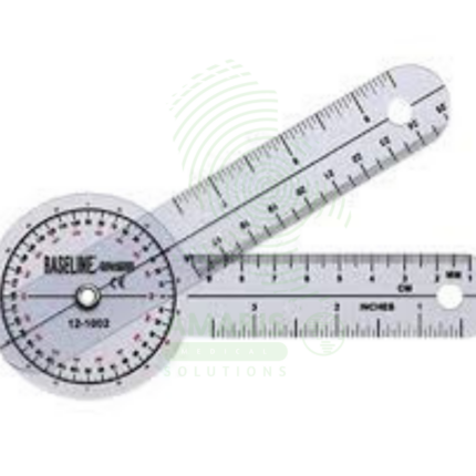
Goniometer
A goniometer is a calibrated, handheld measuring instrument designed to precisely quantify joint range of motion in degrees. Essential for physical and occupational therapy, orthopedic assessment, and rehabilitation medicine, it consists of a protractor-like body with two arms—one stationary, one movable—that align with the patient's limb segments. Available in various sizes for different joints (from fingers to hips) and in materials including transparent plastic, stainless steel, and digital formats, it transforms subjective observations of mobility into objective, reproducible data. This fundamental clinical tool guides diagnosis, documents treatment progress, supports disability determinations, and provides the evidence base for therapeutic interventions.
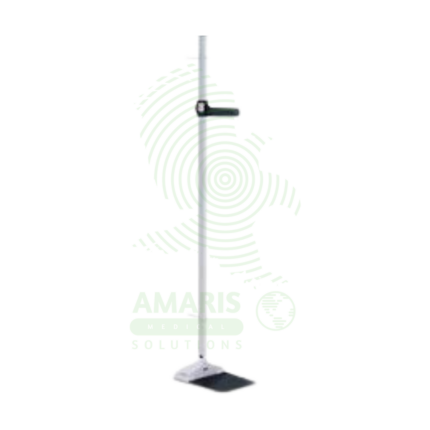
Height Board
A Height Board, or stadiometer, is a precision medical instrument designed for the accurate measurement of human stature. As a wall-mounted or portable device featuring a vertical scale and a sliding horizontal headpiece, it is the clinical standard for obtaining height. This measurement is foundational for calculating Body Mass Index (BMI), tracking growth in children, screening for nutritional issues, and assessing adults for conditions like osteoporosis. Its accuracy, dependent on proper installation and technique, makes it an indispensable tool in pediatrics, primary care, nutrition, and public health
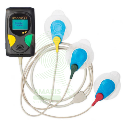
Holter ECG Monitor
A Holter ECG Monitor is a portable, wearable medical device used for continuous, long-term recording of a patient's heart's electrical activity over 24 to 48 hours or more. It is the cornerstone diagnostic tool for detecting intermittent cardiac arrhythmias, evaluating unexplained symptoms like syncope or palpitations, and monitoring the efficacy of antiarrhythmic treatments or implanted cardiac device function. Consisting of a small digital recorder connected to chest electrodes (or an all-in-one adhesive patch), it allows patients to maintain their normal daily routine while capturing a comprehensive electrocardiographic record.
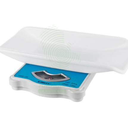
Manual Baby Weighing Scale
A Manual Baby Weighing Scale is a simple, mechanical device used to measure infant weight without the need for electricity or batteries. Operating on a spring or balance principle, it features a suspended sling or seat and a dial display. Its primary advantage is durability and portability for use in community health, outreach programs, and areas with unreliable power. While less precise than digital scales, it provides a reliable and accessible means to monitor growth trends and screen for undernutrition in resource-limited settings, making it a vital tool for basic pediatric care in the field.
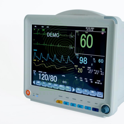
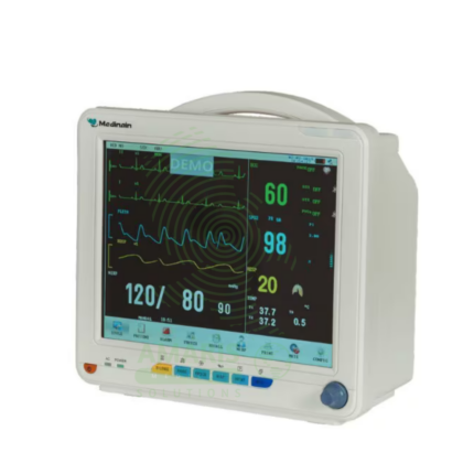
Patient Monitor
The Patient Monitor (6 Parameters BCCMS8000) is a versatile multi-parameter monitor designed for continuous surveillance of core vital signs in various clinical settings. It tracks six essential parameters—ECG, SpO2, Non-Invasive Blood Pressure (NIBP), Respiration, Temperature, and Pulse Rate—providing clinicians with real-time waveforms and numerical data on a clear color display. With its robust alarm system, battery backup for transport, and reliable performance, it is a fundamental tool for ensuring patient safety on general hospital wards, during procedures, and in emergency departments. Its design balances comprehensive monitoring capability with user-friendly operation.