Dermatoscope and Magnifiers
 Diagnostic Kits
 Vital Signs Monitors
 Stethoscopes and Accessories
 Otoscopes, Ophthalmoscopes, and Retinoscopes
 Reflex Hammers and Neurological Tools
 Scales and Measuring Devices
 Spirometers and Pulmonary Function Tests
 Electrosurgical Units and Accessories
 Cutting Instruments
 Grasping and Holding Instruments
 Hemostatic Instruments
 Specialized Surgical Sets
 Single-Use Procedure Trays and Packs
 Surgical Drapes, Gowns, and Covers
 Tissue Unifying Instruments
 Radiation Protection
 X-Ray Machines and Accessories
 Ultrasound Systems and Probes
 MRI and CT Scanners
 Radiology Consumables
 Bone Densitometers
 Fluoroscopy Equipment
 Imaging Tables and Positioning Aids
 Microscopes and Accessories
 Centrifuges and Separators
 Analyzers
 Incubators and Ovens
 Pipettes, Dispensers, and Lab Glassware
 Refrigerators, Freezers, and Storage Units
 Lab Consumables
 Sterilizers and Autoclaves for Lab Use
 Multi-Parameter Monitors
 Ventilators and Respiratory Support Devices
 Defibrillators and AEDs
 Infusion Pumps and IV Systems
 Patient Warmers and Cooling Devices
 Central Monitoring Stations
 Accessories
 Anesthesia Machines and Workstations
 Oxygen Concentrators and Delivery Systems
 Nebulizers and Inhalers
 CPAP/BiPAP Machines
 Airway Management
 Anesthesia Masks, Circuits, and Bags
 Humidifiers and Heaters
 Respiratory Therapy Accessories
 First Aid Kits and Cabinets
 Emergency Resuscitation Equipment
 Trauma Supplies
 Emergency Carts and Crash Carts
 Burn Care Products
 Bleeding Control
 Automated External Defibrillators (AEDs)
 Transport and Evacuation
 Wheelchairs and Accessories
 Walkers, Crutches, and Canes
 Prosthetics and Orthotics
 Physical Therapy Equipment
 Transfer Devices
 Bathroom Safety
 Orthopedic Traction and Tables
 Hot/Cold Therapy Packs and Units
 Beds and Mattresses
 Chairs and Stools
 Tables
 Cabinets and Storage
 Privacy Screens & Curtains
 Stands and Racks
 Linens and Textiles
 Lighting
 Autoclaves and Sterilizers
 Ultrasonic Cleaners
 Disinfectant Solutions and Wipes
 Sterilization Pouches, Wraps, and Indicators
 Instrument Trays and Containers
 UV and Ozone Disinfection Devices
 Washer Disinfectors
 Wound Care
 Gloves
 Masks and Respirators
 Catheters and Tubing
 Swabs, Applicators, and Sponges
 Incontinence Products
 Personal Protective Equipment (PPE)
 Dental Chairs and Units
 Handpieces and Burs
 Instruments
 Consumables
 Sterilization for Dental Use
 Orthodontic Supplies
 Endodontic Tools
 Slit Lamps and Tonometers
 Lensometers and Phoropters
 Ophthalmic Surgical Instruments
 Eyewear Frames and Lenses
 Contact Lens Supplies
 Vision Testing Charts and Devices
 Eye Care Consumables
 Laser Systems for Eye Care
 ENT Exam Chairs and Tables
 Endoscopes
 Audiometers and Hearing Tests
 ENT Instruments
 Nasal and Throat Packs
 Hearing Aids and Accessories
 Otology Supplies
 Fetal Dopplers and Monitors
 Delivery Beds and Tables
 Gynecological Instruments
 Neonatal Incubators and Warmers
 Breast Pumps and Accessories
 Contraceptive Devices
 Maternity Supports and Pads
 Neonatal Consumables
 Cystoscopes and Urethroscopes
 Dialysis Machines and Supplies
 Urological Catheters and Bags
 Lithotripters
 Prostate Treatment Devices
 Urinary Incontinence Products
 Kidney Stone Management Tools
 Consumables & Disposables
 EEG and EMG Machines
 Neurosurgical Instruments
 Nerve Stimulators
 Headrests and Positioning Aids
 Lumbar Puncture Kits
 Seizure Monitoring Devices
 Consumables
 Rehabilitation for Neurological Conditions
 ECG Machines and Accessories
 Holter Monitors
 Stress Test Systems
 Pacemakers and Defibrillator Accessories
 Vascular Access Devices
 Cardiac Catheters and Guidewires
 Blood Flow Meters
 Consumables
 Orthopedic Instruments
 Casts, Splints, and Padding
 Joint Replacement Supplies
 Prosthetic Limbs and Components
 Bone Grafts and Substitutes
 Traction Devices
 Orthopedic Braces and Supports
 Rehabilitation Aids for Orthopedics
 Home Oxygen Therapy
 Hospital Beds for Home Use
 Mobility Aids
 Bathroom and Daily Living Aids
 Wound Care for Home
 Monitoring Devices
 Enteral Feeding Pumps and Tubes
 Hand Sanitizers and Dispensers
 Face Shields and Goggles
 Isolation Gowns and Suits
 Biohazard Waste Containers
 Air Purifiers and HEPA Filters
 Surface Disinfectants
 Sharps Containers
 Protective Barriers
 Cardiovascular & Endurance Training
 Strength Training & Weightlifting
 Functional Training & Core Conditioning
 Physical Therapy & Rehabilitation
 Sports & Outdoor Recreation
 Gym Flooring & Facility Equipment
 Fitness Monitoring & Accessories
 Kids & Novelties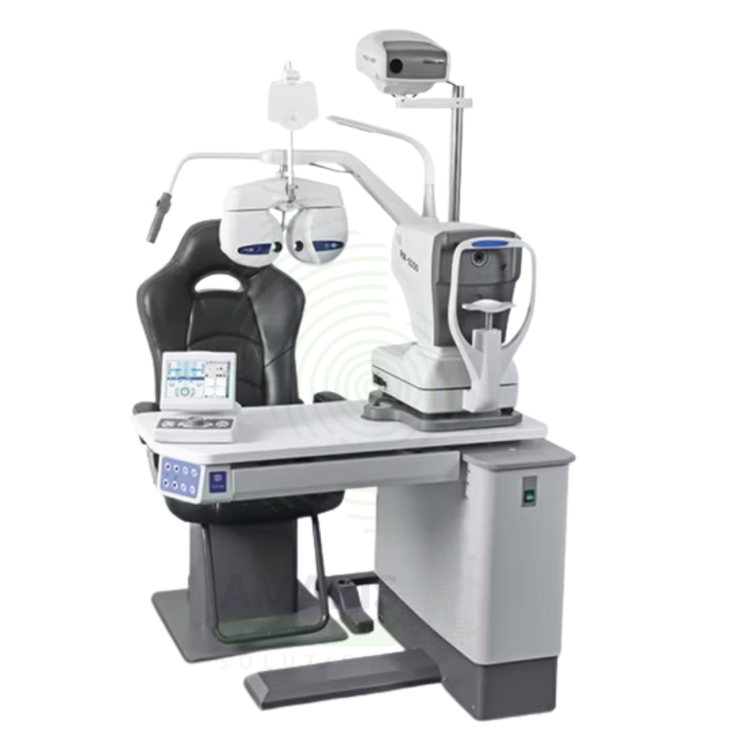
Optometry Unit
$1.00
WhatsApp Order
An Optometry Unit is an integrated workstation for comprehensive eye examinations, combining a phoropter for refraction, visual acuity chart, slit lamp for anterior segment examination, and retinoscope for objective refraction. Used in optometry clinics, ophthalmology practices, and eye care centers, it provides the essential tools for determining corrective lens prescriptions and assessing ocular health. The integrated design streamlines the examination process, enabling efficient, thorough eye care.
Description
Optometry Unit
PRIMARY CLINICAL & DIAGNOSTIC USES
1. Comprehensive Eye Examination and Refraction
-
Primary Use: Provides an integrated workstation for comprehensive eye examinations, including visual acuity testing, refraction (determining prescription for glasses or contact lenses), and assessment of binocular vision. The unit combines a phoropter, trial lens set, and other diagnostic instruments in a single, organized system.
-
How it helps: For the optometrist and ophthalmologist, the integrated optometry unit streamlines the examination process—allowing efficient transition between tests while keeping all necessary instruments within reach. For the patient, this means a more efficient, comfortable eye examination experience with less waiting between tests.
2. Visual Acuity Testing
-
Primary Use: Includes a projected or digital visual acuity chart for measuring distance and near visual acuity. Standardized charts include Snellen, LogMAR, and pediatric optotypes for patients of all ages and literacy levels.
-
How it helps: For the eye care professional, standardized visual acuity testing provides the foundation for assessing visual function—determining the need for corrective lenses and monitoring for changes over time. For the patient, accurate visual acuity measurement ensures appropriate prescription for glasses or contact lenses.
3. Refraction and Prescription Determination
-
Primary Use: The phoropter (refractor) allows the practitioner to perform subjective refraction, determining the precise lens prescription needed to correct refractive errors including myopia (nearsightedness), hyperopia (farsightedness), astigmatism, and presbyopia.
-
How it helps: For the optometrist, the phoropter provides precise control over lens selection during refraction—allowing for accurate determination of spherical, cylindrical, and axis correction. For the patient, accurate refraction ensures optimal visual acuity and comfortable vision correction.
4. Slit Lamp Biomicroscopy
-
Primary Use: Integrated slit lamp allows detailed examination of the anterior segment of the eye including the cornea, conjunctiva, iris, lens, and anterior chamber. The slit lamp provides magnification and illumination for detecting pathology such as cataracts, corneal disease, and conjunctival lesions.
-
How it helps: For the eye care professional, slit lamp examination is essential for detecting anterior segment pathology and monitoring progression of conditions such as cataracts and corneal disease. For the patient, this examination ensures that ocular health issues are detected early and managed appropriately.
5. Retinal Examination and Fundus Imaging
-
Primary Use: The optometry unit often includes a retinoscope for objective refraction and may incorporate fundus cameras or indirect ophthalmoscopes for examination of the retina, optic nerve, and posterior segment. These tools are essential for detecting diabetic retinopathy, glaucoma, and macular degeneration.
-
How it helps: For the optometrist and ophthalmologist, retinal examination is critical for detecting and monitoring sight-threatening conditions such as diabetic retinopathy, glaucoma, and age-related macular degeneration. For the patient, regular retinal examination ensures early detection of conditions that can cause irreversible vision loss.
SECONDARY & SUPPORTIVE USES
1. Tonometry: Measurement of intraocular pressure for glaucoma screening.
2. Color Vision Testing: Assessment of color vision deficiencies.
3. Stereopsis Testing: Evaluation of depth perception and binocular vision.
4. Corneal Topography: Mapping of corneal curvature for contact lens fitting and keratoconus detection.
5. Pachymetry: Measurement of corneal thickness.
6. Visual Field Testing: Assessment of peripheral vision for glaucoma and neurological conditions.
KEY PRODUCT FEATURES
1. BASIC IDENTIFICATION ATTRIBUTES
-
Device Type: An integrated workstation for comprehensive eye examinations and refractive testing.
-
Designation: Optometry Unit, Eye Examination Unit, Refraction Unit, Optometric Workstation.
-
Key Components:
-
Phoropter (Refractor): Instrument for subjective refraction.
-
Visual Acuity Chart: Projected or digital chart for vision testing.
-
Slit Lamp: Biomicroscope for anterior segment examination.
-
Retinoscope: Instrument for objective refraction.
-
Trial Lens Set: For verifying prescriptions.
-
Workstation: Integrated desk with instrument mounts.
-
Computer Interface: Integration with electronic health records.
-
2. TECHNICAL & PERFORMANCE PROPERTIES
-
Refraction Range: -20.00 to +20.00 diopters typical.
-
Cylinder Range: 0 to ±6.00 diopters.
-
Axis: 0-180 degrees.
-
Prism: 0-20 prism diopters.
-
Slit Lamp Magnification: 6x-40x adjustable.
-
Visual Acuity Charts: Snellen, LogMAR, pediatric optotypes.
-
Data Integration: Electronic health record connectivity.
3. PHYSICAL & OPERATIONAL PROPERTIES
-
Construction: Modular workstation with adjustable components.
-
Ergonomics: Adjustable chair and instrument positioning.
-
Controls: Electronic or manual controls for phoropter and instruments.
-
Lighting: Integrated room and examination lighting.
4. SAFETY & COMPLIANCE ATTRIBUTES
-
Regulatory Status: Class I and Class II medical devices regulated by FDA.
-
Electrical Safety: Compliant with medical electrical equipment standards.
-
Infection Control: Surfaces designed for easy cleaning; disposable components for slit lamps.
-
Radiation Safety: No ionizing radiation.
5. STORAGE & HANDLING ATTRIBUTES
-
Storage: Stored in the examination room.
-
Cleaning: Clean surfaces with hospital-grade disinfectants; clean phoropter and slit lamp after each patient.
-
Calibration: Regular calibration of instruments per manufacturer schedule.
-
Maintenance: Professional service as recommended.
6. LABORATORY & CLINICAL APPLICATIONS
-
Primary Application: Comprehensive eye examination and refraction for vision correction and ocular health assessment.
-
Clinical Role: Essential equipment in optometry clinics, ophthalmology practices, and eye care centers.
SAFETY HANDLING PRECAUTIONS
1. SAFETY PRECAUTIONS
-
Infection Control: Clean chin rest and forehead rest after each patient; use disposable covers.
-
Instrument Positioning: Adjust instruments for patient comfort; avoid sudden movements.
-
Electrical Safety: Ensure proper grounding; keep cords away from patient areas.
-
Patient Communication: Explain procedures to patients; address anxiety about eye examinations.
2. FIRST AID MEASURES
-
Patient Syncope: If patient faints during examination, lower head, elevate legs; monitor vital signs.
-
Corneal Abrasion: If corneal injury occurs, stop examination; refer for ophthalmologic evaluation.
3. FIRE FIGHTING MEASURES
-
Flammability: Electrical components may pose fire risk.
-
Extinguishing Media: For electrical fire, use CO₂ or dry chemical extinguisher.
Related products
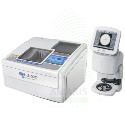
3D Patternless Lens Edger
A 3D Patternless Lens Edger is an automated lens fabrication system that uses digital 3D tracing to cut and shape eyeglass lenses without physical patterns. The system captures the exact frame shape, calculates the optimal lens size and bevel placement, and grinds lenses to precise specifications. Capable of processing multiple lens materials and frame types, it streamlines lens fabrication, reduces errors, and ensures accurate, well-fitted lenses for patients.
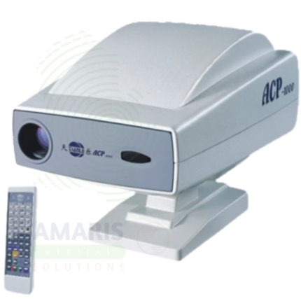
Auto Chart Projector
An Auto Chart Projector is a remote-controlled projection device used in eye examinations to display standardized visual acuity charts for distance and near vision testing. Offering multiple chart options including Snellen, LogMAR, pediatric symbols, and Landolt C, it enables accurate assessment of vision across diverse patient populations. Remote operation streamlines the refraction workflow, allowing efficient chart changes while maintaining patient comfort. Essential for optometry and ophthalmology practices.
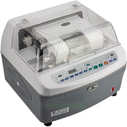
Auto Lens Edger
An Auto Lens Edger is an automated lens fabrication system that cuts and shapes eyeglass lenses to fit into frames using digital or pattern-based tracing technology. Capable of processing multiple lens materials and frame types, it ensures precise lens fitting, accurate optical center placement, and proper bevel alignment. Essential for optical laboratories, optometry practices, and ophthalmology dispensing centers, it streamlines lens fabrication, reduces errors, and ensures patients receive well-fitted, accurately prescribed glasses.
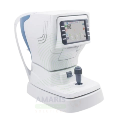
Auto Ref-Keratometer
An Auto Ref-Keratometer combines automated refraction measurement and keratometry in a single instrument, providing comprehensive data for eyeglass prescriptions, contact lens fitting, and assessment of corneal conditions. The device measures spherical and cylindrical refractive error, axis, and corneal curvature, offering essential information for contact lens base curve selection, refractive surgery evaluation, and monitoring of conditions such as keratoconus. Streamlining the pre-examination assessment enhances practice efficiency and patient comfort.
Eye Pads
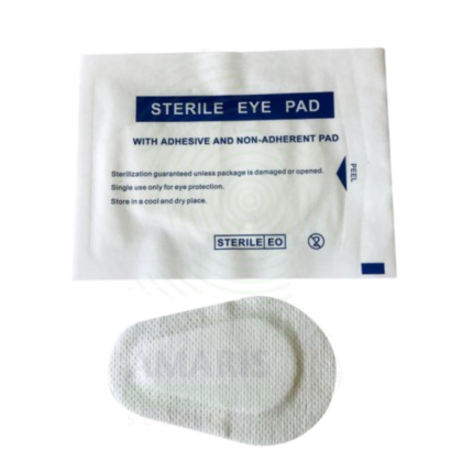
Eye Pads are sterile or non-sterile, single-use, soft, conformable absorbent pads designed specifically for application over the closed eyelid and periorbital region. Constructed from absorbent cotton, rayon, or low-linting non-woven synthetic fibers, they provide cushioning, gentle pressure distribution, and fluid absorption for postoperative eye protection, corneal abrasion management, exposure keratopathy prevention, and periorbital trauma care. Available in standard and pressure-rated thicknesses, with or without integrated adhesive tape tabs, and in sterile (surgical/postoperative) or non-sterile (minor injury/comfort) grades. Critical safety imperatives include absolute contraindication of direct application to an open or ruptured globe, meticulous lint-free construction to prevent corneal foreign body, and single-use-only protocol. An indispensable ophthalmic consumable in surgical, emergency, and outpatient eye care.
Handheld Auto Refractometer
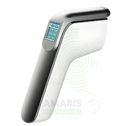
A Handheld Auto Refractometer is a portable, battery-operated instrument for objective measurement of refractive error (sphere, cylinder, axis) in any setting. Ideal for pediatric refraction, bedside examinations, community screenings, and outreach programs, it provides rapid, non-invasive measurements without requiring patient cooperation. The compact, portable design enables eye care professionals to bring refractive assessment to patients who cannot access traditional tabletop equipment, expanding access to essential vision care.
Handheld Vision Screener
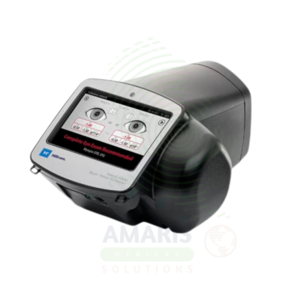
A Handheld Vision Screener is a portable, battery-operated device for rapid screening of refractive errors, amblyopia risk factors, strabismus, and pupillary abnormalities. Designed for use in pediatrics, primary care, schools, and community screening programs, it provides objective measurements without requiring patient cooperation. Early detection of vision problems enables timely intervention, preventing permanent vision loss and ensuring children enter school with the visual skills needed for learning.