Dermatoscope and Magnifiers
 Diagnostic Kits
 Vital Signs Monitors
 Stethoscopes and Accessories
 Otoscopes, Ophthalmoscopes, and Retinoscopes
 Reflex Hammers and Neurological Tools
 Scales and Measuring Devices
 Spirometers and Pulmonary Function Tests
 Electrosurgical Units and Accessories
 Cutting Instruments
 Grasping and Holding Instruments
 Hemostatic Instruments
 Specialized Surgical Sets
 Single-Use Procedure Trays and Packs
 Surgical Drapes, Gowns, and Covers
 Tissue Unifying Instruments
 Radiation Protection
 X-Ray Machines and Accessories
 Ultrasound Systems and Probes
 MRI and CT Scanners
 Radiology Consumables
 Bone Densitometers
 Fluoroscopy Equipment
 Imaging Tables and Positioning Aids
 Microscopes and Accessories
 Centrifuges and Separators
 Analyzers
 Incubators and Ovens
 Pipettes, Dispensers, and Lab Glassware
 Refrigerators, Freezers, and Storage Units
 Lab Consumables
 Sterilizers and Autoclaves for Lab Use
 Multi-Parameter Monitors
 Ventilators and Respiratory Support Devices
 Defibrillators and AEDs
 Infusion Pumps and IV Systems
 Patient Warmers and Cooling Devices
 Central Monitoring Stations
 Accessories
 Anesthesia Machines and Workstations
 Oxygen Concentrators and Delivery Systems
 Nebulizers and Inhalers
 CPAP/BiPAP Machines
 Airway Management
 Anesthesia Masks, Circuits, and Bags
 Humidifiers and Heaters
 Respiratory Therapy Accessories
 First Aid Kits and Cabinets
 Emergency Resuscitation Equipment
 Trauma Supplies
 Emergency Carts and Crash Carts
 Burn Care Products
 Bleeding Control
 Automated External Defibrillators (AEDs)
 Transport and Evacuation
 Wheelchairs and Accessories
 Walkers, Crutches, and Canes
 Prosthetics and Orthotics
 Physical Therapy Equipment
 Transfer Devices
 Bathroom Safety
 Orthopedic Traction and Tables
 Hot/Cold Therapy Packs and Units
 Beds and Mattresses
 Chairs and Stools
 Tables
 Cabinets and Storage
 Privacy Screens & Curtains
 Stands and Racks
 Linens and Textiles
 Lighting
 Autoclaves and Sterilizers
 Ultrasonic Cleaners
 Disinfectant Solutions and Wipes
 Sterilization Pouches, Wraps, and Indicators
 Instrument Trays and Containers
 UV and Ozone Disinfection Devices
 Washer Disinfectors
 Wound Care
 Gloves
 Masks and Respirators
 Catheters and Tubing
 Swabs, Applicators, and Sponges
 Incontinence Products
 Personal Protective Equipment (PPE)
 Dental Chairs and Units
 Handpieces and Burs
 Instruments
 Consumables
 Sterilization for Dental Use
 Orthodontic Supplies
 Endodontic Tools
 Slit Lamps and Tonometers
 Lensometers and Phoropters
 Ophthalmic Surgical Instruments
 Eyewear Frames and Lenses
 Contact Lens Supplies
 Vision Testing Charts and Devices
 Eye Care Consumables
 Laser Systems for Eye Care
 ENT Exam Chairs and Tables
 Endoscopes
 Audiometers and Hearing Tests
 ENT Instruments
 Nasal and Throat Packs
 Hearing Aids and Accessories
 Otology Supplies
 Fetal Dopplers and Monitors
 Delivery Beds and Tables
 Gynecological Instruments
 Neonatal Incubators and Warmers
 Breast Pumps and Accessories
 Contraceptive Devices
 Maternity Supports and Pads
 Neonatal Consumables
 Cystoscopes and Urethroscopes
 Dialysis Machines and Supplies
 Urological Catheters and Bags
 Lithotripters
 Prostate Treatment Devices
 Urinary Incontinence Products
 Kidney Stone Management Tools
 Consumables & Disposables
 EEG and EMG Machines
 Neurosurgical Instruments
 Nerve Stimulators
 Headrests and Positioning Aids
 Lumbar Puncture Kits
 Seizure Monitoring Devices
 Consumables
 Rehabilitation for Neurological Conditions
 ECG Machines and Accessories
 Holter Monitors
 Stress Test Systems
 Pacemakers and Defibrillator Accessories
 Vascular Access Devices
 Cardiac Catheters and Guidewires
 Blood Flow Meters
 Consumables
 Orthopedic Instruments
 Casts, Splints, and Padding
 Joint Replacement Supplies
 Prosthetic Limbs and Components
 Bone Grafts and Substitutes
 Traction Devices
 Orthopedic Braces and Supports
 Rehabilitation Aids for Orthopedics
 Home Oxygen Therapy
 Hospital Beds for Home Use
 Mobility Aids
 Bathroom and Daily Living Aids
 Wound Care for Home
 Monitoring Devices
 Enteral Feeding Pumps and Tubes
 Hand Sanitizers and Dispensers
 Face Shields and Goggles
 Isolation Gowns and Suits
 Biohazard Waste Containers
 Air Purifiers and HEPA Filters
 Surface Disinfectants
 Sharps Containers
 Protective Barriers
 Cardiovascular & Endurance Training
 Strength Training & Weightlifting
 Functional Training & Core Conditioning
 Physical Therapy & Rehabilitation
 Sports & Outdoor Recreation
 Gym Flooring & Facility Equipment
 Fitness Monitoring & Accessories
 Kids & Novelties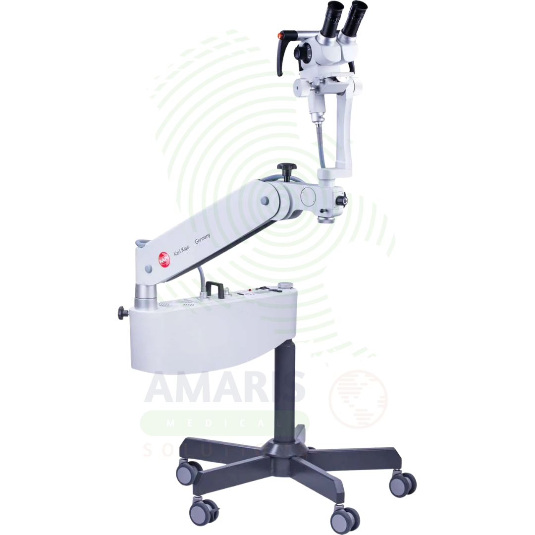
Colposcope
$1.00
WhatsApp Order
A Colposcope is a binocular or video microscope used for magnified examination of the cervix, vagina, and vulva following abnormal cervical cancer screening results. It enables identification of acetowhite lesions, atypical vascular patterns, and other signs of cervical intraepithelial neoplasia or cancer, and guides precise biopsy of suspicious areas. Essential for gynecology clinics and women’s health centers, it provides the visualization needed for accurate diagnosis, treatment planning, and patient education.
Description
Colposcope
PRIMARY CLINICAL & DIAGNOSTIC USES
1. Cervical Cancer Screening and Diagnosis
-
Primary Use: Provides magnified, illuminated visualization of the cervix, vagina, and vulva following an abnormal Pap smear or HPV test. The colposcope is used to identify, locate, and characterize acetowhite lesions, vascular patterns, and other signs of cervical intraepithelial neoplasia or cancer.
-
How it helps: For the gynecologist and colposcopist, the colposcope transforms an abnormal screening result into a visual map of the cervix—revealing subtle changes in color, contour, and blood vessels that indicate precancerous transformation. For the patient who has just learned her Pap smear is abnormal, colposcopy provides the next critical step: determining whether that abnormality represents a transient infection, a precancerous lesion needing treatment, or something more serious requiring immediate intervention.
2. Guided Cervical Biopsy
-
Primary Use: Provides precise visual targeting for taking tissue samples from the most suspicious areas identified during colposcopic examination, which are then sent for histopathological diagnosis. The colposcope’s magnification ensures accurate sampling of abnormal tissue.
-
How it helps: For the gynecologist, the colposcope’s magnification and illumination ensure that biopsies are taken from the areas most likely to yield diagnostic information—targeting the whitest epithelium, the most atypical vessels, the areas closest to the transformation zone. For the patient, this precision means that if precancer is present, it will be found; if the cervix is normal, she can be reassured—avoiding both missed diagnoses and unnecessary treatment.
3. Evaluation of Lower Genital Tract Disorders
-
Primary Use: Used to assess benign conditions such as cervicitis, cervical polyps, Nabothian cysts, genital warts, vaginal adenosis, and lichen sclerosus. The colposcope provides detailed visualization of these conditions.
-
How it helps: For the women’s health provider, the colposcope provides detailed visualization of conditions that may cause symptoms like discharge, bleeding, or pain—distinguishing between benign findings that require only reassurance and those that need treatment. For the patient with persistent symptoms, seeing the magnified image of her own cervix can provide explanation and reassurance, demystifying what’s happening inside her body.
4. Pre-Treatment Planning and Mapping
-
Primary Use: Used to delineate the extent of pre-cancerous lesions (e.g., CIN II/III, VIN, VAIN) before excisional treatments like LEEP or cold knife conization. The colposcope provides a detailed map of lesion extent.
-
How it helps: For the gynecologic surgeon planning an excisional procedure, the colposcope provides a detailed map of lesion extent, ensuring that treatment removes all abnormal tissue while preserving as much healthy cervix as possible. For the patient undergoing treatment for cervical precancer, this mapping means their procedure is precisely targeted, maximizing cure while preserving future fertility.
5. Post-Treatment Follow-Up
-
Primary Use: Used to monitor the cervix after treatment for cervical intraepithelial neoplasia to check for recurrence and assess healing. Serial colposcopic examinations document the healing process and detect any residual or recurrent disease.
-
How it helps: For the gynecologist monitoring a patient after treatment for cervical precancer, the colposcope provides detailed assessment of healing and surveillance for recurrence. For the patient who has already undergone one treatment for precancer, regular colposcopic follow-up offers reassurance that her cervix remains healthy and early detection if additional intervention becomes necessary.
SECONDARY & SUPPORTIVE USES
1. Vulvar and Vaginal Assessment: Diagnosis of vulvar intraepithelial neoplasia, vaginal intraepithelial neoplasia, lichen sclerosus, and other vulvovaginal pathologies.
2. Forensic Examination: Used in sexual assault examinations to document traumatic injuries to the genitalia with high-resolution imaging.
3. Patient Education and Counseling: The live video display allows the physician to show and explain findings to the patient in real-time, improving understanding and compliance with treatment plans.
4. Documentation: Digital imaging systems capture still images and video for patient records, referral documentation, and quality assurance.
5. Teaching and Training: Digital images and videos are used for creating teaching archives and training colposcopists.
6. Research: Used in clinical research studies investigating cervical pathology, HPV infection, and treatment outcomes.
KEY PRODUCT FEATURES
1. BASIC IDENTIFICATION ATTRIBUTES
-
Device Type: A binocular or video microscope with a light source designed for examining the cervix, vagina, and vulva.
-
Designation: Colposcope, Digital Colposcope, Video Colposcope, Colposcopy System.
-
Key Components:
-
Binocular Head: Stereoscopic viewing system with adjustable eyepieces.
-
Magnification: Variable magnification typically 3-25x.
-
Light Source: Halogen, LED, or fiberoptic illumination.
-
Filter: Green filter for vascular pattern visualization.
-
Camera: Integrated digital camera for image capture.
-
Stand: Adjustable floor stand or wall mount.
-
Monitor: For video colposcopy systems.
-
2. TECHNICAL & PERFORMANCE PROPERTIES
-
Magnification: 3-25x variable; stereoscopic for depth perception.
-
Working Distance: 250-300 mm for comfortable positioning.
-
Light Source: High-intensity LED or halogen with green filter.
-
Field of View: Wide field for comprehensive visualization.
-
Camera Resolution: HD or 4K for digital systems.
-
Documentation: Still image and video capture capabilities.
3. PHYSICAL & OPERATIONAL PROPERTIES
-
Mounting: Floor stand, wall mount, or ceiling mount.
-
Portability: Mobile base with locking casters.
-
Controls: Foot pedal or hand controls for magnification and focus.
-
Ergonomics: Adjustable height and angle for operator comfort.
4. SAFETY & COMPLIANCE ATTRIBUTES
-
Regulatory Status: Class II medical device regulated by FDA.
-
Electrical Safety: Compliant with IEC 60601-1.
-
Light Safety: LED light sources are safe for mucosal examination.
5. STORAGE & HANDLING ATTRIBUTES
-
Storage: Stored in the examination room or colposcopy suite.
-
Cleaning: Wipe with hospital-grade disinfectants.
-
Maintenance: Regular calibration and light source maintenance.
6. LABORATORY & CLINICAL APPLICATIONS
-
Primary Application: Cervical cancer screening, diagnosis, and treatment planning.
-
Clinical Role: Essential equipment in gynecology clinics and women's health centers.
SAFETY HANDLING PRECAUTIONS
1. SAFETY PRECAUTIONS
-
Infection Control: Clean oculars and patient contact surfaces between patients.
-
Light Safety: Use green filters for vascular assessment; avoid prolonged intense light exposure.
-
Patient Positioning: Ensure patient comfort and modesty during examination.
2. FIRST AID MEASURES
-
Patient Syncope: If patient faints during procedure, lower head, elevate legs; monitor vital signs.
3. FIRE FIGHTING MEASURES
-
Flammability: Plastic components are combustible.
-
Extinguishing Media: Use water, foam, or CO₂ as appropriate for surrounding materials.
Related products
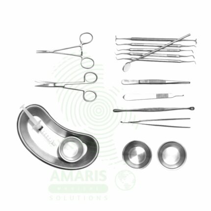
Caesarean Section Set
A Caesarean Section Set is a sterile instrument tray containing all instruments required for Caesarean delivery, including instruments for abdominal incision, uterine incision, fetal delivery, placental removal, and multi-layer closure. Essential for labor and delivery units and obstetric operating rooms, the set ensures that all necessary instruments are sterile, organized, and immediately available for safe and efficient surgical delivery. Proper instrument counting, sterile technique, and careful tissue handling are critical for optimal maternal and neonatal outcomes.
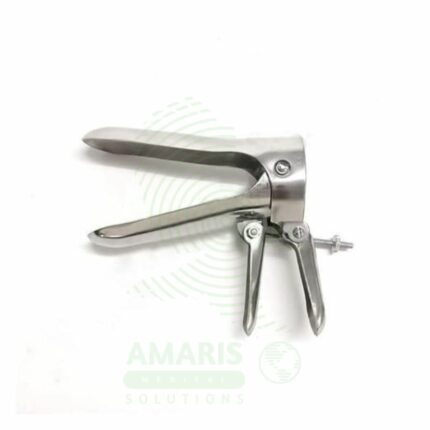
Cusco Speculum
A Cusco speculum is a hinged, bivalve instrument used to separate the vaginal walls for examination of the vagina and cervix. It is the standard tool for performing Pap smears, visual diagnostics, and various gynecological procedures. Available in reusable metal or single-use plastic forms and in multiple sizes, its self-retaining lock allows for hands-free operation. Safe and effective use requires gentle insertion with lubrication, selection of the correct size, careful unlocking before removal, and strict adherence to high-level disinfection or sterilization protocols for reusable models. It is an indispensable instrument in women's health care.

Evacuation Set
An Evacuation Set is a sterile instrument tray containing the instruments necessary for uterine evacuation procedures, including dilation and curettage, suction aspiration, and management of incomplete miscarriage. The set includes cervical dilators, curettes, suction cannulas, ovum forceps, and supporting instruments. Used in obstetrics and gynecology for therapeutic abortion, management of pregnancy loss, retained products of conception, and diagnostic curettage, it ensures that all required instruments are sterile, organized, and immediately available for safe and effective uterine evacuation procedures.
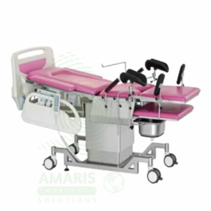
Gynecological examination couch (Parturition Bed)
A Gynecological Examination Couch (Parturition Bed) is a specialized medical couch designed for gynecologic examinations, obstetric delivery, and related procedures. Featuring adjustable backrest, removable leg sections, and adjustable stirrups for lithotomy positioning, it supports pelvic examinations, Pap smears, IUD procedures, and all stages of labor and delivery. The couch provides Trendelenburg and reverse Trendelenburg positioning, height adjustment, and fluid-resistant upholstery for easy cleaning. Essential for gynecology clinics, labor and delivery units, and women's health centers, it combines examination and delivery functions in a single versatile unit.
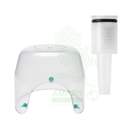
Infant Oxygen Hood
An Infant Oxygen Hood is a Class II medical device used to deliver controlled concentrations of supplemental oxygen to spontaneously breathing newborns and infants. Made of transparent medical-grade acrylic or polycarbonate, the hood fits over the infant's head with a soft foam or rubber neck seal, creating an oxygen-enriched environment while allowing easy access for monitoring and care. Available in premature/neonatal, infant, and older infant sizes with gas inlet ports (22 mm) for connection to air/oxygen blenders (FiO2 21-100%), access ports for monitoring leads and IV lines, and outlet vents to prevent CO2 accumulation. Requires minimum flow rate of 5 L/min to ensure adequate CO2 washout. Used with heated humidifiers for prolonged therapy to prevent airway drying. Primary clinical applications include management of respiratory distress syndrome (RDS) in premature infants, neonatal pneumonia, post-extubation oxygen support, congenital heart disease, and palliative care. Critical safety considerations include maintaining adequate flow rate to prevent rebreathing, monitoring FiO2 at hood level, ensuring proper neck seal without constriction, and preventing hyperoxia to reduce retinopathy of prematurity (ROP) risk. Essential equipment in NICUs and special care nurseries for controlled oxygen delivery to vulnerable infants.
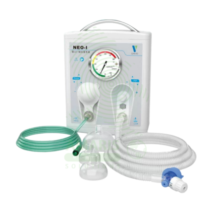
Infant T-Piece Resuscitator
An Infant T-piece Resuscitator is a Class II medical device designed for neonatal resuscitation and controlled positive pressure ventilation, delivering precise, consistent peak inspiratory pressures (PIP) and positive end-expiratory pressure (PEEP) as recommended by Neonatal Resuscitation Program (NRP) guidelines. The T-piece assembly connects to compressed air/oxygen source (5-15 L/min) with adjustable PIP (10-40 cmH₂O) and PEEP (0-10 cmH₂O) controls, built-in pressure manometer, and pressure relief valve (30-40 cmH₂O). Operator occludes port to deliver breaths (typically 40-60/min), with consistent pressures maintained regardless of technique. Essential advantages over bag-valve-mask devices include operator-independent pressure delivery, ability to provide PEEP (critical for preterm lung protection), reduced risk of barotrauma, and hands-free PEEP allowing two-handed mask seal. Primary clinical applications include delivery room resuscitation, stabilization of extremely preterm infants (<1,000 g), CPAP/PEEP delivery, meconium aspiration management, and neonatal transport ventilation. Critical safety considerations include verifying gas supply (5-15 L/min), monitoring manometer pressures, ensuring proper mask seal, observing chest rise, and titrating FiO2 to SpO2 targets. Indispensable resuscitation equipment in every delivery room, NICU, and neonatal transport setting worldwide.
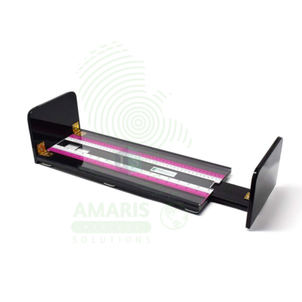
Infantometer
An Infantometer is a medical measuring device (typically 0-100 cm range, 1 mm graduation) designed for accurate recumbent length measurement of neonates, infants, and children up to 2 years of age. Features include rigid base board, fixed headboard, movable perpendicular footboard with smooth-gliding mechanism, and measurement scale (analog engraved or digital display). Constructed from medical-grade materials (wood, acrylic, ABS plastic, aluminum/stainless steel) with non-porous, easy-to-clean surfaces and rounded safety edges. Accuracy ±1-2 mm meets WHO and CDC requirements for growth monitoring. Primary clinical applications include accurate length measurement for growth assessment (WHO/CDC growth charts), early detection of growth abnormalities (failure to thrive, growth retardation), nutritional status monitoring (weight-for-length), premature infant follow-up, endocrine disorder evaluation (growth hormone deficiency), genetic syndrome assessment (syndrome-specific charts), and research studies requiring precise anthropometrics. Class I medical device. Critical safety considerations include two-person measurement technique, proper infant positioning (neutral head, gentle leg extension), infection control (cleaning between infants), stability on level surface, and regular calibration verification. Essential equipment in pediatric clinics, well-child visits, NICU, and child health research.