Dermatoscope and Magnifiers
 Diagnostic Kits
 Vital Signs Monitors
 Stethoscopes and Accessories
 Otoscopes, Ophthalmoscopes, and Retinoscopes
 Reflex Hammers and Neurological Tools
 Scales and Measuring Devices
 Spirometers and Pulmonary Function Tests
 Electrosurgical Units and Accessories
 Cutting Instruments
 Grasping and Holding Instruments
 Hemostatic Instruments
 Specialized Surgical Sets
 Single-Use Procedure Trays and Packs
 Surgical Drapes, Gowns, and Covers
 Tissue Unifying Instruments
 Radiation Protection
 X-Ray Machines and Accessories
 Ultrasound Systems and Probes
 MRI and CT Scanners
 Radiology Consumables
 Bone Densitometers
 Fluoroscopy Equipment
 Imaging Tables and Positioning Aids
 Microscopes and Accessories
 Centrifuges and Separators
 Analyzers
 Incubators and Ovens
 Pipettes, Dispensers, and Lab Glassware
 Refrigerators, Freezers, and Storage Units
 Lab Consumables
 Sterilizers and Autoclaves for Lab Use
 Multi-Parameter Monitors
 Ventilators and Respiratory Support Devices
 Defibrillators and AEDs
 Infusion Pumps and IV Systems
 Patient Warmers and Cooling Devices
 Central Monitoring Stations
 Accessories
 Anesthesia Machines and Workstations
 Oxygen Concentrators and Delivery Systems
 Nebulizers and Inhalers
 CPAP/BiPAP Machines
 Airway Management
 Anesthesia Masks, Circuits, and Bags
 Humidifiers and Heaters
 Respiratory Therapy Accessories
 First Aid Kits and Cabinets
 Emergency Resuscitation Equipment
 Trauma Supplies
 Emergency Carts and Crash Carts
 Burn Care Products
 Bleeding Control
 Automated External Defibrillators (AEDs)
 Transport and Evacuation
 Wheelchairs and Accessories
 Walkers, Crutches, and Canes
 Prosthetics and Orthotics
 Physical Therapy Equipment
 Transfer Devices
 Bathroom Safety
 Orthopedic Traction and Tables
 Hot/Cold Therapy Packs and Units
 Beds and Mattresses
 Chairs and Stools
 Tables
 Cabinets and Storage
 Privacy Screens & Curtains
 Stands and Racks
 Linens and Textiles
 Lighting
 Autoclaves and Sterilizers
 Ultrasonic Cleaners
 Disinfectant Solutions and Wipes
 Sterilization Pouches, Wraps, and Indicators
 Instrument Trays and Containers
 UV and Ozone Disinfection Devices
 Washer Disinfectors
 Wound Care
 Gloves
 Masks and Respirators
 Catheters and Tubing
 Swabs, Applicators, and Sponges
 Incontinence Products
 Personal Protective Equipment (PPE)
 Dental Chairs and Units
 Handpieces and Burs
 Instruments
 Consumables
 Sterilization for Dental Use
 Orthodontic Supplies
 Endodontic Tools
 Slit Lamps and Tonometers
 Lensometers and Phoropters
 Ophthalmic Surgical Instruments
 Eyewear Frames and Lenses
 Contact Lens Supplies
 Vision Testing Charts and Devices
 Eye Care Consumables
 Laser Systems for Eye Care
 ENT Exam Chairs and Tables
 Endoscopes
 Audiometers and Hearing Tests
 ENT Instruments
 Nasal and Throat Packs
 Hearing Aids and Accessories
 Otology Supplies
 Fetal Dopplers and Monitors
 Delivery Beds and Tables
 Gynecological Instruments
 Neonatal Incubators and Warmers
 Breast Pumps and Accessories
 Contraceptive Devices
 Maternity Supports and Pads
 Neonatal Consumables
 Cystoscopes and Urethroscopes
 Dialysis Machines and Supplies
 Urological Catheters and Bags
 Lithotripters
 Prostate Treatment Devices
 Urinary Incontinence Products
 Kidney Stone Management Tools
 Consumables & Disposables
 EEG and EMG Machines
 Neurosurgical Instruments
 Nerve Stimulators
 Headrests and Positioning Aids
 Lumbar Puncture Kits
 Seizure Monitoring Devices
 Consumables
 Rehabilitation for Neurological Conditions
 ECG Machines and Accessories
 Holter Monitors
 Stress Test Systems
 Pacemakers and Defibrillator Accessories
 Vascular Access Devices
 Cardiac Catheters and Guidewires
 Blood Flow Meters
 Consumables
 Orthopedic Instruments
 Casts, Splints, and Padding
 Joint Replacement Supplies
 Prosthetic Limbs and Components
 Bone Grafts and Substitutes
 Traction Devices
 Orthopedic Braces and Supports
 Rehabilitation Aids for Orthopedics
 Home Oxygen Therapy
 Hospital Beds for Home Use
 Mobility Aids
 Bathroom and Daily Living Aids
 Wound Care for Home
 Monitoring Devices
 Enteral Feeding Pumps and Tubes
 Hand Sanitizers and Dispensers
 Face Shields and Goggles
 Isolation Gowns and Suits
 Biohazard Waste Containers
 Air Purifiers and HEPA Filters
 Surface Disinfectants
 Sharps Containers
 Protective Barriers
 Cardiovascular & Endurance Training
 Strength Training & Weightlifting
 Functional Training & Core Conditioning
 Physical Therapy & Rehabilitation
 Sports & Outdoor Recreation
 Gym Flooring & Facility Equipment
 Fitness Monitoring & Accessories
 Kids & Novelties
Electrophoresis Bath
WhatsApp Order
An Electrophoresis Bath is a laboratory instrument used to separate charged molecules (proteins, nucleic acids) through a gel medium using an electric current, based on size and charge differences. Available in horizontal (submarine) formats for agarose gel electrophoresis of DNA/RNA and some proteins, and vertical formats for polyacrylamide gel electrophoresis (PAGE) of proteins. Constructed of polycarbonate or acrylic tanks with platinum or carbon electrodes, safety-interlocked lids, and buffer reservoirs (200-2,000 mL). Designed for use with external power supplies (100-500 V). Primary clinical applications include serum protein electrophoresis (SPEP) for diagnosis of multiple myeloma and monoclonal gammopathies, hemoglobin electrophoresis for sickle cell disease and thalassemias, urine protein electrophoresis for Bence Jones proteins, immunofixation electrophoresis (IFE) for monoclonal protein characterization, isoenzyme analysis (CK-MB, LDH) for myocardial infarction and liver disease, and DNA/RNA electrophoresis for molecular diagnostics. Essential equipment in clinical pathology, hematology, molecular diagnostics, and research laboratories requiring separation and analysis of proteins and nucleic acids for diagnostic and research purposes.
Description
Electrophoresis Bath
PRIMARY CLINICAL & DIAGNOSTIC USES
1. Serum Protein Electrophoresis (SPEP):
-
Primary Use: Electrophoresis baths are essential for separating serum proteins into albumin, alpha-1, alpha-2, beta, and gamma globulin fractions, aiding in diagnosis of multiple myeloma, monoclonal gammopathies, inflammatory conditions, and protein disorders.
-
How it helps: Reveals the distinctive patterns of protein abnormalities that signal cancer, inflammation, and other diseases, helping doctors identify multiple myeloma and other serious conditions before they cause severe symptoms.
2. Hemoglobin Electrophoresis for Hemoglobinopathies:
-
Primary Use: Used to separate and identify normal and abnormal hemoglobin variants (HbS, HbC, HbE, HbF, HbA2) for diagnosis of sickle cell disease, thalassemias, and other hemoglobin disorders.
-
How it helps: Detects the abnormal hemoglobin that causes sickle cell disease and thalassemia, providing families with answers about inherited blood disorders and guiding lifelong management.
3. Urine Protein Electrophoresis:
-
Primary Use: Employed to detect and quantify Bence Jones proteins and other abnormal proteins in urine for diagnosis and monitoring of multiple myeloma and other plasma cell dyscrasias.
-
How it helps: Finds the telltale protein fragments in urine that signal multiple myeloma, helping doctors diagnose and monitor treatment for this serious blood cancer.
4. Immunofixation Electrophoresis (IFE):
-
Primary Use: Used to confirm and characterize monoclonal proteins identified on SPEP, providing definitive diagnosis of monoclonal gammopathies including multiple myeloma, Waldenström’s macroglobulinemia, and MGUS.
-
How it helps: Confirms whether a suspicious protein band is truly a monoclonal protein and identifies exactly what type, guiding diagnosis and treatment decisions for patients with plasma cell disorders.
5. Lipoprotein Electrophoresis:
-
Primary Use: Separates lipoproteins into chylomicrons, VLDL, LDL, and HDL fractions for assessment of dyslipidemias and cardiovascular risk in specialized lipid laboratories.
-
How it helps: Provides detailed information about different types of cholesterol in the blood, helping doctors identify patients at risk for heart disease who might be missed by standard cholesterol tests.
6. Isoenzyme Analysis:
-
Primary Use: Used for separation of creatine kinase (CK-MB), lactate dehydrogenase (LDH) isoenzymes, and alkaline phosphatase isoenzymes for diagnosis of myocardial infarction, liver disease, and bone disorders.
-
How it helps: Pinpoints the exact source of elevated enzymes in the blood—whether from heart muscle during a heart attack, from the liver in hepatitis, or from bone in Paget’s disease—guiding rapid, accurate diagnosis.
7. DNA and RNA Electrophoresis:
-
Primary Use: Employed in molecular diagnostics for separation of DNA fragments, PCR products, and RNA samples for genetic testing, infectious disease diagnostics, and research applications.
-
How it helps: Allows scientists and clinicians to see and measure the genetic material that holds the secrets of inherited diseases, infections, and cancer, enabling personalized diagnosis and treatment.
SECONDARY & SUPPORTIVE USES
1. Research and Molecular Biology: Used in research laboratories for nucleic acid analysis, protein studies, and proteomics research, advancing our understanding of disease at the molecular level.
2. Veterinary Diagnostics: Serum and hemoglobin electrophoresis for animal health applications, extending diagnostic capabilities to veterinary patients.
3. Forensic DNA Analysis: Separation of DNA fragments for forensic identification and paternity testing, helping solve crimes and establish family relationships.
4. Pharmaceutical Quality Control: Analysis of protein-based pharmaceuticals and biosimilars, ensuring the safety and effectiveness of biologic medications.
5. Educational and Teaching Laboratories: Teaching electrophoresis principles and techniques in medical and graduate education, training the next generation of scientists and clinicians.
6. Food Science and Quality Control: Protein analysis in food products and authenticity testing, ensuring food quality and safety.
7. Biotechnology and Bioprocessing: Monitoring of protein products during manufacturing and purification, supporting the production of life-saving biologic drugs.
KEY PRODUCT FEATURES
1. BASIC IDENTIFICATION ATTRIBUTES
-
Product Type: Laboratory instrument that uses electric current to separate charged molecules (proteins, nucleic acids) through a gel medium based on size and charge.
-
Common Names: Electrophoresis Bath, Electrophoresis Apparatus, Electrophoresis Chamber, Gel Electrophoresis System, Horizontal Electrophoresis Cell, Vertical Electrophoresis System, Submarine Electrophoresis Unit.
-
Electrophoresis Types:
-
Horizontal (Submarine) Electrophoresis: Gel submerged in buffer; used for agarose gel electrophoresis of DNA, RNA, and some proteins.
-
Vertical Electrophoresis: Gel positioned vertically; used for polyacrylamide gel electrophoresis (PAGE) of proteins.
-
-
Gel Types:
-
Agarose Gels: For DNA/RNA separation and larger proteins.
-
Polyacrylamide Gels: For higher resolution protein separation (SDS-PAGE, native PAGE).
-
Cellulose Acetate: For serum protein and hemoglobin electrophoresis.
-
Agarose for Immunofixation: Specialized gels for IFE.
-
-
Components:
-
Electrophoresis Tank/Chamber: Buffer reservoir with electrodes.
-
Gel Tray/Casting Tray: Holds gel during casting and electrophoresis.
-
Sample Combs: Create wells in gel for sample loading.
-
Electrodes: Platinum or carbon electrodes (anode and cathode).
-
Safety Lid: Interlocked lid to prevent electrical shock during operation.
-
Power Cables: Connect to power supply.
-
-
Buffer Capacity: 200-2,000 mL depending on chamber size.
-
Gel Size: 5×5 cm to 25×25 cm depending on application.
-
Sample Capacity: 10-100+ samples per run depending on comb configuration.
2. TECHNICAL & PERFORMANCE PROPERTIES
-
Maximum Voltage: 100-500 V depending on chamber design (power supply is separate).
-
Maximum Current: 100-500 mA depending on chamber design.
-
Electrode Material: Platinum (premium) or carbon (economical).
-
Buffer Recirculation: Some systems include buffer recirculation ports for even ion distribution.
-
Heat Dissipation: Built-in heat exchangers or buffer recirculation for high-voltage runs.
-
Gel Casting: Integrated casting system or separate casting trays.
-
Well Format: 8, 10, 12, 15, or 20 wells standard; multichannel pipette compatible formats available.
-
Resolution: Depends on gel type, voltage, and buffer system; capable of single-base resolution for DNA sequencing.
-
Run Time: 30 minutes to several hours depending on application and voltage.
-
Buffer Cooling: Some systems include cooling plates or buffer cooling for protein electrophoresis.
3. PHYSICAL & OPERATIONAL PROPERTIES
-
Dimensions (Horizontal): 30-50 cm L × 15-30 cm W × 5-15 cm H.
-
Dimensions (Vertical): 20-30 cm L × 15-25 cm W × 15-25 cm H.
-
Weight: 2-10 kg depending on size and materials.
-
Construction Materials:
-
Tank: Polycarbonate, acrylic, or UV-transparent plastic.
-
Electrodes: Platinum wire or carbon rods.
-
Gel Trays: UV-transparent plastic for DNA visualization.
-
Lid: Safety-interlocked polycarbonate.
-
-
Color Coding: Red for anode (+), black for cathode (-) standard.
-
Cable Connections: Standard banana plugs or specialized connectors.
-
Safety Interlock: Lid must be properly seated to complete circuit; prevents shock.
-
Leveling Feet: Adjustable feet for leveling on the work surface.
-
Certifications: CE marked; meets laboratory safety standards.
4. SAFETY & COMPLIANCE ATTRIBUTES
-
Electrical Safety: Designed for use with compatible power supplies; safety interlock on lid prevents operation when open.
-
Buffer Hazards: Electrophoresis buffers may contain hazardous materials (TBE, TAE, Tris-glycine, MOPS); follow chemical safety protocols.
-
Chemical Safety: Acrylamide (for PAGE) is a neurotoxin; handle with care; use PPE.
-
UV Safety: If using UV-transparent gels, UV light may be used for visualization; use UV-protective shields or eyewear.
-
Heat Generation: High-voltage runs generate heat; ensure proper cooling to prevent gel melting.
-
Stability: Place on level, stable surface to prevent tipping.
-
Spill Management: Electrophoresis buffers can damage equipment and create slip hazards; clean spills immediately.
-
Ethidium Bromide: Common DNA stain is mutagenic; handle with care and dispose properly.
-
Quality Management: Manufactured under ISO 9001 certified processes.
5. STORAGE & HANDLING ATTRIBUTES
-
Storage: Store clean and dry; protect from UV light (can degrade plastics); avoid stacking heavy items on top.
-
Cleaning: Rinse thoroughly with deionized water after each use; remove buffer residues that can corrode electrodes.
-
Electrode Care: Inspect electrodes regularly for corrosion or damage; clean with soft brush if needed.
-
Gel Trays and Combs: Clean after use; remove residual agarose or polyacrylamide; store in dust-free container.
-
Buffer Disposal: Dispose of used buffers according to institutional hazardous waste guidelines.
-
Inspection: Check for cracks in tank or lid; verify electrode integrity; test safety interlock function.
-
Annual Maintenance: Professional inspection recommended for high-use systems.
6. LABORATORY & CLINICAL APPLICATIONS
-
Primary Application: Separation of proteins and nucleic acids for clinical diagnosis and research.
-
Clinical Protein Applications:
-
Serum Protein Electrophoresis (SPEP): 5-7 samples per gel; run time 30-60 minutes; visualizes albumin and globulin fractions.
-
Immunofixation Electrophoresis (IFE): 2-4 samples per gel; identifies monoclonal proteins.
-
Hemoglobin Electrophoresis: Separates HbA, HbA2, HbF, HbS, HbC; critical for hemoglobinopathy diagnosis.
-
Urine Protein Electrophoresis: Concentrated urine samples; detects Bence Jones proteins.
-
CK Isoenzymes: CK-MM (skeletal muscle), CK-MB (cardiac), CK-BB (brain); for MI diagnosis.
-
LDH Isoenzymes: LDH1-5 patterns for tissue-specific injury assessment.
-
-
Clinical Nucleic Acid Applications:
-
PCR Product Analysis: Confirm amplification and size of PCR products.
-
RFLP Analysis: Restriction fragment length polymorphism for genetic testing.
-
DNA Fragment Analysis: Sizing of DNA fragments for molecular diagnostics.
-
RNA Analysis: Assessing RNA integrity before Northern blot or RT-PCR.
-
-
Hemoglobinopathy Diagnosis:
-
Sickle Cell Disease: Presence of HbS; quantification of HbS, HbF, HbA2.
-
Thalassemia: Elevated HbA2 (β-thalassemia trait) or HbF (β-thalassemia major).
-
HbC Disease: Detection of HbC variant.
-
-
Monoclonal Gammopathy Diagnosis:
-
Multiple Myeloma: Monoclonal spike (M-spike) in gamma region; confirmed by IFE.
-
MGUS: Small monoclonal spike without end-organ damage.
-
Waldenström's Macroglobulinemia: IgM monoclonal gammopathy.
-
SAFETY HANDLING PRECAUTIONS
1. SAFETY PRECAUTIONS
-
Electrical Safety: Never operate with lid open; ensure power supply is off before connecting/disconnecting cables; use only with compatible power supplies.
-
Chemical Hazards: Acrylamide (neurotoxin), ethidium bromide (mutagen), and electrophoresis buffers may be hazardous; use PPE (gloves, lab coat, eye protection); work in fume hood for acrylamide.
-
Heat Management: High-voltage runs generate heat; monitor for overheating; use cooling if available.
-
Buffer Handling: Avoid spills; clean immediately; dispose properly.
-
UV Safety: If using UV transilluminator with UV-transparent gels, use UV-protective face shield or safety glasses.
-
Glassware: Use caution with glass plates (vertical electrophoresis); they can break and cause cuts.
-
Training: Users should be trained on specific electrophoresis techniques and hazards.
-
First Aid: Know location of eyewash and safety shower.
2. FIRST AID MEASURES
-
Electrical Shock: Disconnect power; do not touch victim if still in contact with electrical source; call emergency services; begin CPR if trained.
-
Chemical Splash (Eye): Flush eyes with copious water for 15 minutes; seek medical attention.
-
Chemical Splash (Skin): Wash affected area with soap and water; remove contaminated clothing.
-
Acrylamide Exposure: If skin contact, wash thoroughly; if ingested, seek immediate medical attention.
-
Ethidium Bromide Exposure: Wash skin thoroughly; if eye contact, flush with water; seek medical attention.
-
Buffer Spill: Contain and absorb; dispose as hazardous waste per institutional policy.
3. FIRE FIGHTING MEASURES
-
Flammability: Plastic components are combustible; buffers are typically aqueous and non-flammable.
-
Extinguishing Media: Use CO₂, dry chemical, or foam for surrounding fire.
-
Power Off: Disconnect power if safe to do so.
Related products
Brucella Set
A Brucella Set is a comprehensive serological test system (Class II medical device, CE-marked/FDA-cleared) for qualitative and quantitative detection of antibodies against Brucella species (B. abortus, B. melitensis, B. suis, B. canis) using multiple methodologies including tube agglutination, slide agglutination, and 2-mercaptoethanol (2-ME) testing. The set includes killed, stained Brucella antigens, positive and negative controls, dilution tubes, reaction cards, and 2-ME reagent for IgM/IgG differentiation. Diagnostic titer ≥1:160 or fourfold rise confirms brucellosis. Primary clinical applications include diagnosis of brucellosis in endemic regions (undulant fever, night sweats, arthralgia), fever of unknown origin investigation, occupational health screening in high-risk workers (veterinarians, farmers, slaughterhouse workers), diagnosis of focal complications (sacroiliitis, spondylitis, meningitis, endocarditis), treatment monitoring (fourfold decline indicates response), outbreak investigation, and public health surveillance. Critical safety precautions include handling all samples in biosafety cabinet (Brucella is highly infectious via aerosol), 2-ME reagent toxicity (use fume hood), awareness of prozone phenomenon (serial dilutions essential), cross-reactions with other Gram-negative bacteria, and mandatory reporting to public health authorities. Essential test for brucellosis diagnosis in endemic regions and occupational health settings.
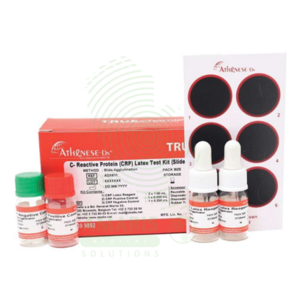
CRP Test Kit
CRP Test Kit (C-Reactive Protein) is a quantitative or semi-quantitative immunoassay (latex agglutination, turbidimetry, nephelometry, ELISA, or rapid immunochromatographic) for detecting C-reactive protein in blood, an acute-phase reactant produced in response to inflammation. Rapid rise within 6-12 hours, peaks at 24-48 hours, half-life 19 hours. Normal <5-10 mg/L; mild elevation 10-100 mg/L (viral infections, autoimmune flares); moderate 100-200 mg/L (bacterial infections); marked >200 mg/L (sepsis, major trauma). High-sensitivity CRP (hs-CRP) detects 0.1-10 mg/L for cardiovascular risk stratification (<1 mg/L low risk, 1-3 mg/L moderate, >3 mg/L high). Primary clinical applications include detection and monitoring of inflammation, diagnosis and management of infections (differentiating bacterial from viral), assessment of autoimmune disease activity, post-surgical monitoring for complications, cardiovascular risk screening (hs-CRP), monitoring rheumatic fever and inflammatory conditions, and neonatal sepsis evaluation. Critical safety precautions include proper sample handling (universal precautions), quality control verification, awareness of non-specific elevation, prozone phenomenon in very high concentrations, and clinical correlation for interpretation. Essential test for evaluating inflammation across all medical specialties.
ESR Tubes Glass
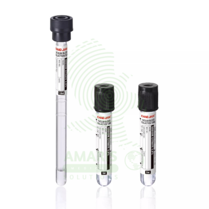
ESR Tubes Glass are specialized glass tubes (300 mm length, 2.55 mm bore diameter, graduated 0-200 mm in 1 mm intervals) meeting ICSH and CLSI Westergren specifications for measuring erythrocyte sedimentation rate, a non-specific marker of inflammation. Used with citrated or diluted EDTA blood, filled to exactly 200 mm, placed vertically in Westergren stand, and read at 60 minutes. Primary clinical applications include determination of ESR for diagnosing and monitoring inflammatory conditions (infections, autoimmune disorders, malignancies), monitoring disease activity in rheumatoid arthritis, assessment of temporal arteritis and polymyalgia rheumatica, evaluation of infection and inflammatory response, screening for occult inflammatory conditions, and monitoring chronic inflammatory diseases (SLE, IBD, vasculitis). Critical safety precautions include handling glass tubes carefully to avoid breakage (sharps hazard), proper disposal in sharps containers as biohazardous waste, ensuring vertical alignment and constant temperature (18-25°C), reading at exactly 60 minutes, and avoiding air bubbles and vibration. Essential consumable for reference Westergren ESR testing in clinical laboratories.
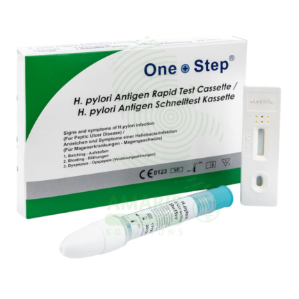
H.pylori test kits (Antigen)
H.pylori test kits (Antigen) are Class II medical devices (FDA-cleared/CE-marked) for rapid immunochromatographic or enzyme immunoassay detection of Helicobacter pylori antigens in stool specimens, enabling non-invasive diagnosis of active infection and post-treatment eradication confirmation. Test principles use monoclonal or polyclonal antibodies specific to H. pylori antigens, producing visible results in 10-20 minutes (rapid tests) or 1-2 hours (EIA). Sensitivity 90-98%, specificity 92-99% compared to gold standard methods. Primary clinical applications include rapid diagnosis of H. pylori infection in patients with dyspepsia, initial screening in uncomplicated dyspepsia (test-and-treat strategy), confirmation of active infection before therapy, post-treatment eradication confirmation (4-6 weeks after therapy), pediatric testing (preferred non-invasive method), screening in high-risk populations (family history of gastric cancer), and monitoring in patients with peptic ulcer disease or MALT lymphoma. Critical safety precautions include mandatory washout period (4 weeks off antibiotics, 2 weeks off PPIs) to avoid false negatives, proper specimen handling (universal precautions), refrigerated storage of kits (2-30°C), quality control verification, and disposal as biohazardous waste. Essential non-invasive tool for H. pylori management in gastroenterology and primary care.
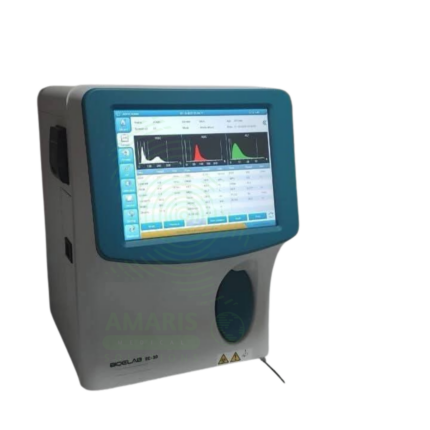
Hematology Analyzer
A Hematology Analyzer is a compact, automated benchtop analyzer designed to perform Complete Blood Counts (CBC) with a 3-part white blood cell differential. Utilizing impedance and photometric methods, it delivers rapid, precise results for red blood cells, white blood cells, platelets, and key indices from a small blood sample. Ideal for physician offices, small clinics, and satellite laboratories, it enables in-house hematology testing with walk-away operation. Its automated flagging system highlights abnormal results that require further manual smear review. As a core instrument for initial hematological screening, it combines efficiency with essential diagnostic capability for routine patient management.
Hepatitis B Test Kit
The Hepatitis B Test Kit is an in vitro diagnostic device for the detection of Hepatitis B virus markers in human blood samples, available in rapid, ELISA, and molecular formats. It detects specific antigens (HBsAg) and antibodies (anti-HBc, anti-HBs) to diagnose acute and chronic infection, monitor treatment response, screen blood donations, and determine immunity status. With high sensitivity (>98%) and specificity (>99%), it provides results in 15 minutes to 4 hours depending on format. Essential applications include prenatal screening to prevent mother-to-child transmission, evaluation of acute hepatitis, pre-immunosuppression screening, and occupational health testing. All patient samples must be handled with universal precautions, and testing must include appropriate quality controls.
Rota virus
Rota virus rapid tests are immunochromatographic assays (CE-marked, FDA-cleared for some brands) for qualitative detection of rotavirus group A antigens in stool specimens, providing results in 10-20 minutes. The test principle uses monoclonal or polyclonal antibodies against rotavirus antigens (typically VP6 protein) immobilized on membranes, capturing antigens that form a visible line. Sensitivity 90-98%, specificity 95-99% compared to ELISA or PCR. The kit includes individually foil-wrapped test devices, sample diluent, and collection applicators. Primary clinical applications include rapid diagnosis of acute gastroenteritis in children (rotavirus is the leading cause of severe diarrhea in under-5 children), outbreak investigation in daycare centers and schools, hospital infection control and isolation precautions, differential diagnosis of acute diarrhea (viral vs. bacterial vs. parasitic), vaccine effectiveness monitoring and surveillance, assessment of dehydration risk, and evaluation of immunocompromised patients. Critical safety precautions include proper specimen collection (clean container, avoid contamination), biological hazard precautions (all stool potentially infectious), quality control verification, and awareness that a negative test does not rule out other causes of gastroenteritis. Essential tool for pediatric gastroenteritis management, infection control, and public health surveillance.