Dermatoscope and Magnifiers
 Diagnostic Kits
 Vital Signs Monitors
 Stethoscopes and Accessories
 Otoscopes, Ophthalmoscopes, and Retinoscopes
 Reflex Hammers and Neurological Tools
 Scales and Measuring Devices
 Spirometers and Pulmonary Function Tests
 Electrosurgical Units and Accessories
 Cutting Instruments
 Grasping and Holding Instruments
 Hemostatic Instruments
 Specialized Surgical Sets
 Single-Use Procedure Trays and Packs
 Surgical Drapes, Gowns, and Covers
 Tissue Unifying Instruments
 Radiation Protection
 X-Ray Machines and Accessories
 Ultrasound Systems and Probes
 MRI and CT Scanners
 Radiology Consumables
 Bone Densitometers
 Fluoroscopy Equipment
 Imaging Tables and Positioning Aids
 Microscopes and Accessories
 Centrifuges and Separators
 Analyzers
 Incubators and Ovens
 Pipettes, Dispensers, and Lab Glassware
 Refrigerators, Freezers, and Storage Units
 Lab Consumables
 Sterilizers and Autoclaves for Lab Use
 Multi-Parameter Monitors
 Ventilators and Respiratory Support Devices
 Defibrillators and AEDs
 Infusion Pumps and IV Systems
 Patient Warmers and Cooling Devices
 Central Monitoring Stations
 Accessories
 Anesthesia Machines and Workstations
 Oxygen Concentrators and Delivery Systems
 Nebulizers and Inhalers
 CPAP/BiPAP Machines
 Airway Management
 Anesthesia Masks, Circuits, and Bags
 Humidifiers and Heaters
 Respiratory Therapy Accessories
 First Aid Kits and Cabinets
 Emergency Resuscitation Equipment
 Trauma Supplies
 Emergency Carts and Crash Carts
 Burn Care Products
 Bleeding Control
 Automated External Defibrillators (AEDs)
 Transport and Evacuation
 Wheelchairs and Accessories
 Walkers, Crutches, and Canes
 Prosthetics and Orthotics
 Physical Therapy Equipment
 Transfer Devices
 Bathroom Safety
 Orthopedic Traction and Tables
 Hot/Cold Therapy Packs and Units
 Beds and Mattresses
 Chairs and Stools
 Tables
 Cabinets and Storage
 Privacy Screens & Curtains
 Stands and Racks
 Linens and Textiles
 Lighting
 Autoclaves and Sterilizers
 Ultrasonic Cleaners
 Disinfectant Solutions and Wipes
 Sterilization Pouches, Wraps, and Indicators
 Instrument Trays and Containers
 UV and Ozone Disinfection Devices
 Washer Disinfectors
 Wound Care
 Gloves
 Masks and Respirators
 Catheters and Tubing
 Swabs, Applicators, and Sponges
 Incontinence Products
 Personal Protective Equipment (PPE)
 Dental Chairs and Units
 Handpieces and Burs
 Instruments
 Consumables
 Sterilization for Dental Use
 Orthodontic Supplies
 Endodontic Tools
 Slit Lamps and Tonometers
 Lensometers and Phoropters
 Ophthalmic Surgical Instruments
 Eyewear Frames and Lenses
 Contact Lens Supplies
 Vision Testing Charts and Devices
 Eye Care Consumables
 Laser Systems for Eye Care
 ENT Exam Chairs and Tables
 Endoscopes
 Audiometers and Hearing Tests
 ENT Instruments
 Nasal and Throat Packs
 Hearing Aids and Accessories
 Otology Supplies
 Fetal Dopplers and Monitors
 Delivery Beds and Tables
 Gynecological Instruments
 Neonatal Incubators and Warmers
 Breast Pumps and Accessories
 Contraceptive Devices
 Maternity Supports and Pads
 Neonatal Consumables
 Cystoscopes and Urethroscopes
 Dialysis Machines and Supplies
 Urological Catheters and Bags
 Lithotripters
 Prostate Treatment Devices
 Urinary Incontinence Products
 Kidney Stone Management Tools
 Consumables & Disposables
 EEG and EMG Machines
 Neurosurgical Instruments
 Nerve Stimulators
 Headrests and Positioning Aids
 Lumbar Puncture Kits
 Seizure Monitoring Devices
 Consumables
 Rehabilitation for Neurological Conditions
 ECG Machines and Accessories
 Holter Monitors
 Stress Test Systems
 Pacemakers and Defibrillator Accessories
 Vascular Access Devices
 Cardiac Catheters and Guidewires
 Blood Flow Meters
 Consumables
 Orthopedic Instruments
 Casts, Splints, and Padding
 Joint Replacement Supplies
 Prosthetic Limbs and Components
 Bone Grafts and Substitutes
 Traction Devices
 Orthopedic Braces and Supports
 Rehabilitation Aids for Orthopedics
 Home Oxygen Therapy
 Hospital Beds for Home Use
 Mobility Aids
 Bathroom and Daily Living Aids
 Wound Care for Home
 Monitoring Devices
 Enteral Feeding Pumps and Tubes
 Hand Sanitizers and Dispensers
 Face Shields and Goggles
 Isolation Gowns and Suits
 Biohazard Waste Containers
 Air Purifiers and HEPA Filters
 Surface Disinfectants
 Sharps Containers
 Protective Barriers
 Cardiovascular & Endurance Training
 Strength Training & Weightlifting
 Functional Training & Core Conditioning
 Physical Therapy & Rehabilitation
 Sports & Outdoor Recreation
 Gym Flooring & Facility Equipment
 Fitness Monitoring & Accessories
 Kids & Novelties
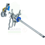
Hysteroscope
WhatsApp Order
A Hysteroscope is a specialized endoscope used for visualization and treatment of the uterine cavity. Diagnostic hysteroscopy provides direct examination of the endometrium for evaluation of abnormal bleeding, infertility, and recurrent pregnancy loss. Operative hysteroscopy enables minimally invasive treatment of polyps, fibroids, adhesions, and uterine septa through the natural orifice of the cervix. Essential for gynecology and reproductive medicine, hysteroscopy offers accurate diagnosis and effective treatment with preservation of fertility and rapid recovery.
Description
Hysteroscope
PRIMARY CLINICAL & DIAGNOSTIC USES
1. Diagnostic Visualization of the Uterine Cavity
-
Primary Use: Provides direct, magnified visualization of the uterine cavity, endometrium, and tubal ostia for diagnostic evaluation of abnormal uterine bleeding, infertility, recurrent pregnancy loss, and suspected intrauterine pathology. The hysteroscope is inserted through the cervix to examine the endometrial cavity.
-
How it helps: For the gynecologist and reproductive endocrinologist, the hysteroscope transforms the uterine cavity from a hidden, inaccessible space into a clearly visualized structure—revealing polyps, fibroids, adhesions, septa, and other abnormalities that cause bleeding, pain, and infertility. For the patient with abnormal bleeding or recurrent miscarriage, hysteroscopy provides the definitive diagnosis that guides treatment and offers hope for successful pregnancy.
2. Operative Hysteroscopy for Intrauterine Pathology
-
Primary Use: Enables minimally invasive surgical treatment of intrauterine pathology including polypectomy, myomectomy, adhesiolysis, and septum resection. The operative hysteroscope includes a working channel for passage of resectoscopes, graspers, scissors, and other surgical instruments.
-
How it helps: For the gynecologic surgeon, the operative hysteroscope provides the ability to treat intrauterine pathology through the natural orifice of the cervix—removing polyps, resecting fibroids, dividing adhesions, and correcting uterine anomalies without abdominal incisions. For the patient, hysteroscopic surgery means preservation of the uterus, avoidance of open surgery, faster recovery, and improved fertility outcomes.
3. Endometrial Biopsy and Tissue Sampling
-
Primary Use: Allows directed biopsy of suspicious endometrial lesions under direct visualization. Biopsy forceps passed through the hysteroscope obtain tissue samples for histopathological examination, ensuring accurate sampling of abnormal areas.
-
How it helps: For the gynecologist, directed hysteroscopic biopsy provides precise sampling of suspicious lesions—increasing diagnostic accuracy compared to blind biopsy techniques. For the patient with suspected endometrial pathology, targeted biopsy ensures that abnormalities are sampled and diagnosed accurately.
4. Tubal Cannulation and Patency Assessment
-
Primary Use: Used to assess tubal patency and perform tubal cannulation for proximal tubal obstruction. The hysteroscope allows visualization of the tubal ostia and passage of catheters or guidewires to open obstructed tubes.
-
How it helps: For the reproductive endocrinologist, hysteroscopic tubal cannulation offers a minimally invasive approach to treat proximal tubal obstruction—potentially restoring fertility without the need for more invasive procedures. For the patient with tubal factor infertility, this procedure offers hope for natural conception.
5. Foreign Body Removal
-
Primary Use: Used to visualize and remove intrauterine foreign bodies including retained intrauterine devices, retained products of conception, and surgical debris. The hysteroscope provides direct visualization for grasping and retrieval instruments.
-
How it helps: For the gynecologist, hysteroscopic foreign body removal enables safe, complete extraction of retained IUD fragments or products of conception—minimizing the risk of infection, bleeding, and future fertility impairment. For the patient, hysteroscopic removal offers a minimally invasive solution to a potentially serious problem.
SECONDARY & SUPPORTIVE USES
1. Office Hysteroscopy: Miniaturized hysteroscopes for office-based diagnostic procedures without anesthesia.
2. Resectoscopy: Hysteroscopic resection of submucous fibroids, endometrial polyps, and uterine septa.
3. Endometrial Ablation: Hysteroscopic ablation of the endometrium for treatment of menorrhagia.
4. Sterilization Procedures: Hysteroscopic sterilization techniques such as Essure placement.
5. Preoperative Assessment: Mapping of intrauterine pathology before surgical planning.
6. Postoperative Evaluation: Assessment of uterine cavity after myomectomy, septum resection, or adhesiolysis.
KEY PRODUCT FEATURES
1. BASIC IDENTIFICATION ATTRIBUTES
-
Device Type: A specialized endoscope designed for visualization and treatment of the uterine cavity.
-
Designation: Hysteroscope, Diagnostic Hysteroscope, Operative Hysteroscope, Resectoscope.
-
Types:
-
Diagnostic Hysteroscope: Small diameter (2-4 mm) for office-based examination.
-
Operative Hysteroscope: Larger diameter (5-9 mm) with working channel for instruments.
-
Resectoscope: Specialized operative hysteroscope for resection of fibroids and polyps.
-
Flexible Hysteroscope: Flexible shaft for improved navigation of the uterine cavity.
-
-
Key Components:
-
Telescope: Rod lens system with angled optics.
-
Sheath: Outer sleeve with inflow/outflow channels.
-
Light Source: Fiberoptic or LED illumination.
-
Working Channel: Passage for instruments and fluid.
-
Camera: High-definition camera for image capture.
-
2. TECHNICAL & PERFORMANCE PROPERTIES
-
Diameter: 2-9 mm depending on type.
-
Viewing Angle: 0°, 12°, 30°, or 70° optics.
-
Working Channel: 1-3 mm for instrument passage.
-
Fluid Management: Continuous flow or passive irrigation.
-
Resolution: HD and 4K options.
-
Sterilization: Steam autoclave or low-temperature sterilization.
3. PHYSICAL & OPERATIONAL PROPERTIES
-
Construction: Precision-machined stainless steel or flexible fiberoptic.
-
Ergonomics: Designed for comfortable single-handed operation.
-
Sterilization: Autoclavable.
-
Portability: Office-based systems for outpatient procedures.
4. SAFETY & COMPLIANCE ATTRIBUTES
-
Regulatory Status: Class II medical device regulated by FDA.
-
Biocompatibility: Materials safe for intrauterine use.
-
Fluid Management: Systems to prevent fluid overload.
-
Electrical Safety: Compliant for use with electrosurgical generators.
5. STORAGE & HANDLING ATTRIBUTES
-
Storage: Store in protective cases; protect lenses from damage.
-
Cleaning: Thorough cleaning after each use; ultrasonic cleaning recommended.
-
Sterilization: Steam autoclave per manufacturer instructions.
-
Inspection: Inspect for lens damage, scratches, or misalignment before use.
6. LABORATORY & CLINICAL APPLICATIONS
-
Primary Application: Diagnosis and treatment of intrauterine pathology.
-
Clinical Role: Essential in gynecology, reproductive endocrinology, and infertility.
SAFETY HANDLING PRECAUTIONS
1. SAFETY PRECAUTIONS
-
Fluid Management: Monitor fluid deficit to prevent fluid overload syndrome.
-
Cervical Dilation: Gentle dilation to prevent cervical injury.
-
Sterility: Ensure sterility before intrauterine use.
-
Distension Medium: Use appropriate distension medium for procedure.
2. FIRST AID MEASURES
-
Uterine Perforation: If perforation occurs, stop procedure; assess for bleeding; consult accordingly.
-
Fluid Overload: If signs of fluid overload, stop procedure; administer diuretics; monitor.
3. FIRE FIGHTING MEASURES
-
Flammability: Metal components are non-flammable; light cables may burn.
-
Extinguishing Media: Use water, foam, or CO₂ as appropriate for surrounding materials.
Related products
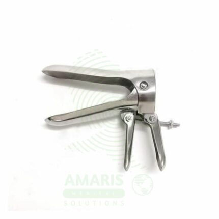
Cusco Speculum
A Cusco speculum is a hinged, bivalve instrument used to separate the vaginal walls for examination of the vagina and cervix. It is the standard tool for performing Pap smears, visual diagnostics, and various gynecological procedures. Available in reusable metal or single-use plastic forms and in multiple sizes, its self-retaining lock allows for hands-free operation. Safe and effective use requires gentle insertion with lubrication, selection of the correct size, careful unlocking before removal, and strict adherence to high-level disinfection or sterilization protocols for reusable models. It is an indispensable instrument in women's health care.
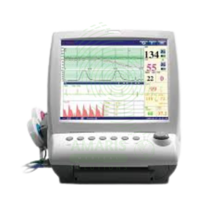
Fetal & Maternal Monitor
A Fetal & Maternal Monitor is an electronic monitoring system that simultaneously records fetal heart rate (via external ultrasound or internal scalp electrode), uterine contractions (via external tocodynamometer or intrauterine pressure catheter), and maternal vital signs (blood pressure, heart rate, oxygen saturation). Used for antepartum and intrapartum fetal assessment, non-stress tests, contraction stress tests, and monitoring of high-risk pregnancies including gestational diabetes, hypertensive disorders, and multiple gestations. Features high-resolution display, continuous paper recording, alarm systems for fetal distress, and data storage for medical records. External monitoring is non-invasive for routine use; internal monitoring provides more accurate data but requires ruptured membranes and cervical dilation. Class II medical device requiring FDA clearance and qualified interpretation of fetal heart rate patterns. Essential for assessing fetal well-being and guiding obstetric interventions.
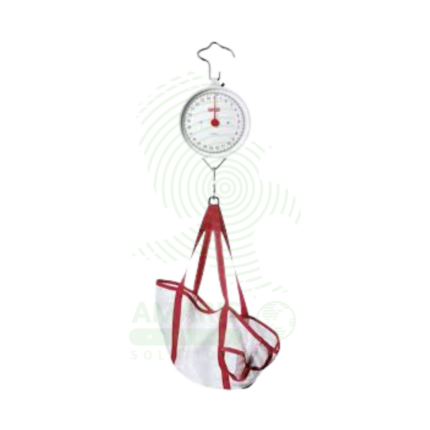
Infant Hanging Scale
An Infant Hanging Scale is a Class II medical device (mechanical spring/balance or digital load cell) designed for accurate weight measurement of neonates and infants up to 2-3 years (capacity 10-25 kg, accuracy ±5-10 g) using a suspended sling or pan. Features include soft, washable fabric sling with head support and leg openings, tare function to zero out sling weight, mechanical dial or backlit digital display, hold function for active infants, test-weighing mode for breastfeeding assessment (digital models), and portable carrying case. Primary clinical applications include newborn weight monitoring in hospital nurseries (detecting excessive weight loss), NICU daily weights (tracking growth, calculating fluids), breastfeeding adequacy assessment (test-weighing), pediatric outpatient and well-child visits, home healthcare and visiting nurse services, malnutrition screening in developing world, and research studies requiring precise infant weights. Critical safety considerations include secure hanging from sturdy support (never ceiling tiles), thorough sling inspection before each use, proper infant positioning with head support, tare verification with sling attached, never leaving infants unattended, and strict infection control with clean sling per patient. Essential equipment for accurate infant growth monitoring and nutritional assessment in all healthcare settings serving pediatric populations.
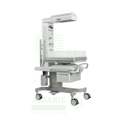
Infant radiant warmer (resuscitaire)
An Infant Radiant Warmer (Resuscitaire) is an open-bassinet, overhead radiant heating system designed for neonatal thermoregulation and resuscitation. It provides servo-controlled infrared warmth to maintain infant body temperature during the critical transition from intrauterine to extrauterine life. The integrated resuscitation platform features built-in oxygen and air sources, suction apparatus, cardiorespiratory monitoring, and storage for resuscitation equipment, ensuring all necessary tools are immediately accessible during newborn stabilization. Essential equipment for every delivery room and neonatal intensive care unit, the radiant warmer supports thermoregulation, resuscitation, and ongoing care while allowing unrestricted access to the infant. Proper temperature probe placement, pre-warming, and alarm response are critical for safe operation.
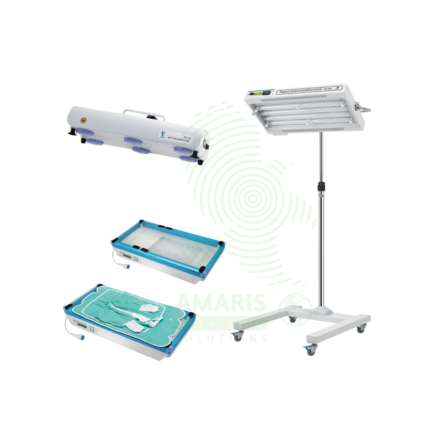
Neonatal Jaundice Treatment Device
A Neonatal Jaundice Treatment Device (phototherapy unit) is a Class II medical device that delivers therapeutic blue light (450-470 nm) to treat hyperbilirubinemia in newborns, preventing bilirubin-induced neurological damage (kernicterus). Available as overhead units (mobile stands with LED or fluorescent lamps), fiberoptic systems (Biliblanket) with illuminated pads placed against the infant's skin, and combination systems for intensive therapy. Irradiance ranges from 10-50+ µW/cm²/nm at treatment distance. LED units offer specific wavelength output, minimal heat, long life (10,000-50,000 hours), and energy efficiency. Treatment indications follow AAP hour-specific nomograms based on gestational age, birth weight, postnatal age, and risk factors. Essential safety requirements include opaque eye shields to protect neonatal retinas, temperature monitoring to prevent hyperthermia, hydration maintenance, and regular irradiance verification. Used in hospital NICUs, newborn nurseries, and (select devices) home care settings for mild jaundice. Critical for preventing permanent neurological sequelae of untreated severe neonatal jaundice.
Phototherapy Machine
A Phototherapy Machine is a therapeutic light delivery system used to treat neonatal hyperbilirubinemia by converting toxic, fat-soluble bilirubin into water-soluble isomers that can be excreted. Using blue-spectrum light from LED, fluorescent, or fiber optic sources, it reduces bilirubin levels non-invasively, preventing kernicterus and permanent neurological damage. Available in overhead, ceiling-mounted, and fiber optic blanket configurations, it is standard equipment in hospital nurseries and NICUs, with portable units available for home use. Safe operation requires proper eye protection, temperature monitoring, and verification of adequate light output.

Pinnard Fetoscope (plastic/metal)
A Pinnard Fetoscope (Pinard horn) is a mechanical acoustic horn (150-200 mm length, 40-60 mm bell) for auscultation of fetal heart sounds through the maternal abdomen, available in traditional metal (stainless steel/chrome-plated, 150-300g, autoclavable, durable, requires warming) and modern plastic (medical-grade polymer, 30-80g, lightweight, unbreakable, economical, not autoclavable) versions. The flared bell collects fetal heart sounds, concentrating them through the tapered horn to the single earpiece for mechanical amplification (10-20 dB). Primary clinical applications include intermittent fetal heart rate monitoring during prenatal care and labor in low-risk pregnancies, confirming fetal viability, determining fetal presentation and position through point of maximal intensity, monitoring multiple gestations, and providing non-interventive fetal assessment in natural childbirth, home birth, and resource-limited settings. Class I medical device. Requires significant practitioner training and experience; reliable after 18-20 weeks gestation; limited by maternal obesity, anterior placenta, and ambient noise. Critical safety considerations include infection control (metal autoclavable, plastic chemical disinfection only), gentle abdominal pressure, maternal positioning, and recognition that Pinnard is not a replacement for electronic monitoring in high-risk pregnancies.